With 6 text-figura Printed in Great Britain
THREE PARALLEL CONDUCTING SYSTEMS IN THE
STALK OF A HYDROID
BY ROBERT K. JOSEPHSON
Department of Zoology, University of Minnesota, Minneapolis
(Received 30 May 1964)
INTRODUCTION
The concept of a single conducting system in coelenterates, represented by 'the nerve net', is being replaced by the realization that there are often multiple conducting systems co-existing in the same tissue. Romanes, in 1877, demonstrated two kinds of conduction in the bell of the jellyfish Aurelia which were distinguished by having different thresholds, conduction velocities, and behavioural correlates. More recent work by Bozler (1926a, b) and Horridge (1955, 1956a, b) make it appear that multiple conducting systems in the subumbrellar epithelium is a general feature of coelenterate medusae. Multiple conducting systems are also known for coelenterate polyps. Anemones, the most studied coelenterate polyps, show rapidly conducted responses, which can be ascribed to activity in a nerve net, as well as very slowly conducted waves of muscle contraction (e.g. Batham & Pantin, 1954). Behavioural evidence indicates that there are two conducting systems in the column and oral disk of
Cerianthus (Horridge, 1958), in the oral disk of the alcyonarian Heteroxenia (Horridge,
1956c), and in the coenosarc of the hydroid Hydractinia echinata (Josephson, 1961 a). Mackie (i960) has clearly demonstrated two histologically distinct nerve plexuses in the same epithelium in the chondrophore Velella which should be regarded as a polyp (Mackie, 1959). Electrical recording indicates that there are at least two conducting systems in the column of Hydra (Passano & McCullough, 1962, 1963), even though histologically there appears to be but a single complete nerve plexus (Semal-Van Gansen, 1952). This paper will give evidence from electrical recording that there are three parallel but distinct conducting systems in the stalk of the hydroid
Tubularia.
MATERIALS AND METHODS
The animals used in this study were Tubularia larynx Ellis & Solander,* collected from logs and pilings in the northern end of the Cape Cod Canal. Tubularia is a large hydroid; the hydranth is 2-3 mm- long and the stalk can be several cm. long. The hydranth and stalk are joined by a short, contractile neck region. The hydranth has two sets of tentacles, the large proximal tentacles and the smaller distal tentacles which surround the mouth. The distal tentacles and mouth are borne on a contractile proboscis.
Tubularia polyps were fastened to a cork platform on the bottom of a dish of sea
water with staples made from small insect pins. Cold water was passed through a
glass coil in the dish to maintain the sea water surrounding the polyp at 190 C. The stalk was stimulated electrically through silver wires, insulated to the tips and placed one on each side of the stalk. Unless otherwise stated, the stimuli were square pulses of 1 msec, duration. Electrical potentials were recorded between one or two electrodes in or on the polyp and an indifferent electrode in the sea water surrounding the polyp. The potentials were amplified with capacitor-coupled amplifiers having long time-constants, and the final display was on an oscilloscope or pen-writer.
The stalk of Tubularia is covered with a thick perisarc, so recording electrodes which penetrate the stalk must be strong and rigid. Micro-electrodes of platinum-indium or stainless steel, fixed in a manipulator, were found to be satisfactory. The micro-electrodes were etched electrolytically (Green, 1958; Wolbarsht, MacNichol & Wagner, i960) and insulated with lacquer. The indifferent electrode used with steel micro-electrodes was a coil of silver wire; the indifferent electrode used with platinum-iridium micro-electrodes was also of platinum-indium. The platinum-platinum-iridium electrodes seemed to offer no advantages over the steel, and as the latter are easier to make they were used in most of the experiments. Freshly made metal micro-electrodes were often too noisy to record potentials of the small size found in the Tubularia stalk. Pushing the tip of the electrode into a piece of cork usually reduced this noise, pre-sumably by removing some of the insulation from the tip and reducing the electrode resistance.
The hydranth is quite mobile and recording from it presents some special difficulties. Electrodes fixed in a manipulator are not satisfactory as the animal either pulls off the electrodes or they tear the hydranth tissue. Some successful recordings were made with electrodes etched from small insect pins (minuten nadeln) soldered to a loop of very thin copper wire. These flexibly mounted electrodes were inserted into the hy-dranth by hand. But the most satisfacory method found for recording from the hydranth was by means of suction electrodes, similar to the ' minute' suction electrodes used for recording from the toad heart by Kanno (1963). These electrodes were made by drawing plastic tubing (Tygon) over a small flame until it ended in a flexible, hollow filament. A silver wire was mounted in the tubing near the drawn end and the other end of the tubing was attached to a small syringe. Sea water was drawn into the tubing with the syringe until it reached the silver wire. The tip of the filament was then placed against a portion of the hydranth and the plunger of the syringe was drawn back until the suction was just great enough to hold the tip in place. The suction generally used was 40-50 mm. Hg, as calculated from the change in the gas volume of the syringe and tubing as the plunger was withdrawn. Potentials were recorded between the silver wire in the tubing and an indifferent electrode in the bath. The suction electrodes used had internal diameters at the tip of 30-90fi. With such electrodes it is possible to record from any part of the hydranth, including bases, sides and tips of the proximal and distal tentacles. Because of their flexibility, the electrodes freely follow the hydranth as it contracts.
when the suction is reduced. Such potentials can be troublesome in long-term recordings, but were not a nuisance in the present study as attention was given only to evoked responses, coming in a short period after a stimulus.
RESULTS
A. The distal opener system
Stimuli of sufficient intensity to the stalk of Tubularia evoke a synchronous de-pression or opening of the distal tentacles of the hydranth. The conducting system in the stalk which mediates this response will be termed the distal opener system (DOS). Often potentials are recorded from an electrode in the stalk or hydranth which have precisely the same threshold as does the tentacle opening, even if the stimulus duration is varied from o-oi to 50 msec. These potentials are an indication of activity in the DOS, and will be termed DOS pulses (DOSP's).
Distal tentacle opening and DOSP's are very stable responses. The threshold may change considerably during a long experiment but several threshold determinations made within a short interval, for example 1 min. apart, seldom vary from each other by more than 10%. Repetitive firing of the DOS has been seen only twice. In each case the animals were being stimulated at a low frequency, 1 per 5 and 1 per 10 sec., with shocks of not much above threshold intensity. After a number of stimuli had been given DOSP's began appearing in short bursts following each stimulus rather than the previous one pulse per stimulus. This repetitive firing lasted for only a few stimuli. Repetitive firing was seen at no other time, even when the stimulus intensity used was twice or three times threshold. When the stimulus frequency is greater than about 1 per 2 sec., the distal tentacles remain strongly depressed and responses to each stimulus are not seen. DOSP's follow each stimulus at frequencies up to 2 per sec. in most animals and in some animals the DOSP's will follow at least a few stimuli at 10 per sec, after which they appear after every other stimulus and still later erratically.
The DOS in the stalk
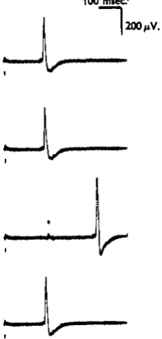
DOSP's, recorded with a metal micro-electrode in the stalk, can be as large as 200 /iV., but are more generally less than 50 /iV. (Fig. 1 A, B). They are usually larger in the stalk just below the hydranth than more proximally. Successful recording of DOSP's often depends on critical electrode placement; a small change in the electrode position can greatly diminish or enhance the size of the recorded potential. The durations of the larger DOSP's recorded were between 5 and 15 msec. The durations of the usual small DOSP's can not be determined with any certainty, but appear to be in this range.
electrode more removed from the stimulating electrodes. These results suggest that the DOS in the stalk, rather than being a single conducting system, is a series of parallel conducting systems. The double peaks then would be due to activity in two adjacent subsystems which were slightly out of phase, and the changing intervals between peaks at different points in the stalk would be a result of different conduction velocities in the parallel subsystems.
nftn
•
.U
iMJ
J
LMJ
Fig. 1. DOSP's in the stalk and hydranth. (A) DOSP's as usually recorded in the stalk. (B) Unusually large DOSP recorded from the neck region. (C) Single DOSP's in one recording channel and double pulses in the other. (D) DOSP's from the proximal tentacles. A-C were recorded with metal micro-electrodes and each is several superimposed sweeps. D is a single sweep, recorded with suction electrodes. The dots (•) in this and following figures indicate the position of DOSP's where these are not obvious. Calibration: 200/*V. and 100 msec.
The absolute value of the difference in conduction velocities in the two directions averaged 0-4 cm./sec. It would seem, from this admittedly small sample, that con-duction occurs equally well in either direction. In the animal showing double DOSP's, the conduction velocity was computed separately for the first and second pulse to appear at each recording site. The conduction velocities for the faster pulse were 16-8 and 17-5 cm./sec. in the directions toward and away from the hydranth respec-tively, and those for the slower pulse 15-1 and 14-9 cm./sec. The parallel subsystems postulated to account for the double pulses also appear to conduct essentially equally well in either direction.
The DOS in the hydranth
The DOS is found throughout the hydranth. DOSP's have been recorded with suction electrodes from the proboscis and from the sides and tips of both proximal and distal tentacles (Fig. 1D). They have even been recorded from an electrode inserted through the mouth and ending somewhere on the inside of the proboscis. The DOSP's are usually between 50 and 100 fiV. when recorded with a suction electrode and, as in the stalk, are 5-15 msec, in duration.
When the recording electrode is on a distal tentacle or on the proboscis at the base of the distal tentacles, the DOSP is followed by a slow, usually positive potential, up to 800 fiV. in amplitude and 200 msec, in duration. This slow wave shows marked antifacilitation. If the stimulus frequency is greater than about 1 per 15 sec, successive potentials become progressively smaller (Fig. 2 A). It had been earlier noticed that distal tentacle opening was also an antifacilitating response. When using stimulus frequencies between 1 per 5 and 1 per 20 sec. the tentacle opening is most pronounced to the first stimulus and is less vigorous to subsequent stimuli. This similar behaviour of the slow wave and the distal tentacle opening was a first indication that the slow wave might be a result of activity in the tentacle-opening musculature.
Some experiments were carried out with excess Mg2"1" to further establish the relation between the slow wave and the tentacle-opening response. A first effect of excess Mg2+ in coelenterates appears to be the blockage of neuromuscular transmission (Ross & Pantin, 1940; Bullock, 1943). A polyp was stimulated at 1 shock per 20 sec. After several minutes of stimulation isosmotic MgCl2 at 19° C. was added to the sea water surrounding the polyp until it made 25 % of the final solution. This was accom-plished in about 10 sec. The stimulation was continued, and the DOSP, slow wave, and tentacle opening were all observed. This experiment was repeated three times, each time with essentially the same result. Tentacle opening and the slow wave began to decline immediately after the addition of MgCl2; after 60-80 sec. neither could be detected (Fig. 2 B). The DOSP's continued unchanged for 6-8 min. and then suddenly stopped appearing. Excess Mg2* rather effectively separates the DOSP from the following slow wave. The most likely explanation for the sensitivity of the slow wave and the relative insensitivity of the DOSP to excess Mg2* is that the DOSP is a result of nerve activity and the slow wave a muscle action potential, with excess magnesium blocking the link between the two.
contracts. Conduction times between the hydranth base and the proboscis were measured in five animals. Five determinations were made on each animal. The distances between the recording electrodes were measured when the proboscis was fully extended. The average conduction velocity through the hydranth was 10 cm./sec. and the range in the means for individual animals was 8—13 cm./sec. The fact that the conduction velocity computed for the hydranth is lower than that for the stalk does not necessarily mean that conduction in the DOS is slower in the hydranth. It is not known where the DOS lies in the hydranth. If it is parallel to the hydranth surface, as is most likely, it must move laterally, away from the polyp midline, as it approaches the proximal tentacles and back toward the midline above the proximal tentacles. Thus the distance between the hydranth base and the tip of the proboscis is probably less than the conduction path length, and using this distance would result in a low value for conduction velocity.
in in
j
200 msec200/iV.
360 380
500/iV. 500 msec.
Fig. 2. DOSP's and the following slow potentials recorded near the base of the distal tentacles. (A) Successive potentials recorded with metal micro-electrodes during stimulation at i per 2-5 sec. Note the constancy of the DOSP and the antifacilitation of the slow potential. (B) Potentials recorded with a suction electrode 20 sec. before and 40, 100, 360, and 380 sec. after the addition of MgClt to the bathing solution. Note the rapid decline of the slow potential and the constancy of the DOSP until it suddenly stops appearing.
the stalk from the base of the distal tentacles. The average latency determined in this manner was 280 msec., with a range in the individual means of 260-310 msec. DOSP's were not recorded in these experiments, but some estimate of the proportion of this latency which is due to conduction time can be made by using conduction velocities determined from other animals. If it is assumed that 2 mm. of the total length represents conduction through the hydranth and 8 mm. conduction through the stalk, the total conduction time should be about 75 msec. This leaves about 200 msec, for neuromuscular delay and the actual mechanical latency. It is clear, even from these rather crude figures, that the tentacle movement is just beginning at the time when the slow wave following the DOSP is subsiding or has completely subsided. It is presumably the relative long latent period which led to the low estimate for the DOS conduction velocity of 3-5 cm./sec, based on the stimulus to response time, in an earlier study (Josephson, 1961a).
B. The slow system
A strong stimulus to a Tubularia stalk often evokes both a DOSP and a second type of potential which is conducted at a considerably lower velocity than the DOSP (Figs. 3, 4). This second potential will be called a slow pulse (SP) and the system in which it is conducted will be termed the slow system (SS). SP's are typically bi-phasic, having a prominent negative phase followed by a usually smaller positive phase. SP's are much larger and longer than DOSP's; the initial negative phase is usually between o-i and 1 mV. in amplitude and 20—70 msec, in duration. The amplitude and form of recorded SP's are not altered by small changes in the position of the recording electrode. SS conduction must be non-decremental, for the size of SP's is not changed as the stimulating electrodes are moved toward or away from the recording electrode. Occasionally SP's show notches or double peaks (Fig. 4), indicating that they are of compound origin. Despite repeated attempts to find a function for the SS, neither behavioural responses of the polyp nor changes in the spontaneous electrical activity of the hydranth have been seen following SS activation.
The SS is very labile. SP's can be predictably demonstrated only in a fresh, unstimulated animal, and usually fail to appear after a few supra-threshold stimuli, even if the stimuli are separated by five minutes or more. The SP threshold in a fresh animal is two to three times that of the DOSP and increases with stimulation. The SS often fires repetitively if the stimulus intensity is increased above its threshold; as many as four SP's have been seen following a single stimulus (Fig. 3). The minimum interval between SP's in a burst is about 100 msec. Because of the lability of the SS it has not been possible to examine the relation between stimulus strength and the number of pulses evoked. Experiments with two recording electrodes in the stalk have shown that not all SP's are conducted throughout the stalk. Single SP's or the later SP's of a burst are sometimes recorded with an electrode near the stimulating electrodes but not at a more distal recording electrode.
The conduction velocity of the SS has been determined from the difference in the time of SP appearance at two recording sites in the stalk. In six animals it was possible to make five or more conduction velocity measurements before the SS stopped
146 ROBERT K. JOSEPHSON
15-9mm,
UGJ
200 msec
Fig. 3. Repetitive firing and non-polarized conduction in the slow system. The stimulus intensity in each case was about 25 % above threshold. The fourth SP in the upper trace of the first record never reached the second recording electrode. Note the decline in SP amplitude and the increasing conduction tune between the recording electrodes through the bursts. Faster portions of the SP's have been retouched.
""1
200 MV.Fig. 4. SP with notch on the falling phase, indicating a compound origin for the pulse.
SS fires repetitively, the second pulse has a lower amplitude and is conducted more slowly than the first pulse (Fig. 3). During conduction velocity measurements, two or more SP's were recorded at both recording sites a total of eighteen times in experi-ments on five different animals. The conduction velocities for the first pulse in this group averaged 5-6 cm./sec. and those for the second pulse 4-4 cm./sec.
[image:9.451.168.285.137.381.2]200 pV.
Fig. 5. Triggered NP's. Judging from the long latency, the NP in the third sweep is spon-taneous and not triggered. Note that the triggered NP's in the other sweeps precede the DOSP seen in the third sweep.
C. The triggering system
148 ROBERT K. JOSEPHSON
triggering is 10 to 30% lower than that of the DOSP's. There must be a third con-ducting system in the stalk, one which triggers NP's. This system will be called the triggering system (TS). No electrical potentials have been recorded which can be definitely correlated with TS activity.
The system which produces the NP's is sensitive to triggering immediately following either a spontaneous NP or a triggered NP. This makes it possible to drive the NP system with a burst of stimuli if these are at a high enough frequency. For example, a burst of stimuli at 1 per sec. can trigger ten to twenty NP's before the NP system ceases to respond, while a burst at 1 per 5 sec. will usually not trigger more than a single NP. Not all bursts of stimuli of appropriate frequency will drive the NP system; the stimuli must begin immediately after a spontaneous NP. If the stimulation is started as late as 5-10 sec. after an NP, usually neither the first nor the following stimuli will trigger NP's. NP's can be driven at frequencies up to 5 per sec. with stalk stimulation, showing that the TS will follow to at least this frequency. The NP bursts produced by such stimulation closely resemble the NP bursts which appear spontaneously, except of course that the driven NP's are at a constant frequency while the NP frequency usually declines during the course of a spontaneous burst.
Often in a cluster of Tubularia, polyps can be found whose stalks appear to fuse proximally, giving a small Y-shaped colony of two hydranths, one on each of the arms of the Y. Such colonies might have arisen through branching during growth, or through the settling and subsequent development of an actinula larva upon an already formed stalk. In many but not all branched preparations there is tissue continuity through the intersection. Whether this continuity is primary, or formed secondarily through the fusion of tissues from two individuals is not known. When there is tissue continuity there is often continuity of the triggering systems of each polyp. A stimulus to one of the arms of the Y can trigger NP's in the hydranth on the other arm of the Y. This shows that conduction in the TS can occur in either direction, for the TS conduction is away from the hydranth on the stimulated arm and toward the second hydranth on the other arm. The occurrence of branched colonies is fortunate for the experimenter for otherwise, short of recording electrical activity from the TS, there would be no way to study TS conduction in both directions. The DOS can also be continuous between two hydranths on a branched preparation, but apparently DOS continuity is less common than TS continuity. In several cases stimulation of one arm of a Y preparation triggered NP's in the hydranth on the other arm even though strong stimulation did not give rise to DOSP's in the opposite arm. The converse, DOS continuity without TS continuity, has not been seen.
one near the point of branching and one on each of the branches somewhat below the recording electrode in each neck region. Further, the conduction velocity away from each hydranth can be determined using the same three stimulating electrode positions by measuring the decrease in triggered NP latency in the opposite hydranth as the stimulating electrodes are moved from a branch to the junction. Single conduction-velocity determinations were made for each polyp, but these are based on the dif-ferences between averages of eight to thirty latency determinations at each stimulating electrode position. The conduction velocity toward the hydranth for the six polyps averaged 17 cm./sec. (range: 13-21 cm./sec.) and the conduction velocity away from the hydranth for the four polyps of the branched preparations also averaged 17 cm./ sec. (range: 15-21 cm./sec).
The triggered NP latency is usually rather variable. The coefficients of variation ( = 100 (standard deviation)/mean) for the twelve sets of latency measurements used in determining the conduction velocity toward the hydranth averaged 7-0. In several animals the latencies have not varied continuously, but have fallen into two discrete groups. For example, fifteen latency determinations were made with the stimulating electrodes 27-9 mm. below the recording electrode in one of the single polyps of the conduction-velocity experiments. Of these, seven were between 157 and 160 msec, and the other eight between 176 and 182 msec. The reasons for this discontinuous distribution are not known.
32 12 28 32
1200 jiV.
200 msec.
Fig. 6. Summation of DOSP's and triggered NP's. The distance (mm.) between the stimu-lating electrodes and the fixed recording electrode is given below each trace. Judging by the short latency, the NP in the first trace is not triggered but spontaneous. When the stimulating electrodes are 12 mm. from the recording electrode the DOSP just precedes the NP; at the greater distances the NP begins before the DOSP.
150 ROBERT K. JOSEPHSON
based on the TS conduction velocity and the distance between the stimulating elec-trodes and the recording electrode, from the actual onset of the NP. This has been done for the six polyps for which conduction velocities were obtained. The delay to the triggered NP, other than that due to conduction in the stalk, averaged 15 msec, (range: 7-33 msec.).
DISCUSSION
It is quite clear that there are at least three conducting systems in the stalk of
Tubularia. But what are the cellular elements of these conducting systems?
Ap-parently there is no information available on the types and distribution of nerve cells in the stalk of Tubularia. Leghissa (1950) reports that there is a single, widely distributed nerve plexus in the hydranth of Tubularia, based on results with staining by silver impregnation. Mackie (1963, personal communication) also finds a single nerve plexus in Tubularia hydranths with reduced methylene blue staining. The effects of excess Mg2"1" on the DOSP indicate that it is of nervous origin, and the distribution of DOSP's throughout the hydranth is in agreement with the wide distribution of the epidermal nerve net. It therefore seems quite likely that the system which conducts DOSP's in the hydranth, and by extension in the stalk as well, is nervous. I can only speculate on the substrates of the other two conducting systems. The TS is similar to the DOS in its conduction velocity, threshold and ability to-follow repetitive stimuli. These similarities suggest that the TS is also nervous. The SS, on the other hand, is rather different from the other two systems. Its threshold is much higher, its conduction velocity is much lower, and it is far more labile. SP's differ from DOSP's in their size, long duration, and insensitivity to small changes in the position of the recording electrode. If the DOS is nervous, it seems likely that the SS is not. Mackie (i960, 1964) gives several examples of tissues in coelenterates in which conduction can be demonstrated but in which nerve fibres have not been found. He suggests that conduction in these cases is by way of cells of the epithelial layer or the muscular bases of epithelial cells. The possibility of epithelial conduction should be considered for the SS of Tubularia, but this problem cannot be solved until the histology of the stalk is better known.
Electrical potentials have been recorded from two other hydroid genera, Hydra (Passano & McCullough, 1962, 1963) and Cordylophora (Josephson, 19616). Two conducting systems have been found in the column of Hydra. One, termed the RP system, conducts small amplitude potentials which appear to be very similar in form to the DOSP and following slow wave as these are recorded from an electrode near the distal tentacles of Tubularia. In Hydra the fast initial component has a duration of 50 msec, or less while the following slow component lasts about 500 msec. It is tempting to regard the RP system of Hydra and the DOS of Tubularia as homologous. It would be most interesting to know if the slow component of the Hydra potentials could be selectively reduced by anaesthetics. The second conducting system of
Hydra produces potentials of large amplitude which immediately precede contraction
of the longitudinal muscles. It is difficult to compare the second Hydra system with those of the Tubularia stalk, as the Tubularia stalk apparently lacks muscle elements and it is likely that part or all of the potential associated with activity in the second
from the stalk of Cbrdylophora although others may be present, for example one which maintains the synchrony of peristaltic waves in the individual hydranths of a colony as found by Fulton (1963). The conducting system of Cordylophora is quite like the slow system of Tubularia, for it conducts slowly (27 cm./sec.), produces large potentials with a long duration, fires repetitively to stimuli whose intensity is not much above its threshold, and is very labile. These similarities put some doubt on the proposed nervous nature of the Cordylophora conducting system.
Perhaps the best parallel to the conducting systems in the Tubularia stalk is found in the subumbrellar epithelium of the scyphozoan Cyanea (Horridge, 19566). Here there are two nerve nets; one, the giant net, controls the symmetrical contraction of the bell and the other, the diffuse net, can increase the frequency of the swimming beat by acting on the marginal ganglia. But there are slow contraction waves as well, which sweep over the muscles with a velocity of about 2 cm./sec. These should be considered as representing a conducting system distinct from the diffuse net for, according to Horridge, activation of the diffuse net is not always accompanied by a slow wave of contraction. Thus there appear to be three conducting systems in the bell of Cyanea, two nervous and one presumably not nervous; just the situation which is here postulated fo r the stalk of Tubularia.
SUMMARY
1. There are three, non-polarized conducting systems in the stalk of Tubularia. These are termed the distal opener system (DOS), the triggering system (TS), and the slow system (SS).
2. The DOS controls opening of the distal tentacles of the hydranth. Activation of the DOS produces a small electric pulse in the stalk which is conducted at about 15 cm./sec. At the base of the distal tentacles this pulse is followed by a slow potential. The slow potential but not the pulse shows antifacilitation and is quickly blocked by excess Mg24", as are distal tentacle movements. This is taken to indicate that the pulse and slow potential are a result of nerve and muscle activity respectively.
3. The TS can trigger the potentials which normally appear spontaneously in the neck region of the hydranth. The TS conducts at about 17 cm./sec and has a lower threshold than the DOS. No electrical correlate of TS activity has been found.
4. Activation of the SS produces large (up to 1 mV.), slowly propagated (about 6 cm./sec.) potentials in the stalk. The SS is very labile. In a fresh animal the SS threshold is two to three times that of the DOS and the SS often fires repetitively to stimuli above its threshold. SS activity has apparently no effect on polyp behaviour or on spontaneous electrical activity in the hydranth.
This work was supported by grant G 23822 from the NSF. I wish to thank Dr G. O. Mackie for assistance and helpful discussions during the course of this study.
REFERENCES
BATHAM, E. J. & PANTIN, C. F. A. (1954). Slow contraction and its relation to spontaneous activity in the sea-anemone Metridtum temle (L.). J. Exp. Biol. 31, 84-103.
BOZLER, E. (1926a). Sinnes- und nervenphysiologisches Untersuchungen an Scyphomedmen. Z.
BOZLER, E. (19266). Weitere Untersuchungen zur Sinnes- und Nervenphysiologie der Medusen: Erregungsleitung, Funktion der RandkSrper, Nahrungsaufnahme. Z. vergl. Physiol. 4, 797—817. BULLOCK, T. H. (1943). Neuromuscular facilitation in scyphomedusae. J. Cell. Comp. Phystol. 23,
251-72.
FULTON, C. (1963). Rhythmic movements in Cordylophora. J. Cell Comp. Phynol. 61, 39—51. GREEN, J. D (1958). A simple micrcelectrode for recording from the central nervous system. Nature,
Lond., 182, 962.
HORRIDGE, G. A (1955) The nerves and muscles of medusae. II. Geryoma probotcidalis Eschscholtz.
J Exp. Bwl. 32, 555-68.
HORRIDGE, G. A. (1956a). The nervous system of the ephyra larva of Aurellia aurita. Quart. J. Micr.
Sci. 97,
59-74-HORRIDGE, G. A. (19566). The nerves and muscles of medusae. V. Double innervation in scyphozoa.
J. Exp. Bwl. 33, 366-83.
HORRIDGE, G. A. (1956c). The responses of Heteroxenia (Alcyonaria) to stimulation and to some in-organic ions. J. Exp. Bwl. 33, 604-14.
HORRIDGE, G. A. (1958) The co-ordination of the responses of Cerianthus (Coelenterata). J Exp. BioL 35. 369-82.
JOSEPHSON, R. K. (1961a). Colonial responses of hydroid polyps. J Exp Biol. 38, 559-77.
JOSEPHSON, R. K. (19616). Repetitive potentials following brief electric stimuli in a hydroid. J. Exp.
Biol. 38,
579-93-JOSEPHSON, R K. (1962). Spontaneous electrical activity in a hydroid polyp. Comp. Biochem Physiol.
s, 45-58.
KANNO, T. (1963). Electrical activity of the atnoventncular conducting tissue of the toad, studied by a minute suction electrode. Jap. J. Physiol. 13, 97—m.
LEGHISSA, S. (1955). L'evoluzione morfologica del tessuto nervoso nei Celenterati fissi. Boll. Zool. 17,. Suppl., 213-53.
MACKIE, G. O. (1959). The evolution of the Chondrophora (Siphonophora-Disconanthae): new evidence from behavioural studies. Trans Roy. Soc Can. 53, Sect. 5, 7-20.
MACKIE, G. O. (i960) The structure of the nervous system in Velella. Quart. J. Micr. Set 101,119—31. MACKIE, G. O. (1964). Analysis of locomotion in a siphonophore colony. Proc. Roy. Soc. B, 159,
366-91.
PASSANO, L. M. & MCCULLOUGH, C. B. (1962). The light response and the rhythmic potentials of hydra. Proc. Nat. Acad. Sri , Wash., 48, 1376-82.
PASSANO, L. M. & MCCULLOUGH, C. B. (1963). Pacemaker hierarchies controlling the behaviour of hydras. Nature, Lond., 199, 1174-75.
ROMANES, G. J. (1877). Further observations on the locomotor system of medusae. Phil. Trans 167, 659-752.
Ross, D. M. & PANTIN, C. F. A. (1940). Factors influencing facilitation in Actinozoa. The action of certain ions. J. Exp. Biol. 17, 61-73.
SEMAL-VAN GANSEN, P. (1952). Note sur le systeme nerveux de l'hydre. Bull. acad. Belg. Cl Set. 38,
718-35-WOLBARSCHT, M. L., MACNICHOL, E. F. Jr. & WAGNER, H. G. (i960). Glass insulated platinum