Original Article
Activation of sonic hedgehog signaling attenuates
oxidized low-density lipoprotein-stimulated brain
microvascular endothelial cells dysfunction in vitro
Xiu-Long Jiang, Ting Chen, Xu Zhang
Department of Neurology, Provincial Clinic College of Fujian Medical University, Fujian Provincial Hospital, Fuzhou 350001, China
Received August 27, 2015; Accepted September 28, 2015; Epub October 1, 2015; Published October 15, 2015
Abstract: The study was performed to investigate the role of sonic hedgehog (SHH) in the oxidized low-density lipo-protein (oxLDL)-induced blood-brain barrier (BBB) disruption. The primary mouse brain microvascular endothelial cells (MBMECs) were exposed to oxLDL. The results indicated that treatment of MBMECs with oxLDL decreased the cell viability, and oxidative stress was involved in oxLDL-induce MBMECs dysfunction with increasing intracel-lular ROS and MDA formation as well as decreasing NO release and eNOS mRNA expression. In addition, SHH
signaling components, such as SHH, Smo and Gli1, mRNA and protein levels were significantly decreased after incubation with increasing concentrations of oxLDL. Treatment with oxLDL alone or SHH loss-of-function significantly
increased the permeability of MBMECs, and overexpression of SHH attenuated oxLDL-induced elevation of perme-ability in MBMECs. Furthermore, SHH gain-of-function could reverse oxLDL-induced apoptosis through inhibition caspase3 and caspase8 levels in MBMECs. Taken together, these results demonstrated that the suppression of SHH in MBMECs might contribute to the oxLDL-induced disruption of endothelial barrier. However, the overexpres-sion of SHH could reverse oxLDL-induced endothelial cells dysfunction in vitro.
Keywords: Sonic hedgehog, brain microvascular endothelial cells, oxidized low-density lipoprotein, blood-brain barrier
Introduction
Brain microvascular endothelial cells (BMECs) have a crucial role in maintaining brain vascular homeostasis, and endothelial dysfunction is recognized as an early event in the pathogene-sis of cerebrovascular diseases such as isch-emic stroke and subarachnoid hemorrhage [1-3]. Moreover, cerebral endothelial cells form-ing the blood-brain barrier (BBB) facilitate the exchange of nutrients and proteins between these compartments, and endothelial cells injury can influence the permeability of BBB [4, 5]. However, a variety of intrinsic and extrinsic factors or disease conditions may affect the physiologies and pathophysiologies of BMECs. A growing body of evidence suggests that oxi-dized low-density lipoprotein (oxLDL) causes atherosclerotic lesions via a process of binding to macrophage scavenger receptors to form lipid-laden foam cells [6]. In vascular smooth
muscle cells, oxLDL has been shown to sup-press cell proliferation and induce cell apopto-sis and DNA damage [7, 8]. Previous reports have shown that oxLDL induces mouse cerebral endothelial cells apoptosis via regulating Bax-mitochondria-caspase protease pathway [9]. However, the molecular mechanisms of oxLDL-induced injury in BMECs have not been clearly delineated.
12821 Int J Clin Exp Pathol 2015;8(10):12820-12828 upregulating tight junction (TJ) proteins in
capil-lary endothelial cells [13]. SHH loss-of-function, its receptor Patched-1 (Ptch-1) suppresses a G-coupled-protein receptor Smoothened (Smo), which is critical for the activation of a transcrip-tion factor Gli-1 that is an important regulator of TJ protein expression and BBB formation. SHH binds and inactivates Ptch-1, which allows Smo to activate Gli-1, subsequently, the TJ protein expression is upregulated and enhances BBB integrity [14]. Intriguingly, interleukin-1β induc -es BBB disruption by downregulating SHH in Astrocytes [15], and recombinant human SHH protein upregulates the expression of ZO-1 and occludin to repair the tight junction and amelio-rate the brain edema and BBB permeability [16].
oxLDL is a proinflammatory cytokine that acts on both endothelial cells and astrocytes to increase BBB permeability [17-20]. However, the mechanisms of BBB disruption by oxLDL have not been fully elucidated. The present study was designed to elucidate the role of SHH in oxLDL-induced impairment to endothelial permeability in primary mouse brain microvas-cular endothelial cells (MBMECs).
Materials and methods
Cell culture
The mouse brain microvascular endothelial cells (MBMECs) were isolated from gender-matched 6-week old C57BL/6 mice and main-tained in RPMI-1640 (Invitrogen, USA) supple -mented with 10% FBS (Invitrogen, USA) at 37°C in a humidified incubator (Thermo, USA), 5% CO2, 95% air atmosphere.
Cell viability detection by CCK8
MBMECs (5.0 × 10³/well) were plated and
treated in 96-well plates (three wells per group) with oxLDL for 24 h. 10 μL of Cell Counting Kit-8 (CCK-8) was added to the cells, and the OD value of the cells was measured at 450 nm using an ELISA reader (BioTek, USA) according to the manufacturer’s instructions.
Caspase3 and caspase8 activity and cell apoptosis assay
MBMECs lysates were prepared and incubated with anti-caspase3 and anti-caspase8. Im- munocomplexes were incubated with peptide substrate in assay buffer for 2 h at 37°C.
Release of p-nitroaniline was measured at 405 nm using an ELISA reader (MD SpectraMax M5, USA) according to the manufacturer’s instruc -tions. Cell apoptosis was assessed by the ter-minal deoxynucleotidyl transferase-mediated deoxyuridine triphosphate nick endlabeling (TUNEL) method, which examines DNA-strand breaks during apoptosis by using BD ApoAlertTM DNA Fragmentation Assay Kit.
Nitric Oxide, ROS, LDH and MDA quantification
MBMECs were plated and treated in 96-well plates and were stimulated with oxLDL for 24 h. Centrifugate to obtain the supernatant, and the level of nitric oxide was measured by nitrite pro-duction using the Griess reagent (Invitrogen, USA) at 540 nm using an ELISA reader (BioTek, USA) according to the manufacturer’s protocol. ROS released by cells was determined by the 2,7-dichlorofluorescin diacetate (DCFH-DA) method using an ROS assay kit according to the manufacturer’s protocol. Cellular cytotoxicity was detected by the LDH assay using a cytotox-icity detection kit (Invitrogen, USA) according to the manufacturer’s protocol. The Biochemical Analysis Kit (Jiancheng Biotechnology Co., Nanjing, China) was used for the measurement of MDA according to manufacturer’s protocol.
Overexpression and small interfering RNA
For the transfection of MBMECs, lentiviral vec-tors harboring SHH was constructed, and the MBMECs were infected. Briefly, MBMECs were cultured in McCoy’s 5α medium containing 10% FBS and when they reached the exponen-tial growth phase, 1.0 × 10⁵ cells per well were
plated in 96 plates. Next, 300 μl complete cul -ture medium, containing recombinant lentivi-ruses, control lentiviruses or McCoy’s 5α medi -um (all containing 6 μg/ml polybrene; Sigma) was added into the plates when the cells reached 50-60% confluence. Two days later, the virus-containing medium was replaced with fresh complete medium.
Real-time PCR
MBMECs RNA extraction was performed according to the TRIzol manufacturer’s protocol (Invitrogen, Carlsbad, CA, USA). Synthesis of cDNAs was performed by reverse transcription reactions with 4 μg of total RNA using moloney
[image:3.612.96.520.74.549.2]murine leukemia virus reverse transcriptase (Invitrogen) with oligo dT (15) primers (Fer-mentas) as described by the manufacturer. The first strand cDNAs served as the template for the regular polymerase chain reaction (PCR) performed using a DNA Engine (ABI 7300). PCR with the following primers: eNOS, forward Figure 1. MBMECs were incubated with oxLDL for 24 h, and the cell viability was examined by CCK8 assay (A). Cellular cytotoxicity was assessed by LDH production when the MBMECs were exposed to oxLDL for 24 h (B). MDA (C) and NO (D) concentrations were measured with ELISA assay. ROS concentration was analyzed by fluorescence
intensity values (E). eNOS mRNA expression was determined by real-time PCR (F). Values were expressed as mean ± SD, n = 3 in each group. *P < 0.05, **P < 0.01, ***P < 0.001 versus control group. LDH lactate dehydrogenase, MDA
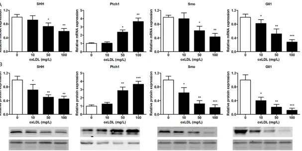
12823 Int J Clin Exp Pathol 2015;8(10):12820-12828 Figure 2. MBMECs were incubated with oxLDL (100 mg/L) for 24 h, SHH, Ptch1, Smo and Gli1 mRNA expression were determined by real-time PCR (A). SHH, Ptch1,
Smo and Gli1 protein expression were determined by Western blotting (B). Values were expressed as mean ± SD, n = 3 in each group. *P < 0.05, **P < 0.01, ***P <
5’-CTTCAAGTTGCCCAT-3’, reverse 5’-ATGGGCA- ACTTGAAG-3’; SHH, forward 5’-TCCAGAAACT- CCGAGCGATTTAAG-3’, reverse 5’-CACTCCTGG- CCACTGGTTCA-3’; Ptch1, forword 5’-CTGCGTC- AGCAGAGTGATTC-3’, reverse 5’-AGCTGAGGGT- GTCCTGTGTC-3’; Smo, forward 5’-CCTGCTCAC- CTGGTCACTC-3’, reverse 5’-CACGGTATCGGTA- GTTCTTGTAG-3’; Gli1, forword 5’-AGGGAGTG- CAGCCAATAC-3’, reverse 5’-CCGGAGTTGATGT- AGCTGGT-3’; GAPGH, forward 5’-GGTGGAGGT- CGGGAGTCAACGGA-3’, reverse 5’-GAGGGATC-
TCGCTCCTGGAGGA-3’. Glyceraldehyde 3-phos-phate dehydrogenase (GAPDH) as an internal control was used to normalize the data to deter-mine the relative expression of the target genes. The reaction conditions were set accord-ing to the kit instructions.
Western blotting
MBMECs were homogenized and extracted in NP-40 buffer, followed by 5-10 min boiling and centrifugation to obtain the supernatant. Samples containing 50 μg of protein were sepa -rated on 10% SDS-PAGE gel, transferred to nitrocellulose membranes (Bio-Rad Laborato- ries, Hercules, CA, USA). After saturation with 5% (w/v) non-fat dry milk in TBS and 0.1% (w/v) Tween 20 (TBST), the membranes were incu-bated with the following antibodies: SHH, Ptch1, Smo and Gli1 (Santa Cruz Biotechnoogy, CA, USA). After three washes with TBST, the membranes were next incubated with the appropriate HRP (horseradish peroxidase)-con-jugated antibody visualized with chemilumines-cence (Thermo, USA).
Statistical analysis
The data from these experiments were report-ed as mean ± standard errors of mean (SEM) for each group. All statistical analyses were performed by using PRISM version 4.0 (Gra-phPad). Inter-group differences were analyzed by one-way ANOVA, and followed by Tukey’s multiple comparison test as a post test to com-pare the group means if overall P < 0.05. Differences with P value of < 0.05 were consid-ered statistically significant.
Results
oxLDL induces MBMECs dysfunction in vitro
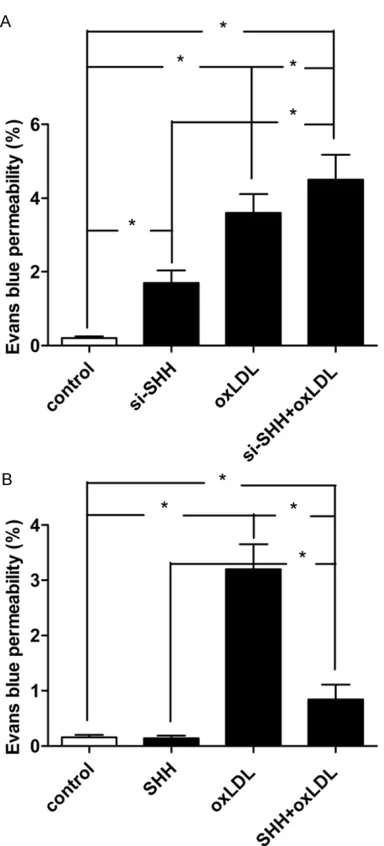
[image:5.612.92.284.67.490.2]To explore oxLDL-induced MBMECs viability, the CCK8 assay was performed to evaluate the cytotoxicity of oxLDL toward cells. Administration of MBMECs with oxLDL at the concentrations of 10 mg/L for 24 h did not affect cell survival. When the concentrations reached 50 mg/L and 100 mg/L, oxLDL decreased the viability of MBMCs by 20% and 33% respectively (Figure 1A). Moreover, we further analyzed the cytotox-icity of MBMECs induced by oxLDL, the LDH production was measured. LDH release was increased to 159% and 207% at the concentra-tion of 50 mg/L and 100 mg/L respectively Figure 3. Effect of SHH loss-of-function on
endo-thelial permeability in MBMECs when treated with
ox-LDL (100 mg/L) for 24 h (A). Effect of SHH
gain-of-function on endothelial permeability in MBMECs
when treated with ox-LDL (100 mg/L) for 24 h (B).
12825 Int J Clin Exp Pathol 2015;8(10):12820-12828 (Figure 1B). To indicate oxLDL-induced
oxida-tive stress of MBMECs, we analyzed the MDA and NO release, intracellular ROS concentra-tion, and eNOS mRNA expression by ELISA and real-time PCR, respectively. Results showed that MBMECs exposure to oxLDL led to increase the MDA level and decrease the NO level in a time-dependent manner (Figure 1C and 1D). Furthermore, treatment with 50 mg/L and 100 mg/L oxLDL significantly increased the ROS level and decreased the eNOS mRNA expres-sion level in MBMECs (Figure 1E and 1F). Therefore, these results demonstrated that
oxi-dative stress was involved in oxLDL-induce MBMECs dysfunction with increasing intracel-lular ROS and MDA formation as well as decreasing NO release and eNOS mRNA expression.
oxLDL regulates mRNA and protein expression of SHH signalling components in endothelial cells
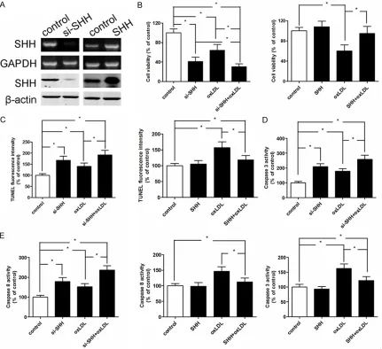
[image:6.612.92.523.72.466.2]Real-time PCR was performed to evaluate the effect of oxLDL on the mRNA expression of SHH signalling components in MBMECs. As shown in Figure 4. The mRNA and protein expression of SHH were measured by real-time PCR and western blotting respec-tively when MBMECs were treated with SHH-siRNA and SHH-transfected (A). Effect of SHH loss-of-function or
gain-of-function on cell viability in MBMECs when treated with ox-LDL (100 mg/L) for 24 h (B). Effect of SHH loss-of-gain-of-function or gain-of-function on cell apoptosis in MBMECs when treated with ox-LDL (100 mg/L) for 24 h (C). Effect of SHH loss-of-function or gain-of-function on caspase3 levels in MBMECs when treated with ox-LDL (100 mg/L) for 24 h
(D). Effect of SHH loss-of-function or gain-of-function on caspase8 levels in MBMECs when treated with ox-LDL (100
Figure 2A, SHH, Smo and Gli1 mRNA levels were significantly decreased after incubation with increasing concentrations of oxLDL. In contrast, Ptch1 mRNA levels were upregulated following oxLDL treatment. Based on the mRNA expression of SHH signalling components in MBMECs, we further analyzed the protein of SHH signalling components in MBMECs in- duced by oxLDL. In accordance with the result of mRNA expression levels, MBMECs stimulat-ed with oxLDL showstimulat-ed a remarkable decrease in SHH, Smo and Gli1 protein and increase in Ptch1 as compared to untreated cells (Figure 2B).
SHH loss-of-function or gain-of-function regu-lates oxLDL-induced permeability transition in MBMECs
First, we confirmed the effects of oxLDL on BBB integrity using MBMECs as an in vitro BBB model. Treatment with oxLDL alone or SHH loss-of-function significantly increased the per -meability of MBMECs, and the per-meability in SHH loss-of-function combination with oxLDL treatment group was markedly higher than those of single treatment by either oxLDL or SHH loss-of-function group and untreatment group (Figure 3A). Intriguingly, we found that overexpression of SHH attenuated oxLDL-induced elevation of permeability in MBMECs (Figure 3B). These results demonstrated that the suppression of SHH in MBMECs might con-tribute to the oxLDL-induced disruption of endo-thelial barrier.
SHH loss-of-function or gain-of-function regu-lates oxLDL-induced apoptosis in MBMECs
To investigate the function of SHH in oxLDL-induced MBMECs apoptosis, SHH loss-of-func-tion by small interfering RNA was dramatically inhibited the mRNA and protein expression, and MBMECs transfected with SHH obvious increased the mRNA and protein expression (Figure 4A). The inhibiting effect of cell viability was significantly suppressed in the combined treatment group compared with single treat-ment group or untreattreat-ment group, however, SHH overexpression resulted in a restored cell viability (Figure 4B). In addition, we examined whether SHH abnormal expression regulated oxLDL-induced apoptosis in MBMECs through an apoptotic mechanism. Annexin V-PI double-labeling, caspase3 and caspase8 activity assay
were measured when MBMECs were exposed to oxLDL for 24 h. The results indicated that treatment with oxLDL alone or SHH loss-of-function showed significant cell apoptosis as compared to that of the control group (Figure 4C), simultaneously, the caspase3 and cas-pase8 activity were increased (Figure 4D and 4E). However, SHH gain-of-function could reverse oxLDL-induced apoptosis in MBMECs. Discussion
It is well known that oxLDL induces endothelial cells dysfunction through various signal path-ways, such as activation of NF-κB [21], GSK3β/ β-catenin [22], mitochondrial-dependent apop -totic pathway [9] and sodium-dependent glu-cose transporter (SGLT1) [23]. Intriguingly, SHH signalling pathway regulates apoptosis through Smo protein in endothelial cells [12], and topi-cal SHH gene therapy accelerates wound heal-ing in diabetes by enhancheal-ing endothelial pro-genitor cell-mediated microvascular remodel-ing [24]. However, for all we know, no literature has been reported that SHH signal pathway mediates oxLDL-induced cell dysfunction in MBMECs. In the present study, we revealed that oxidative stress was involved in oxLDL-induce MBMECs dysfunction with increasing intracellular ROS and MDA formation as well as decreasing NO release and eNOS mRNA expres-sion. Next, treatment with oxLDL significantly increased the permeability and induced apop-tosis in MBMECs, however, overexpression of SHH could reverse oxLDL-induced apoptosis and permeability transition in MBMECs. In addi-tion, SHH signalling components were signifi -cantly decreased after incubation with increas-ing concentrations of oxLDL. These results indi-cated that oxLDL increased ROS and induced apoptosis and permeability transition in MBMECs, the underlying mechanism was medi-ated, at least partially, through regulation SHH signalling components expression.
12827 Int J Clin Exp Pathol 2015;8(10):12820-12828 astrocytes, which are composed of BBB [13]. In
this work, we first demonstrated that SHH gain-of-function attenuated oxLDL-induced eleva-tion of endothelial permeability in MBMECs, and downregulation of SHH in MBMECs might contribute to the oxLDL-induced disruption of endothelial barrier.
It has been reported that excessive ROS can accelerate proinflammatory cytokine injury to human brain microvascular endothelial barrier properties and cell apoptosis [9, 20, 27]. In the current study, we found that oxLDL induced ROS formation and cell apoptosis. Overex- pression SHH attenuated oxLDL-induced endo-thelial cell apoptosis, indicating that SHH was involved in the oxLDL-induced cytotoxicity for-mation in MBMECs. Cascade activation of cas-pase3 and caspase9 plays a critical role in oxLDL-induced apoptosis of mouse endothelial cells. Administration of oxLDL in mouse endo-thelial cells increased caspase-9 activity [28]. Our present results further demonstrated that suppression of caspase3 and caspase8 activa-tion significantly lowered oxLDL-induced cell apoptosis through the overexpression of SHH. In conclusion, this study showed that oxLDL could damage MBMECs through increasing intracellular ROS and MDA formation as well as decreasing NO release and eNOS expression, and oxLDL-treated significantly increased the permeability of MBMECs. Interestingly, the overexpression of SHH could reverse oxLDL-induced endothelial cells dysfunction in vitro.
Disclosure of conflict of interest
None.
Address correspondence to: Dr. Xiu-Long Jiang, Department of Neurology, Provincial Clinic College
of Fujian Medical University, Fujian Provincial
Hospital, Fuzhou 350001, China. Tel: (86) 591-87557768; Fax: (86) 591-591-87557768; E-mail: xl_ [email protected]
References
[1] Ma J, Zhao S, Gao G, Chang H, Ma P and Jin B. Probucol Protects Against Asymmetric Dimethylarginine-Induced Apoptosis in the Cultured Human Brain Microvascular En-
dothelial Cells. J Mol Neurosci 2015; 57:
546-53.
[2] Fang L, Li X, Zhong Y, Yu J, Yu L, Dai H and Yan M. Autophagy protects Human Brain Micro- vascular Endothelial Cells against Methylgl-
yoxal-induced Injuries: in parallel with Cerebral Ischemic Model in Diabetic Rats. J Neurochem 2015.
[3] Xiao X, Zhang C, Ma X, Miao H, Wang J, Liu L, Chen S, Zeng R, Chen Y and Bihl JC. An- giotensin-(1-7) counteracts angiotensin II-induced dysfunction in cerebral endothelial
cells via modulating Nox2/ROS and PI3K/NO
pathways. Exp Cell Res 2015; 336: 58-65.
[4] Dejana E. Endothelial cell-cell junctions: happy
together. Nat Rev Mol Cell Biol 2004; 5:
261-270.
[5] Liu N, Li AL, Zhou XP, Chen Q and Cao W. P120 catenin attenuates lipopolysaccharide-indu-
ced blood-brain barrier dysfunction and inflam -matory responses in human brain microvascu-lar endothelial cells. Int J Clin Exp Pathol 2015;
8: 4204-4212.
[6] Daugherty A, Rateri DL and King VL. IL-5 links
adaptive and natural immunity in reducing
ath-erosclerotic disease. J Clin Invest 2004; 114:
317-319.
[7] Jovinge S, Crisby M, Thyberg J and Nilsson J. DNA fragmentation and ultrastructural chang-es of degenerating cells in atherosclerotic le-sions and smooth muscle cells exposed to oxi-dized LDL in vitro. Arterioscler Thromb Vasc Biol 1997; 17: 2225-2231.
[8] Liu S, Hou W, Qin H and Wang Y. Oxidized LDL stimulates lipid peroxidation-derived DNA and protein adducts in human vascular endothelial
and smooth muscle cells. J Huazhong Univ Sci
Technolog Med Sci 2015; 35: 200-205. [9] Chen TG, Chen TL, Chang HC, Tai YT, Cherng
YG, Chang YT and Chen RM. Oxidized low-den-sity lipoprotein induces apoptotic insults to mouse cerebral endothelial cells via a Bax-mitochondria-caspase protease pathway.
Toxicol Appl Pharmacol 2007; 219: 42-53.
[10] He QW, Xia YP, Chen SC, Wang Y, Huang M, Huang Y, Li JY, Li YN, Gao Y, Mao L, Mei YW and Hu B. Astrocyte-derived sonic hedgehog con-tributes to angiogenesis in brain microvascular
endothelial cells via RhoA/ROCK pathway after
oxygen-glucose deprivation. Mol Neurobiol
2013; 47: 976-987.
[11] Han BW, Layman H, Rode NA, Conway A, Schaffer DV, Boudreau N, Jackson WM and
Healy KE. Multivalent conjugates of Sonic
hedgehog ccelerate diabetic wound healing. Tissue Eng Part A 2015; 21: 2366-78.
[12] Zhu SL, Luo MQ, Peng WX, Li QX, Feng ZY, Li ZX, Wang MX, Feng XX, Liu F and Huang JL. Sonic hedgehog signalling pathway regulates apop-tosis through Smo protein in human umbilical vein endothelial cells. Rheumatology (Oxford)
2015; 54: 1093-1102.
Bourbonniere L, Bernard M, van Horssen J, de Vries HE, Charron F and Prat A. The Hedgehog pathway promotes blood-brain barrier integrity and CNS immune quiescence. Science 2011;
334: 1727-1731.
[14] Osterlund T and Kogerman P. Hedgehog signal -ling: how to get from Smo to Ci and Gli. Trends Cell Biol 2006; 16: 176-180.
[15] Wang Y, Jin S, Sonobe Y, Cheng Y, Horiuchi H,
Parajuli B, Kawanokuchi J, Mizuno T, Takeuchi
H and Suzumura A. Interleukin-1beta induces blood-brain barrier disruption by downregulat-ing Sonic hedgehog in astrocytes. PLoS One
2014; 9: e110024.
[16] Xia YP, He QW, Li YN, Chen SC, Huang M, Wang Y, Gao Y, Huang Y, Wang MD, Mao L and Hu B. Recombinant human sonic hedgehog protein regulates the expression of ZO-1 and occludin by activating angiopoietin-1 in stroke damage. PLoS One 2013; 8: e68891.
[17] Schreurs MP, Hubel CA, Bernstein IM, Jeya- balan A and Cipolla MJ. Increased oxidized low-density lipoprotein causes blood-brain barrier disruption in early-onset preeclampsia through
LOX-1. FASEB J 2013; 27: 1254-1263.
[18] Lenart N, Walter FR, Bocsik A, Santha P, Toth ME, Harazin A, Toth AE, Vizler C, Torok Z, Pilbat AM, Vigh L, Puskas LG, Santha M and Deli MA. Cultured cells of the blood-brain barrier from apolipoprotein B-100 transgenic mice: effects of oxidized low-density lipoprotein treatment. Fluids Barriers CNS 2015; 12: 17.
[19] Kim JH, Lee SJ, Kim KW, Yu YS and Kim JH.
Oxidized low density lipoprotein-induced se-nescence of retinal pigment epithelial cells is followed by outer blood-retinal barrier
dysfunc-tion. Int J Biochem Cell Biol 2012; 44: 808-814.
[20] Lenart N, Walter FR, Bocsik A, Santha P, Toth ME, Harazin A, Toth AE, Vizler C, Torok Z, Pilbat AM, Vigh L, Puskas LG, Santha M and Deli MA. Cultured cells of the blood-brain barrier from apolipoprotein B-100 transgenic mice: effects of oxidized low-density lipoprotein treatment. Fluids Barriers CNS 2015; 12: 17.
[21] Ji KT, Qian L, Nan JL, Xue YJ, Zhang SQ, Wang
GQ, Yin RP, Zhu YJ, Wang LP, Ma J, Liao LM and Tang JF. Ox-LDL induces dysfunction of endo-thelial progenitor cells via activation of NF-kappaB. Biomed Res Int 2015; 2015: 175291.
[22] Lin P, Liu J, Ren M, Ji K, Li L, Zhang B, Gong Y
and Yan C. Idebenone Protects against Oxidized Low Density Lipoprotein Induced Mitochondrial Dysfunction in Vascular En-
dothelial Cells via GSK3beta/beta-catenin sig -naling pathways. Biochem Biophys Res Commun 2015.
[23] Jin X, Yi L, Chen ML, Chen CY, Chang H, Zhang T, Wang L, Zhu JD, Zhang QY and Mi MT. Delphinidin-3-glucoside protects against oxi-dized low-density lipoprotein-induced mito-chondrial dysfunction in vascular endothelial cells via the sodium-dependent glucose trans-porter SGLT1. PLoS One 2013; 8: e68617.
[24] Asai J, Takenaka H, Kusano KF, Ii M,
Lue-demann C, Curry C, Eaton E, Iwakura A, Tsu-
tsumi Y, Hamada H, Kishimoto S, Thorne T, Kishore R and Losordo DW. Topical sonic
hedgehog gene therapy accelerates wound healing in diabetes by enhancing endothelial progenitor cell-mediated microvascular
remod-eling. Circulation 2006; 113: 2413-2424.
[25] Schreurs MP and Cipolla MJ. Cerebrovascular dysfunction and blood-brain barrier permeabil-ity induced by oxidized LDL are prevented by apocynin and magnesium sulfate in female
rats. J Cardiovasc Pharmacol 2014; 63: 33-39.
[26] Wang J, Sun L, Si YF and Li BM. Overexpression of actin-depolymerizing factor blocks oxidized low-density lipoprotein-induced mouse brain microvascular endothelial cell barrier dysfunc-tion. Mol Cell Biochem 2012; 371: 1-8. [27] Rochfort KD, Collins LE, McLoughlin A and
Cummins PM. Shear-dependent attenuation of
cellular ROS levels can suppress proinflamma -tory cytokine injury to human brain microvas-cular endothelial barrier properties. J Cereb
Blood Flow Metab 2015; 35: 1648-56.
[28] Wu CY, Tang ZH, Jiang L, Li XF, Jiang ZS and Liu
LS. PCSK9 siRNA inhibits HUVEC apoptosis in -duced by ox-LDL via Bcl/Bax-caspase9-cas- pase3 pathway. Mol Cell Biochem 2012; 359: