(12) INTERNATIONAL APPLICATION PUBLISHED UNDERTHEPATENT COOPERATION TREATY (PCT)
(19)World Intellectual Property Organization
International Bureau
(10)International Publication Number
(43)International Publication Date
WO
2017/011450
Al
19 January 2017 (19.01.2017) P O P C T
(51) International Patent Classification: BZ, CA, CH, CL, CN, CO, CR, CU, CZ, DE, DK, DM, A61N 1/05(2006.01) H01B 1/12(2006.01) DO, DZ, EC, EE, EG, ES, FI, GB, GD, GE, GH, GM, GT, HN, HR, HU, ID, IL, IN, IR, IS, JP, KE, KG, KN, KP, KR, (21) International ApplicationNumber: KZ, LA, LC, LK, LR, LS, LU, LY, MA, MD, ME, MG, PCT/US20 16/04 1885 MK, MN, MW, MX, MY, MZ, NA, NG, NI, NO, NZ, OM, (22) International FilingDate: PA, PE, PG, PH, PL, PT, QA, RO, RS, RU, RW, SA, SC,
12July 2016 (12.07.2016) SD, SE, SG, SK, SL, SM, ST, SV, SY, TH, TJ, TM, TN, TR, TT, TZ, UA, UG, US, UZ, VC, VN, ZA, ZM, ZW.
(25) Filing Language: English
(84) Designated States (unless otherwise indicated, for every (26) Publication Language: English kind of regional protection available): ARIPO (BW, GH, (30) PriorityData: GM, KE, LR, LS, MW, MZ, NA, RW, SD, SL, ST, SZ, 62/192,752 15July 2015 (15.07.2015) US TZ, UG, ZM, ZW), Eurasian (AM, AZ, BY, KG, KZ, RU, TJ, TM), European (AL, AT, BE, BG, CH, CY, CZ, DE, (71) Applicant: THE UNIVERSITY OF FLORIDA RE¬ DK, EE, ES, FI, FR, GB, GR, HR, HU, IE, IS, IT, LT, LU, SEARCH FOUNDATION, INC. [US/US]; 233 Grinter LV, MC, MK, MT, NL, NO, PL, PT, RO, RS, SE, SI, SK, Hall, Gainesville, FL 3261 1 (US). SM, TR), OAPI (BF, BJ, CF, CG, CI, CM, GA, GN, GQ,
GW, KM, ML, MR, NE, SN, TD, TG). (72) Inventors:HARDY,John; 4 Warding Drive, Little Com
mon Bexhill-on-sea, East Susse, TN39 4QN (GB). Declarations under Rule 4.17 : SCHMIDT, Christine, E.; 1922 SW 106th Terrace,
— as toapplicant's entitlement toapply for andbegranted a
Gainesville, FL 32607 (US).
patent (Rule4.17(H))
(74) Agents: LINDER, Christopher, B. et al; THOMAS / — as to theapplicant's entitlement toclaimthepriority of the HORSTEMEYER LLP, 400 Interstate North Parkway, SE, earlier application (Rule 4.17(in))
Suite 1500, Atlanta, GA 30339 (US).
Published: (81) Designated States (unless otherwise indicated, for every
kind of national protection available): AE, AG, AL, AM, — withinternational search report(Art.21(3)) AO, AT, AU, AZ, BA, BB, BG, BH, BN, BR, BW, BY,
(54) Title: CONFORMAL CONDUCTIVE SCAFFOLDS AND METHODS OF USING CONFORMAL CONDUCTIVE SCAF FOLDS
o
ρβοστ-s
© Fig. 1. 1
[image:1.595.55.453.497.736.2]CONFORMAL CONDUCTIVE SCAFFOLDS AND METHODS OFUSING
CONFORMAL CONDUCTIVE SCAFFOLDS
CROSS-REFERENCE TO RELATED APPLICATIONS
This application claims the benefit of and priority to U.S. Provisional Application
SerialNo. 62/192,752, having the title "CONFORMAL CONDUCTIVE SCAFFOLDS AND
METHODS OFUSING CONFORMAL CONDUCTIVE SCAFFOLDS," filed on July 15,
2015, the disclosure of which is incorporated herein in by reference in its entirety.
BACKGROUND
Cellsinhabit environments known asthe extracellular matrix (ECM) which consists
of a mixture of different biomolecules, and the precise composition and topographical
properties are different in different tissues (e.g.,bone,brain, muscle, skin). Cellsinteract
intimately with the ECM, not only constructing the biomolecules, but assist its organization
in 3D space, and its degradation (which is important for tissue remodeling); reciprocally,
cellsrespond to the ECM (e.g.,by modifying their size, shape). Cellular alignment is
observed in organs and tissues suchasbones, muscles and skin, and this alignment is
important for the healthy functioning of the organ/tissue.
SUMMARY
Embodiments of the present disclosure provide for structures including a conformal
conductive scaffold, methods of making the structure, method of using structure, and the like.
An embodiment of the present disclosure provides for a structure, among others, that
includes a conformal conductive scaffold including a polyelectrolyte complex, chitosan, and
gelatin, which are crosslinked by genipin. In an embodiment, the polyelectrolyte complex
canbe a PEDOT derivative, polypyrrole-polystyrenesulfonate (PSS), polyaniline-PSS,
polythiophene-PSS, or a combination thereof.
An embodiment of the present disclosure provides for a method of alignment of cells,
among others, that includes: providing a structure having a conformal conductive scaffold
including a polyelectrolyte complex, chitosan, and gelatin, which are crosslinked by genipin;
introducing cells to the structure; periodically providing electrical stimulation to cells; and
a human mesenchymal stemcell. In anembodiment, the electrical stimulation can be a direct
current (DC) or an alternating current (AC).
Other compositions, structures, methods, features, and advantages will be or become
apparent to one with skill in the art upon examination of the following drawings and detailed
description. It is intended that all suchadditional compositions, apparatus, methods, features
and advantages be included within this description, be within the scope of the present
disclosure, and be protected by the accompanying claims.
BRIEFDESCRIPTION OF THE DRAWINGS
Further aspects of the present disclosure will be more readily appreciated upon review
of the detailed description of its various embodiments, described below, when taken in
conjunction with the accompanying drawings.
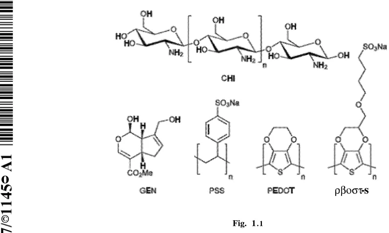
Figure 1.1 illustrates molecules employed in this study. Abbreviations: (CHI)
Chitosan, (GEN) Genipin. (PSS) Polystyrenesulfonate, (PEDOT)
Poly(3,4-ethylenedioxythiophene), and (PEDOT-S) Sulfonated PEDOT derivative.
Figures 1.2A-E show adhesion and proliferation of human dermal fibroblasts on
various surfaces up to 4 days in culture. Fig. 1.2A illustrates tissue culture plate controls. Fig.
1.2Billustrates PEDOT-PSS-based multilayer films. Fig. 1.2Cillustrates PEDOT-S-based
multilayer films. Cells were stained with a LIVE/DEAD® Viability/Cytotoxicity Kit, live
cells were green and dead cells were red. Scalebars represent 250 µ . Fig. 1.2Dillustrates
cell viability after 4 days in culture asdetermined with a LIVE/DEAD®
Viability/Cytotoxicity Kit. Fig. 1.2Eillustrates cell adhesion after 2 days (grey bars) or 4
days (black bars) asdetermined by the AlamarBlue® assay. Error bars represent standard
deviations (n =3).
Figures 1.3A-F show analysis of the morphology of human dermal fibroblasts on
various surfaces with optional electrical stimulation. Figure 1.3A illustrates
PEDOT-PSS-based multilayer films without electrical stimulation, PEDOT-PSS(-). Figure 1.3B illustrates
PEDOT-PSS-based multilayer films with electrical stimulation, PEDOT-PSS(+). Figure
1.3Cillustrates PEDOT-S-based multilayer films without electrical stimulation, PEDOT-S(-).
Figure 1.3Dillustrates S-based multilayer films with electrical stimulation
PEDOT-S(+). DAPI-stained nuclei are blue and Alexa Fluor® 488-stained actin is green. Scale bars
represent 200 µ . Figure 1.3E illustrates assessment of cell alignment. Figure 1.3F
illustrates assessment ofcell length. Error bars represent standard errors of the mean (n =
Figure 1.4 is an example of an experimental setup for dip coating multilayer films
using a Gilson 223 Sample Changer converted for use as a dip coater.
Figure 1.5 is a schematic of an experimental setup for electrical stimulation of
PEDOT-based films (Not to scale). Abbreviations: (CE) counter electrode, (CT) copper tape,
(PCW) polycarbonate well, (RE) reference electrode, and (WE) working electrode.
Figure 1.6 illustrates cell alignment & length assessment.
Figures 1.7A-B show polar plots for multilayer films. Fig. 1.7A illustrates
PEDOT-PSS-based films. Fig. 1.7B illustrates PEDOT-S-based films. Black circles represent cells
without electrical stimulation, whereas red circles represent cells with electrical stimulation;
black and red lines represent the corresponding trend lines.
Figures 1.8A-B areFDVISCson multilayer films after 3 days in culture. Fig. 1.8A
illustrates PEDOT-PSS-based multilayer films. Fig. 1.8B illustrates PEDOT-S-based
multilayer films. DAPI-stained nuclei are blue and Alexa Fluor® 488-stained actin is green.
Scale bars represent 150 µ .
DETAILEDDESCRIPTION
This disclosure is not limited to particular embodiments described, and as such may,
of course, vary. The terminology used herein serves the purpose of describing particular
embodiments only, and is not intended to be limiting, since the scope of the present
disclosure will be limited only by the appended claims.
Where a range of values is provided, each intervening value, to the tenth of the unit of
the lower limit unless the context clearly dictates otherwise, between the upper and lower
limit of that range and any other stated or intervening value in that stated range, is
encompassed within the disclosure. The upper and lower limits of these smaller ranges may
independently be included in the smaller ranges and are also encompassed within the
disclosure, subject to any specifically excluded limit in the stated range. Where the stated
range includes one or both of the limits, ranges excluding either or both of those included
limits are also included in the disclosure.
A s will b e apparent to those of skill in the art upon reading this disclosure, each of the
individual embodiments described and illustrated herein has discrete components and features
which may be readily separated from or combined with the features of any of the other
several embodiments without departing from the scope or spirit of the present disclosure.
Any recited method may be carried out in the order of events recited or in any other order that
Embodiments of the present disclosure will employ, unless otherwise indicated,
techniques of organic chemistry, biochemistry, microbiology, molecular biology,
pharmacology, medicine, and the like, which are within the skill of the art. Suchtechniques
areexplained fully in the literature.
Prior to describing the various embodiments, the following definitions are provided
and should be used unless otherwise indicated.
Unless otherwise defined, alltechnical and scientific terms used herein have the same
meaning as commonly understood by one of ordinary skill in the art of microbiology,
molecular biology, medicinal chemistry, and/or organic chemistry. Although methods and
materials similar or equivalent to those described herein can be used in the practice or testing
of the present disclosure, suitable methods and materials are described herein.
As used in the specification and the appended claims, the singular forms "a," "an,"
and"the" may include plural referents unless the context clearly dictates otherwise. Thus, for
example, reference to "a support" includes a plurality of supports. In this specification and in
the claims that follow, reference will be made to a number of terms that shall be defined to
have the following meanings unless a contrary intention is apparent.
Discussion :
Embodiments of the present disclosure provide for structures including a conformal
conductive scaffold, methods of making the structure, method of using structure, and the like.
Embodiments of the present disclosure can be used to align cells (e.g.,fibroblasts and human
mesenchymal stem cells) on the surface of the conformal conductive scaffold. In a particular
embodiment, cells can be cultured on the surface of the conformal conductive scaffold in an
appropriate medium for the particular type ofcell andthen a current (e.g.,DC) is applied to
the conformal conductive scaffold so that the cells align on the surface of the conformal
conductive scaffold, where the electrical stimulation can be periodically applied to the
electroactive scaffold.
In this regard, embodiments of the disclosure provide for a structure that includes the
conformal conductive scaffold that can be used to aligncells. An embodiment of the present
disclosure includes introducing cells to the conformal conductive scaffold, where the
conformal conductive scaffold and the cells are cultured in an appropriate medium.
Subsequently, electrical stimulation can be periodically applied to the cells to cause the cells
to align on the conformal conductive scaffold. In particular, the cells may align along the
coating used to dispose the polymer film on the structure. As shown in Example 1, electrical
stimulation of the conformal conductive scaffold in the presence of cells shows alignment of
the cells, which is a characteristic of some cells types including bone cells, cardiac cells, and
nerve cells.
The term "conformal" asused herein refers to materials that are typically three
dimensional and may or may not be inherently conductive.
In an embodiment, the structure includes the conformal conductive scaffold that has a
polyelectrolyte complex, chitosan, and gelatin film, where the chitosan and the gelatin film
cancrosslink with genipin. In an embodiment, the polyelectrolyte complex is selected from
the group consisting of: PEDOT derivative, polypyrrole-polystyrenesulfonate (PSS),
polyaniline-PSS, polythiophene-PSS, and a combination thereof. The polymer film can be
coated on the structure using various techniques, including dip coating, drop coating, and spin
coating. In anembodiment, the film can have a thickness of about 1 nm to 1 cm and area of
about 10mm2to 10 m2.
To illustrate the interaction of the components, the following is an example of the
interaction. In an embodiment where the polyelectrolyte complex is a PEDOT derivative
(e.g.,PEDOT-polystyrenesulfonate (PSS)), the negatively charged sulfates on PEDOT-PSS,
(or those on PEDOT-S, shown below), will non-covalently interact with the positively
charged moieties on chitosan or gelatin (e.g., amines),while genipin will covalently crosslink
with chitosan and gelatin.
In an embodiment, the PEDOT derivative can include a sulfonated PEDOT having the
following structure: ∞ - (wherein n is 1 to infinity (e.g., 10,000, 100,000,
(PEDOT-PSS) (where the unit of each be 1 to infinity (e.g., 10,000,
100,000, 1,000,000, or more)). PEDOT-PSS is a mixture of two ionomers that form a
macromolecular salt. In other embodiments, another negatively charged conducting polymer
canbe used suchaspolypyrrole-PSS, polyaniline-PSS, or polythiophene-PSS.
Geni in is a chemical a gardena fluid extract. Genipin has the following structure:
Chitosan is an oligosaccharide including β-linked D-glucosamine and
N-actyl-D-glucosamine that are randomly distributed in the oligomer. In an embodiment, the chitosan
has a molecular weight of about 1 kDa to 20 MDa.
Gelatin is an irreversibly hydrolyzed form of collagen, where the hydrolysis results in
the reduction of protein fibrils into smaller peptides, which will have broad molecular weight
ranges (e.g.,about 1 kDa to 20 MDa) associated with physical and chemical methods of
denaturation, based on the process of hydrolysis.
In an embodiment, the cells can be human mesenchymal stem cells, human dermal
fibroblasts, myoblasts, osteoblasts, osteoclasts, neurons, Schwann cells, pluripotent stem
cells, or the like. In an embodiment where the cells are human dermal fibroblast, the medium
canbe HDF growth medium, composed of: high glucose Dulbecco's Modified Eagle Medium
(DMEM, 440 mL); fetal bovine serum (50 mL); antibiotic-antimycotic (5mL);non-essential
amino acids (5mL), and2 ng mL- 1basic fibroblast growth factor.
Electrical stimulation can include direct contact of the material with a power source
via a wire, wireless energy transfer, magnetic force, and the like. In an embodiment, the
electrical stimulation can be direct current (DC) or alternating current (AC). The term
"periodically" refers to applying the electrical stimulation at established time frames that may
be at regular or irregular time intervals on the time frames of seconds, hours, days, weeks, or
months (e.g.,about 1 s to 2 months, about 1 hour to 1 day, about 1 day to 1 month, or other
the like) depending upon the specific circumstances. In an embodiment, the impulses of the
second to 1 day, about 10 secondsto 1 hour, about 1 minute to 12 hours, about 1 hour to 1
day, or the like) depending upon the specific circumstances. In an embodiment, the electrical
stimulation may only include a single long pulse that may last seconds to minutes, to hours,
to days. In an embodiment, the electrical stimulation can be in the range of millivolts to volts
(e.g., about 10mV to 10volts, about 1 mV to 100mV, or the like). The time frame, duration
of electrical stimulation, and intensity of the electrical stimulation can be designed based on
particular circumstances and requirements of a specific situation.
As stated above, embodiments of the present disclosure provide for a structure having
the conformal conductive scaffold. In an embodiment, the conformal conductive scaffold can
include one or more agents (e.g.,a chemical or biological agent), where the agent can be
disposed indirectly or directly on the conformal conductive scaffold. As described herein, the
agent can include a stemcell suchasa human mesenchymal stem cell or human dermal
fibroblast.
In addition, an additional agent that can be disposed on the conformal conductive
scaffold can include, but is not limited to, a drug, a therapeutic agent, a radiological agent, a
small molecule drug, a biological agent(e.g.,polypeptides (e.g.,proteins such as,but not
limited to, antibodies (monoclonal or polyclonal)), antigens, nucleic acids (both monomelic
andoligomeric), polysaccharides, haptens, sugars, fatty acids, steroids, purines, pyrimidines,
ligands, and aptamers) and combinations thereof, that can be used to image, detect, study,
monitor, evaluate, and the like, the cells. In an embodiment, the agent is included in an
effective amount to accomplish its purpose, where such factors to accomplish the purpose are
well known in the medical arts.
In general, the agent can be bound to the structure by a physical, biological,
biochemical, and/or chemical association directly or indirectly by a suitable means. The term
"bound" can include, but is not limited to, chemically bonded (e.g., covalently or ionically),
biologically bonded, biochemically bonded, and/or otherwise associated with the
electroactive supramolecular polymeric assembly. In an embodiment, being bound can
include, but is not limited to, a covalent bond, a non-covalent bond, an ionic bond, a chelated
bond, aswell asbeing bound through interactions suchas,but not limited to, hydrophobic
interactions, hydrophilic interactions, charge-charge interactions, π-π stacking interactions,
combinations thereof, and like interactions. In an embodiment, cell-electroactive scaffold
interactions could be controlled through the inclusion of cell-adhesive peptides (e.g.,RGD,
indeed oligoalanines that are degraded by elastase), osteoinduction can be enhanced (e.g.,
NSPVNSKIPKACCVPTELSAI), and directing mineralization (e.g.FHRRIKA).
While embodiments of the present disclosure are described in connection with the
Examples and the corresponding text and figures, there is no intent to limit the disclosure to
the embodiments in these descriptions. On the contrary, the intent is to cover allalternatives,
modifications, and equivalents included within the spirit and scope of embodiments of the
present disclosure.
EXAMPLE 1 :
We report the preparation of conducting polymer-based films. Composite biomaterials
based on poly(3,4-ethylenedioxythiophene) derivatives, chitosan and gelatin were prepared
andtheir physicochemical properties were characterized. Fibroblasts adhered to and
proliferated on the films, and were shown to align with a DC current passed through the
films.
Thespecific properties of bodily tissues actascues(individually or in concert) that
determine the behaviour of cells that inhabit them, and are utilised to engineer instructional
tissue scaffolds to facilitate the regeneration of functional tissues.[l] The topographical
properties of tissues may instruct cells to align, asisclearlyobservable within the
anisotropically aligned pores observed in bone, cardiac, nerve and other tissues [2]. Scientists
andengineers have reported novel methodologies of imparting biomimetic porous structures
within biomaterials, including the removal of sacrificial templates embedded within a matrix
(e.g., colloidal crystals, ice crystals, electrospun fibers), and 3D printing technologies that
offer methods of precisely positioning pores with specific geometries in such structures.[2]
Endogenous electric fields are another cue influencing cell behaviour (e.g. processes
suchasembryogenesis and wound healing). Electrical fields are also known to align cells,
with endogenous electric fields playing a role in the alignment of cells,[3] and exogenous
electric fields have been shown to align a variety ofcelltypes (including astrocytes, epithelial
cells,fibroblasts) in vitro,[3] motivating the development of electrically conductive materials
that instruct cell alignment aided by application of an electrical field/current that mimics
endogenous electrical fields/currents.[3]
Conducting biomaterials based on conducting polymers (CPs) such asderivatives of
polyaniline, polypyrrole or polythiophene, have potential for both long term biomedical
tissue engineering).[4] CP-based scaffolds have been developed for the regeneration of bone
andnerve tissues, and organs including the heart andskin. [4]
Layer-by-layer assembly is a method of producing thin multilayer films that was
popularized by Mohwald and others.[5] Conductive multilayer films have been prepared from
a variety of organic electronic components, often for applications in energy, of which a
number of examples based on CPs exist.[5]Wallace and coworkers reported the preparation
of bioerodible CP-based multilayer films based on an anionic sulfonate-displaying
polythiophene derivative (with a w= 13 160Da and = 6682 Daasdetermined by GPC)
andcationic polyethyleneimine (17 kDa),[6] both of which are below the renal filtration limit
of approximately 50kDa; andthe films were shown to be suitable for the growth and
proliferation of cells derived from the connective and muscle tissue of mice (L929 and
C2C12 cells, respectively).[6] They subsequently showed that it was possible to prepare
CP-based multilayer films CP-based solely on anionic and cationic polythiophene derivatives that
could be -disassembled upon the application of an electrochemical trigger (a potential step of
650mV for 19-42hours).[7] Wei and coworkers reported the first example of biologically
functional biodegradable CP-based multilayer films for bone tissue engineering, that were
used for the electrical stimulation of osteoblast precursor MC3T3-E1 cells (derived from
mice) that resulted in increased expression of osteopontin and runt-related transcription factor
2 which are markers of osteogenesis.[8]
Here we describe the preparation of CP-based films composed of
poly(3,4-ethylenedioxythiophene) (PEDOT) derivatives, chitosan and gelatin, that are crosslinked by
genipin (Figure 1.1). These conducting films enabled the electrical stimulation of human
dermalfibroblasts cultured thereon which resulted in the preferential alignment of the cells
with a DC current passed through the films.
Films were deposited on non-conductive glass slides that were previously rendered
positively charged by modification with aminopropyltrimethoxysilane. The polyelectrolyte
complex composed of PEDOT and polystyrenesulfonate (PSS), PEDOT-PSS, or the
sulfonated PEDOT derivative PEDOT-S (Figure 1.1)were used for film preparation because
they are electrochemically stable over periods long enough to facilitate short term electrical
stimulation of tissues in their vicinity, and have been shown to be relatively
non-immunogenic after implantation in various tissues. The polysaccharide chitosan was chosen
asthe cationic polymer to interact with the anionic PEDOTs (PEDOT-PSS and PEDOT-S,
respectively) asit is also known to be relatively non-immunogenic (particularly by
electrochemically reducing the backbone of PEDOT-S makes it less positively charged which
inturn reduces the number of sulfonates necessary to dope the polymer backbone rendering
the surface charge of PEDOT-S films to be predominantly negative; therefore, PEDOT-S can
be employed asa surface coating that enables electrochemically triggered cell desorption.
Therefore we employed gelatin asa surface coating to render the films cell adhesive, and
crosslinked the films with genipin (a natural crosslinker of biopolymers including chitosan,
collagen and gelatin, which is markedly less toxic than the more commonly used
glutaraldehyde), to ensure film stability for the duration of the experiments.
Film preparation by drop casting yielded films with µιη-scale roughnesses, water
contact angles (WCAs) of approximately 50°, and conductivities of the order of 107 S cm 1
(Table 1). By contrast,layer-by-layer assembly via dip-coating (Figure 1.4)yielded
multilayer films that were smoother (nm-scale roughnesses) and more conductive, 104to10 3
S cm 1(Table 1).Film conductivities are on a similar order of magnitude to those of
mammalian tissues (typically > 104 S cm1) .[9]
Table 1 . Surface properties of the films.
Ra) average roughness. Rq) root-mean-square roughness. Error bars represent
standard deviations (the two highlighted numbers were different than in the provisional,
please confirm this are correct)
With a view to the application of the multilayer films as coatings for biomaterials we
cultured human dermal fibroblasts (FIDFs) ontheir surfaces and compared them to
controls. HDFs cultured on TCP controls adhered to and spread on the TCP asexpected
(Figure 1.2A), FIDFshad somewhat more rounded morphologies when cultured on the
PEDOT-PSS-based or PEDOT-S-based multilayer films (Figure 1.2B and 1.2C)which is
indicative of slightly poorer cell adhesion to the conducting substrates despite the gelatin
coating. Interestingly, we found that cell viability for the HDFs cultured on the
PEDOT-based films was comparable, and indeed somewhat better than for HDFs cultured on TCP
controls (Figure 1.2D). We found that HDFs adhered to and proliferated on the surface of the
films over the period of 4 days, although HDFs cultured on the TCP control substrates
proliferated somewhat faster than on the PEDOT-PSS-based or PEDOT-S-based films
(Figure 1.2E), similar to the findings of Wallace and coworkers with mouse derived L929 and
C2C12 cells on polythiophene-based films.[6]
To assess the potential of the PEDOT-based films to actasinstructive coatings for
biomaterials, we investigated four different systems: 1) cells seeded on PEDOT-PSS-based
multilayer films without electrical stimulation, 2) cells seeded on PEDOT-PSS-based
multilayer films with electrical stimulation, 3)cells seeded on PEDOT-S-based multilayer
films without electrical stimulation, and 4) cells seeded on PEDOT-S-based multilayer films
with electrical stimulation using a custom built setup (Figure 1.5). Those samples without
electrical stimulation were cultured for 3 days, whereas those that were electrically stimulated
were cultured for 1 day without stimulation, followed by one period of stimulation at 10 mV
mm1for 4 hours, then 44 hours without stimulation.
We found that both the PEDOT-PSS-based and PEDOT-S-based substrates were
stable to electrical stimulation at 10 mV mm 1for 4 hours, and moreover that HDFs
maintained adherence to the substrates after electrical stimulation. We observed that HDFs
displayed a moderate preference for aligning with the direction in which the slides were
dipped during the dip coating process (Figures 1.3A-E). Cells without any preferential
alignment would be expected to have an average orientation of45° (Figures 1.3A-F, 1.6 and
1.7A-B), however, we saw that HDFs on PEDOT-PSS-based multilayer films had an average
orientation of 38.6° ± 1.5°(Figures 1.3A and 1.3E), whereas HDFs on PEDOT-S-based
multilayer films had an average orientation of35.3° ± 1.8°(Figure 1.3B and 1.3E),which is
likely to be because the polymer chains were aligned during the dip coating process asis
commonly observed for multilayer films prepared in this fashion. Interestingly, electrical
stimulation of HDFs on the conductive substrates led to an increase in their propensity to
align with the direction of the DC current passed through the substrate, and we found that
(Figures 1.3Cand 1.3E), whereas FIDFsonPEDOT-S-based multilayer films had an average
orientation of32.1°± 1.1°(Figures 1.3C and 1.3E). The fact that the increase in cell
alignment was somewhat lower for the cells on the PEDOT-S-based multilayer films than the
PEDOT-PSS-based multilayer films is probably because of minor changes in the surface
chemistry of the films that alter cell-substrate interactions as reported by Berggren and
coworkers for PEDOT-S-based films. Further evidence in support of this hypothesis can be
found in changes in the length of the cells on the PEDOT-S-based films after electrical
stimulation, with their average lengths reduced from approximately 135 µ to 80µ
(Figures 1.3C, 1.3D, 1.3F, 1.6 and 1.7A-B).Nevertheless, the increased cell alignment on
both PEDOT-PSS-based and PEDOT-S-based multilayer films suggests that they may find
application asinstructive biomaterial coatings, potentially for tissue engineering of skin or a
variety of other niches in which cell alignment is an important feature.[2]
Cellalignment within specific tissues is clearly observable within the anisotropically
aligned pores observed in bone, cardiac, nerve and other tissues. This has motivated the
development of novel materials that instruct the cells thereon/therein to align, most
commonly through topological cues engineered into the materials.
In this example we report the preparation of CP-based multilayer films. Films based
onpoly(3,4-ethylenedioxythiophene) derivatives, chitosan and gelatin were prepared by dip
coating and their physicochemical properties characterized. Fibroblasts cultured on their
surfaces of such films were shown to adhere and proliferate in vitro, and moreover, to
respond to a DC current passed through the films by aligning with the current. Such
conductive films have prospects for the development of thin conformal bioactive coatings
that induce cell alignment upon the surface of implantable biomaterials. Furthermore, the fact
that human mesenchymal stem cells adhere to the films (Figure 1.8A-B) highlights their
potential for patient specific applications aspersonalized medical devices.
Manufacturing multilayer films by dip coating is very attractive asit is simple and
industrially scalable. Moreover, the properties of the films could be easily tuned by altering
the contents of the dipping baths. For example, using water soluble CPs with molecular
weights below the renal filtration limit of approximately 50kDa allows the preparation of
bioerodible conductive films, and it is likely that adhesion could be improved by coating with
mixtures of extracellular matrix-derived proteins (e.g. collagen, fibronectin, laminin). We
believe the simplicity of our approach enables us to tailor the properties of the films to
References
1 A .Atala,J .Tissue Eng. Regen. Med., 2007, 1,83; J . O .Hollinger, S .Winn andJ .
Bonadio, Tissue Engineering, 2000, 6,341; C .T. Laurencin, A .M . A .Ambrosio, M .
D .Borden andJ .A .Cooper, Jr.,Annu. Rev. Biomed. Eng., 1999, 1, 19; J .R .Porter, T.
T. Ruckh andK .C .Popat, Biotechnol. Prog, 2009, 25, 1539; M . A .
Fernandez-Yague, S .A .Abbah, L .McNamar, D .I .Zeugolis, A .Pandit andM .J .Biggs,Adv.
DrugDeliv. Rev., 2014, http://dx.doi.Org/10.1016/j.addr.2014.09.005; D .Marolt, M .
Knezevic andG .Vunjak-Novakovic, Stem CellRes. Ther., 2010, 1, 10; W .L .
Grayson, T. P. Martens, G .M .Eng.,M .Radisic and G .Vunjak-Novakovic, Semin.
Cell Dev. Biol., 2009, 20, 665; M .Frohlich, W .L .Grayson, L . Q .Wan, D .Marolt, M .
Drobnic and G .Vunjak-Novakovic, Curr.Stem CellRes. Ther., 2008, 3,254.
2 J .G .Hardy, R . C .Cornelison, R . C .Sukhavasi, R . J .Saballos, P. Vu,D .L .Kaplan, C .
E .Schmidt, Bioengineering, 2015, 2, 15.
3 a)J .Y. Huang, J .Samorajski, R .Kreimer, P .C .Searson, PLoS ONE, 2013, 8,
e59447; b)K .R .Robinson,J . Cell Biol., 1985, 101,2023.
4 B .Guo, L .Glavas andA .C .Albertsson, Prog. Polym. Sci., 2013, 38, 1263; M .
Muskovich and C . J .Bettinger,Adv. Healthcare Mater., 2012, 1,248; M .Berggren
and A .Richter-Dahlfors, Adv. Mater., 2007, 19, 3201; R .Balint, N .J .Cassidy and S .
H .Cartmell,Acta Biomaterialia, 2014, 10, 2341;R .A .Green,N . H .Lovell, G . G .
Wallace andL .A .Poole-Warren, Biomaterials, 2008, 29, 3393; Mihai Irimia-Vladu,
Chem. Soc. Rev., 2014, 43, 588;N . K .Guimard,N .Gomez and C .E .Schmidt, Prog.
Polym. Sci., 2007,32, 876;J .G .Hardy, J .Y. Lee andC .E .Schmidt, Curr.Opin.
Biotechnol., 2013, 24, 847; D .Svirskis, J .Travas-Sejdic, A .Rodgers and S .Garg,J .
Control. Release, 2010, 146, 6;J .G .Hardy, D .J .Mouser, N .Arroyo-Curras, S .
Geissler, J .K .Chow, L .Nguy, J .M .Kim and C .E .Schmidt,J .Mater. Chem. B,
2014, 2,6809; T . F. Otero andJ .G .Martinez, J .Mater. Chem. B, 2013, 1,26; R .
Gracia and D .Mecerreyes, Polym. Chem., 2013, 4,2206; T.H .Qazi, R .Rai and A .R .
Boccaccini, Biomaterials, 2014, 35, 9068; J .Zimmerman, R .Parameswaran andB .
Tian,Biomater. Sci., 2014, 2, 619.
5 T. Boudou, T. Crouzier, K .Ren, G .Blin, C.Picart,Adv. Mater., 2010, 22, 441.
6 D .Mawad, K .Gilmore, P. Molino, K .Wagner, P. Wagner, D .L .Officer, G .G .
7 D . Mawad, P .J .Molino, S .Gambhir, J .M .Locke, D .L .Officer, G . G .Wallace,
Adv.
Fun . Mater.,
2012, 22, 5020.8 H . Cui, Y. Wang, L .Cui, P . X .Wang, Y. Wei, X . Chen, Biomacromolecules,
2014, 15, 3146.
9 YYY a)C .Gabriel, S .Gabriel and E .Corthout, Phys. Med. Biol., 1996, 41,
2231; b) 107 S .Gabriel, R .W . Lau and C .Gabriel, Phys. Med. Biol., 1996, 41, 2251;
c) S .Gabriel, R .W . Lau and C .Gabriel, Phys. Med. Biol., 1996, 41, 2271.
Example 1 Experimental Details
Materials and methods
Unless otherwise stated, allchemicals were of ACS grade, purchased from
Sigma-Aldrich and used asreceived without further purification, e.g.,
Poly(3,4-ethylenedioxythiophene)-poly(styrenesulfonate) 1.1%inH20 , neutral pH, high-conductivity
grade (PEDOT-PSS), chitosan (70 kDa), phosphate buffered saline (PBS) tablets etc.
Hydroxymethyl EDOT was purchased from Sarchem Laboratories, Inc., and ITO slides were
purchased from Ted Pella, Inc.For cell culture experiments, all reagents were purchased from
Invitrogen (Carlsbad, CA) unless otherwise stated. Human dermal fibroblasts (hDFs) were
purchased from Lonza (Gaithersburg, MD).
Film Preparation:
Aminopropyltrimethoxysilane-functionalized glass microscope slides were used as
positively charged substrates for the deposition of conducting polymer-based films.
Negatively charged polymers were either
poly(3,4-ethylenedioxythiophene)-poly(styrenesulfonate) 1.1%inH2O,neutral pH, high-conductivity grade (PEDOT-PSS), or
PEDOT-S; whereas the positively charged polymer was chitosan (70 kDa) that was dissolved
inaqueous acetic acid ( 1% v/v).
Drop cast films were prepared by briefly vortexing aqueous solutions of the polymers
(typically 1 mg mL , adjusted to pH 7 and buffered with 10 mmol PBS), casting on glass
slides, air drying for 24 hours and then drying under vacuum for 48 hours.
Multilayer films were prepared from aqueous solutions of the polymers (typically 1
mg mL 1, adjusted to pH 7 and buffered with 10 mmolPBS) using a Gilson 223 Sample
Changer (Gilson, Inc. Middleton, WI, USA) converted for useasa dip coater controlled by a
script written in LabVIEW (National Instruments, Inc., Austin, TX, USA). Films were
deposited by repetitive sequences of: dipping in negatively charged polymer solution (dip
minute, dipping in chitosan (70 kDa, dip time 15 minutes), air drying 1 minute, rinsing in
water (dip time 30 seconds), air drying 1 minute. The films were air dried for 24 hours and
then dipped in a solution of gelatin ( 1mg/mL, dip time 15 min), air dried for 1 minute, rinsed
in water (dip time 30 seconds), air dried 1 minute, rinsed in water (dip time 30 seconds), air
dried for 24 hours and vacuum dried for48hours.
Profilometry:
Profilometry was carried out using a Veeco Dektak 6M Stylus Profilometer (Veeco
Instruments Inc., NY) fitted with a 12.5 µ stylus tip. The profilometer was isolated on an air
table to reduce ambient vibrations. The profilometer was operated at 10mg of stylus force,
and used to record profiles of distances ofca. 1cm, recording data points every 555 nm. Data
analysis was carried out with the software provided by the manufacturer, which allowed the
determination of the thickness and roughness of the films. The surface roughness parameters
are analyzed and reported in accordance with the ISO 25178 series. The average roughness
(Ra) is the arithmetic average of the deviation from the mean line, and is the most used
international parameter of roughness, and the root-mean-square roughness (Rq) is based upon
this.
Water contact angle measurements:
Measurements were carried out with a high-speed contact angle measurement device
(FTA200 video-based semi-automatic contact angle goniometer supplied by First Ten
Angstroms, Inc., Portsmouth, VA, USA). Images of a drop of deionized water (2 µ ) laid on
the surface of the samples were recorded at a frame rate of 360 frames per second, and the
contact angles for the droplets were recorded after 3 seconds of contact with the film. The
reported values are the average of at least 3 measurements at different positions on a film.
Conductivity Determination:
Resistance (R inΩ) was measured between two silver electrodes using a digital
multimeter (DM-8A, Sperry Instrument, Milwaulkee, WI). The resistivity, p (Ω/cm), of the
films was determined in accordance with equation 1:
p =Rwt/L (1)
The resistance, R ,was recorded in at least ten different positions on the materials, Wis the
width of the film, tcorresponds to the thickness of the film (as determined via profilometry),
andLis the distance between the two silver electrodes. The conductivity (S/cm) of the films
was determined in accordance with equation 2 :
σ=1/p (2)
Samplepreparation and conditions for in vitro cell culture:
Pristine indium tin oxide (ITO) slides (Ted Pella, Inc.) or glass slides coated with
electroactive multilayer films (ofca. 1 cm2) were inserted in tissue culture plates and
sterilized by incubation in70% ethanol solution, followed by exposure to UV for30
minutes. After sterilization, films were incubated for30minutes under 3 mm of HDF growth
medium composed of: high glucose Dulbecco's Modified Eagle Medium (DMEM, 440 mL);
fetalbovine serum (50 mL); antibiotic-antimycotic (5mL);non-essential amino acids(5
mL), and2 ng mL- 1basic fibroblast growth factor. Medium was aspirated and replaced prior
to HDF seeding. Cell viability before starting the experiment was determined by the Trypan
Blue (Sigma, USA) exclusion method, and the measured viability exceeded 95% inall cases.
Cellswere seeded at 5,000 cells per cm2under 3 mm of medium, and incubated at 37 °C,
95%humidity, and a C02content of5%.
Cellproliferation studies:
After 2 days the cells were washed gently with PBS, followed by the addition of fresh
medium containing 10%v/v of the AlamarBlue® reagent. After 2.5 hours of culture, the
medium was aspirated and replaced with fresh medium, and 100 µΐ of the aspirated medium
containing the AlamarBlue® reagent was placed in a96well plate, and the fluorescence was
measured with a fluorimeter (Synergy HT Multi-Mode Microplate Reader, Biotek US,
Winooski, VT). Two controls were considered during the measurement of the fluorescence:
the first was wells containing medium alone(i.e. no cells or AlamarBlue® reagent), which
was not fluorescent; and the second was wells that contained the AlamarBlue® reagent but
no cells (used for baseline correction). Numbers ofcell adhered to the various surfaces
studied herein are reported relative to their initial seeding density of 5,000 cells per cm2,
which was assigned an arbitrary value of 100%. After another 2 days(i.e. at4 days after
initial seeding) this process was repeated. The medium was aspirated and replaced once more
at6 days after initial seeding, and finally after a total of 8 days in culture the viability of the
cellswas evaluated using a LIVE/DEAD® Viability/Cytotoxicity Kit for mammalian cells
(Molecular Probes, Eugene, OR). Themedium was removed and cells on the surfaces were
incubated with 4 µΜ ethidium and 2 µΜ calcein AM in PBS for 15 min at37 °Cinthe dark.
Live cells were stained green because of the cytoplasmic esterase activity, which results in
reduction of calcein AM into fluorescent calcein, and dead cells were stained red by
ethidium,which enters the cells via damaged cell membranes and becomes integrated into the
DNA strands. Fluorescence images of cells were captured using a color CCD camera
Olympus America Inc.). Cells were counted with the cell counter tool (plugin) in the open
sourceprogram ImageJ, allcells onallimages were counted. Results of AlamarBlue® assays
presented are the average of four samples and ethidium/calcein stained images are
representative of 3 samples (typically 3 images per sample).
Cell orientation studies in the absence or presence of electrical stimulation:
Cell orientation studiesemployed a custom built setup (see Figure 1.6).Electroactive
multilayer films supported on glass slides were (width of2.5 cm, length of7.5 cm)were
sterilized by incubation in70% ethanol solution, followed by exposure to UV for30
minutes. Polycarbonate wells (square polycarbonate blocks, thickness of 1cm, sidesof2.5
cm,with square holes with sides of0.9 cmcut out), Dow Corning®high vacuum grease, and
medium binder clips (Staples®, Framingham, MA) were sterilized by autoclaving. Holes were
drilled into the sides of 10 cmpolystyrene Petri dishes using a Dremel saw (Lowes,
Mooresfield, NC, USA), and the plates were sterilized by exposure to UV for 60min.
Adhesive-backed copper tape (5 mm width, Ted Pella, Inc.), waterproof Kapton®tape ( 1cm
width, Fisher Scientific, Waltham, MA, USA), wires and alligator clips were sterilized by
exposure to UV for60min.
Electroactive multilayer films supported on glass slides and secured in position with
two thin strips of adhesive-backed copper tape that were attached to the films, parallel to one
another and separated by a distance ofca. 4cm. Oneface of the polycarbonate wells was
coated with vacuum grease and placed on the electroactive tissue scaffolds, greased side
down,in contact with the glass slide. A binder clip on either side of the well was used to
secure this in position and render it water tight. A strip of copper tape was run between the
parallel copper strips attached to the scaffolds and the ends of the slidesaspoints of contact
for the alligator clip-terminated wires attached to the multipotentiostat (CH Instruments,
Austin, TX, USA). The counter and reference electrodes were connected together and clipped
to copper tape on one side of the slide, and the working electrode was clipped to copper tape
onthe other side of the slide. HDFs were plated and cultured for 1 day asdescribed above. A
potential step of +10 mV mm 1was placed across the substrate for the duration of 4 hours,
after which the wires were disconnected and the substrates cultured asnormalfor a further 40
hours. The medium was aspirated and the samples were washed gently with PBS. Cells were
fixed with 4% paraformaldehyde in PBS for 15 min, permeabilized with 0.1% Triton X-100
(Fluka) and 2% bovine serum albumin (BSA) in PBS buffer for 5 min, followed by blocking
with 2% BSA in PBS buffer for30min at room temperature. Actin filaments and cell nuclei
min and 4',6-diamidino-2-phenylindole (DAPI, Invitrogen, USA) for 5 min. The cells were
then washed three times with PBS buffer and stored at 4°Cuntil images were acquired.
Fluorescence images of cells were captured using a colour CCD camera (Optronics®
MagnaFire, Goleta, CA,USA) attached to a fluorescence microscope (ΓΧ-70; Olympus
America Inc.). Images are representative of 3 samples. Images were analysed using ImageJ.
A line was drawn across cells for measurements, and lengths in pixels were converted to
lengths in µιηusing the measure bar in ImageJ. Angles were determined measuring from the
left side of the image to the right side of the image to ensure that all anglesmeasured would
be within the 1stand 2nd quadrants. In Excel the absolute value of the negative angles were
taken so allangles measured would be converted to the 1stquadrant, andthese data were
converted to cartesian coordinates. Polar plots were generated automatically using a polar
plot add-in (http://www.andypope.info/charts/polarplot.htm) and the data transferred to Excel
for further calculations. A minimum of 150 cellswere counted per experiment, and error bars
represent standard errors.
Human Mesenchymal Stem Cell Adhesion Studies:
HMSCs were supplied by Lonza (Walkersville, MD). Samples were prepared as
described above (in "film preparation"). After sterilization, the samples were incubated for30
minutes in 24 well plates containing HMSC growth medium that was composed of: high
glucose Dulbecco's Modified Eagle Medium (DMEM, 440 mL); fetal bovine serum (50 mL);
antibiotic-antimycotic (5mL); non-essential amino acids (5mL), and2 ng mL-1 basic
fibroblast growth factor. Medium was aspirated and replaced prior to HMSC seeding. Cell
viability before starting the experiment was determined by the Trypan Blue exclusion
method, andthe measured viability exceeded95% in allcases. HMSCs were seeded at 5,000
cells cm-2, andincubated at 37 °C, 95% humidity, and a C02 content of 5% .Sampleswere
fixed and stainedasdescribed above (n =3).
It should be noted that ratios, concentrations, amounts, and other numerical data may
be expressed herein in a range format. It is to be understood that such a range format is used
for convenience and brevity, and thus, should be interpreted in a flexible manner to include
not only the numerical values explicitly recited as the limits of the range, but also to include
allthe individual numerical values or sub-ranges encompassed within that rangeasif each
numerical value and sub-range is explicitly recited. To illustrate, a concentration range of
"about 0 .1%to about 5%" should be interpreted to include not only the explicitly recited
(e.g., 1%, 2%, 3%, and4%) andthe sub-ranges(e.g.,0.5%, 1.1%, 2.2%, 3.3%, and4.4%)
within the indicated range. In an embodiment, the term "about" can include traditional
rounding according to significant figures of the numerical value. In addition, the phrase
"about 'x' to 'y' " includes "about ' ' to about 'y"\
Many variations and modifications may be made to the above-described
embodiments. All such modifications and variations are intended to be included herein
CLAIMS
We claim:
1 . A structure, comprising:
a conformal conductive scaffold including a polyelectrolyte complex, chitosan, and
gelatin, which are crosslinked by genipin.
2 . Thestructure of claim 1,wherein the polyelectrolyte complex is selected from the
group consisting of: PEDOT derivative, polypyrrole-polystyrenesulfonate (PSS),
polyaniline-PSS, polythiophene-PSS, and a combination thereof.
3 . Thestructure of claim2,wherein the PEDOT derivative is a sulfonated PEDOT
having the following structure: - ¾ o the PEDOT derivative is
PEDOT-polystyrenesulfonate (PSS) (PEDOT-PSS).
4 . Thestructure of claims 1,2 or3,wherein the conformal conductive scaffold is a
polymer film.
5 . Thestructure of claim4,wherein the polymer film is a multi-layer film assembled by
dip-coating.
6 . Thestructure of claim 5,wherein the multi-layer film has a conductivity similar to
that of mammalian tissues.
8 . A method of alignment of cells, comprising:
providing a structure having a conformal conductive scaffold including a
polyelectrolyte complex, chitosan, and gelatin, which are crosslinked by genipin;
introducing cells to the structure;
periodically providing electrical stimulation to cells; and
aligning the cells on the structure.
9 . Themethod of claim 8,wherein the cell isa human dermal fibroblast or a human
mesenchymal stem cell.
10. Themethod of claim 8 or9,wherein the electrical stimulation is direct current (DC).
11. Themethod of claim 8 or9,wherein the electrical stimulation is alternating current
(AC).
12. Themethod of claim 8,wherein the alignment is in line with the dip-coating direction.
13. Themethod of claim 8,wherein the polyelectrolyte complex is selected from the
group consisting of: PEDOT derivative, polypyrrole-polystyrenesulfonate (PSS),
polyaniline-PSS, polythiophene-PSS, and a combination thereof.
14. Themethod of claim 13, wherein the PEDOT derivative is a sulfonated PEDOT
having the following structure: - 0r the PEDOT derivative is
PEDOT-polystyrenesulfonate (PSS) (PEDOT-PSS).
16. Themethod of claim 15, wherein the polymer filmisa multi-layer film assembled by
dip-coating.
17. Themethod of claim 16,wherein the multi-layer film has a conductivity similar to
that of mammalian tissues.
INTERNATIONAL SEARCH REPORT International application No.
PCT/US 16/41885
A . CLASSIFICATION O F SUBJECT MATTER
IPC(8) - A61N 1/05; H01B 1/12 (2016.01)
CPC - A61N1/0565; A61N1/0568; A61N1/05; H01B1/124
According to International Patent Classification (IPC) or to both national classification and IPC
B. FIELDS SEARCHED
Minimum documentation searched (classification system followed by classification symbols) IPC(8) - A61N 1/05; H01B 1/12 (2016.01)
CPC - A61N1/0565; A61N1/0568; A61N1/05; H01B1/124
Documentation searched other than minimum documentation to the extent that such documents are included inthe fields searched Patents and NPL (classification, keyword; search terms below)
USPC - 252/500; 607/122; 525/328.5
Electronic data base consulted during the international search (name of data base and, where practicable, search terms used) PatBase (AUBEBR CAC H C N DE DKEP ESF lFR GBINJ PKRS ETHTWUSWO), PubWest, Google Web
search terms: conductive scaffold, polyelectrolyte complex, chitosan, gelatin, genipin, PEDOT, sulfonated, PEDOT/S03Na/PEDOT-PSS, polymerfilm,aligning cells, electrical stimulation
C . DOCUMENTS CONSIDERED T O E RELEVANT
Category* Citation o f document, with indication, where appropriate, of the relevant passages Relevant to claim No.
US 2013/0183352 A 1(Xie)18 July 2013 (13.07.2013), para [0008]-[0009], [0022], [0043], 1-18 [0051], [0071], [0073]; Fig7A-B,8 ,10-11.
US 2012/01491 11A 1(Wegst t al.)14June 2012 (14.06.2012), para [0108],[0114],[0148] 1-18
US 2009/0226757 A 1(Song e t al.) 10 September 2009 (10.09.2009), para [0014] 3,(4-7)/3, 14
US 2002/0034796 A 1(Shastri e t al.)2 1March 2002 (21.03.2002), para [0025], [0029]-[0030], 6-7, 17-18 [0041]; Fig 7
I
Further documents are listed inthe continuation of Box C . | |* Specialcategoriesof citeddocuments: "T" laterdocumentpublishedafter theinternationalfilingdateorpriority
"A" document definingthegeneral stateoftheart whichisnotconsidered dateand notinconflictwith the application but cited to understand tobeofparticularrelevance theprincipleortheoryunderlyingtheinvention
"E" earlier applicationor patent butpublished onor afterthe international "X" documentof particular relevance;theclaimedinventioncannot be filing date considered novelor cannotbeconsidered toinvolveaninventive "L" document which maythrow doubtson priorityclaim(s)orwhichis step when thedocumentistakenalone
cited to establish thepublicationdate of another citation or other
special reason(as specified) "Y" document ofconsidered particularrelevance;theclaimedinventioncannot be
to involve an inventive step when the documentis
"O" documentreferringto anoraldisclosure,use,exhibitionor other combined withoneormoreothersuchdocuments, such combination means beingobvioustoapersonskilledintheart
"P" document published prior totheinternationalfilingdatebutlater than "&" documentmemberof the samepatent family theprioritydateclaimed
Date of the actual completion of the international search Date o f mailing of the international search report
15 September 2016 (15.09.2016)
7
OCT
2016
Name and mailing address o f the ISA/US Authorized officer:MailStop PCT, Attn: ISA/US, Commissioner for Patents Lee W . Young P.O. Box 1450, Alexandria, Virginia 22313-1450
PCTHelpdesk:571-272-4300
Facsimile No. 571-273-8300 PCTOSP:571-272-7774