Investigation of zeolitic imidazolate frameworks using
13
C and
15
N
solid-state NMR spectroscopy
Scott Sneddon, Jürgen Kahr, Angelica F. Orsi, David J. Price, Daniel M. Dawson, Paul A. Wright,
Sharon E. Ashbrook
*School of Chemistry, EaStCHEM and Centre of Magnetic Resonance, University of St Andrews, St Andrews KY16 9ST, United Kingdom
A R T I C L E I N F O
Keywords:
Zeolitic imidazolate frameworks Microporous materials ZIFs
DFT
Solid-state NMR 13C NMR
15N NMR
CSA-amplified PASS
A B S T R A C T
Zeolitic imidazolate frameworks (ZIFs) are a subclass of metal-organic frameworks (MOFs) with extended three-dimensional networks of transition metal nodes (bridged by rigid imidazolate linkers), with potential applications in gas storage and separation, sensing and controlled delivery of drug molecules. Here, we investigate the use of 13C and15N solid-state NMR spectroscopy to characterise the local structure and disorder in a variety of
single-and dual-linker ZIFs. In most cases, a combination of a basic knowledge of chemical shifts typically observed in solution-state NMR spectroscopy and the use of dipolar dephasing NMR experiments to reveal information about quaternary carbon species are combined to enable spectral assignment. Accurate measurement of the anisotropic components of the chemical shift provided additional information to characterise the local environment and the possibility of trying to understand the relationships between NMR parameters and both local and long-range structure. First-principles calculations on some of the simpler, ordered ZIFs were possible, and provided sup-port for the spectral assignments, while comparison of these model systems to more disordered ZIFs aided interpretation of the more complex spectra obtained. It is shown that13C and15N NMR are sufficiently sensitive to detect small changes in the local environment,e.g., functionalisation of the linker, crystallographic inequivalence and changes to the framework topology, while the relative proportion of each linker present can be obtained by comparing relative intensities of resonances corresponding to chemically-similar species in cross polarisation experiments with short contact times. Therefore, multinuclear NMR spectroscopy, and in particular the mea-surement of both isotropic and anisotropic parameters, offers a useful tool for the structural study of ordered and, in particular, disordered ZIFs.
1. Introduction
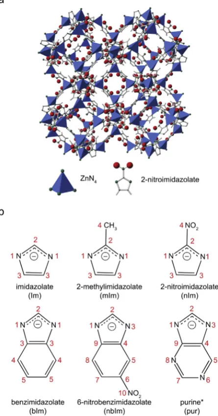
Zeolitic imidazolate frameworks (ZIFs) are a relatively new subclass of metal-organic frameworks (MOFs) with extended three-dimensional networks of transition metal nodes (predominantly Zn2þ or Co2þ) bridged by rigid imidazolate linkers[1,2]. The observed Zn-Im-Zn bond angles are similar to that of the ideal Si–O–Si bond angle (~145) found in zeolites, so that many ZIFs exhibit the same framework topol-ogies as zeolites, although ZIFs with unique framework topoltopol-ogies have also been synthesised[2]. As an example,Fig. 1a shows the structure of ZIF-65, which is synthesised with 2-nitroimidazolate as the linker, and exhibits the well-known sodalite (SOD) framework topology[3]. ZIFs have attracted considerable recent attention, owing to their potential applications in gas storage and separation, fluid separation, sensing, encapsulation and controlled delivery of drug molecules [2,4–9]. The
range of applications can be widened by using functionalised imidazolate linkers, altering the chemical nature of the pores produced, while maintaining the framework topology[10]. Additionally, more than one type of imidazolate linker can be included into the framework struc-ture[2].
ZIFs are typically synthesised by reacting a hydrated transition metal salt (e.g., Zn(NO2)2⋅6H2O) with the desired imidazole (or functionalised
imidazole) in a polar amide solvent (e.g., N,N-dimethylformamide (DMF)). In their as-prepared forms, many ZIFs contain solvent molecules in the pores, which can usually be removed by exchange or heating[1]. The linker/metal ratio and the concentration of the metal ion in solution are important factors for achieving the desiredfinal product, and it has been shown that by varying these it is possible to target specific frame-work topologies[2]. As described above, a range of linkers can be used to prepare ZIFs–those used in this work are given (along with the naming
* Corresponding author.
E-mail address:[email protected](S.E. Ashbrook).
Contents lists available atScienceDirect
Solid State Nuclear Magnetic Resonance
journal homepage:www. elsevi er.com/ locat e/ssnmr
http://dx.doi.org/10.1016/j.ssnmr.2017.09.001
Received 8 August 2017; Received in revised form 4 September 2017; Accepted 5 September 2017 Available online 9 September 2017
and numbering conventions used) inFig. 1b, and the ZIFs synthesised from them outlined inTable 1. (Note that the numbering scheme used for the purine (pur) linker is not that used conventionally in organic chem-istry, but has been chosen to facilitate comparison with other linkers). It has been shown that linkers with“bulkier”functional groups, such as benzimidazolate (bIm), produce framework materials with larger pores
and/or windows, whereas linkers with smaller functional groups, such as 2-nitroimidazolate (nIm), typically produce smaller pores with narrower apertures[2,11]. Using a combination of more than one type of linker, more complex structural frameworks, which exhibit a wider range of chemical or physical properties, can be synthesised. For example, ZIF-68 (GME topology), which is synthesised with 2-nitroimidazolate and ben-zimidazolate (in a 1:1 ratio), has both hydrophilic and hydrophobic pores
[11]. There are a few examples of ZIFs that have been prepared with more than one metal centre[12–14], which can result in a material that possesses different metal coordination environments, with a unique chemistry that can be exploited for various applications, such as catalysis. The characterisation of ZIFs commonly uses diffraction-based exper-iments, such as single-crystal or powder X-ray diffraction (XRD). Such experiments are able to provide the average (over both time and space) positions of the atoms in an average unit cell. Although single-crystal diffraction is an extremely powerful technique for structure solution, this approach is limited by the need for a suitably large and stable single crystal. Solving structures from powder XRD data is much more chal-lenging, particularly if the sample is poorly crystalline, owing to the presence of fewer resolved reflections in the diffraction pattern. Diffi -culties can also occur in refinements when lighter atoms, such as C, N and H, or isoelectronic species, are present in the material. Furthermore, if there is any disorder present, be it compositional (resulting from the use of more than one type of linker/metal in the synthesis), positional (arising from a variation in the position/orientation of a functional group, linker or solvent molecule), or temporal (resulting from the dy-namics of linkers, adsorbed solvent or guest molecules), structure solu-tion using XRD is further complicated. As many of the interesting properties of solids arise from deviations in long-range periodicity, spectroscopic techniques that probe the local, atomic-scale structure can often be a useful and complementary approach for characterising ZIFs.
[image:2.595.52.275.59.484.2] [image:2.595.38.289.655.742.2]NMR spectroscopy has been widely used to investigate the structure of many solids, owing to its sensitivity to the local environment, without the need for any long-range order[15–17]. This is a suitable approach for studying ZIFs, owing to the variety of NMR-active nuclides present
[17,18].1H,13C and15N NMR spectroscopy can, in principle, be used to investigate the number, type and relative proportions of the framework linkers. The strong dipolar couplings present for1H result in a significant spectral broadening that limits resolution (and the extraction of struc-tural information) unless very fast magic-angle spinning (MAS) is used
[15,16]. For both13C and15N, the low sensitivity (resulting primarily from their low natural abundances of 1% and 0.34%, respectively) can be overcome by cross polarisation (CP)[15,16], where magnetisation is transferred from spatially close spins with highγand high abundance (typically1H). While resulting in a considerable sensitivity enhancement (the maximum depending upon γH/γX), this approach does have the
disadvantage of being non quantitative (as the transfer depends on the spatial proximity of the two species), and direct integration of the spec-tral resonances does not necessarily result in accurate relative pro-portions of chemical species unless they have a similar environment. In principle, spectroscopic study of the metal centres in ZIFs would provide information on the coordination environment and the framework to-pology [17,18]. However, many typical framework metals have rela-tively unfavourable NMR properties. For example, Zn (used in this work) has only one NMR-active isotope (67Zn, I¼5/2) with a relatively lowγ and low natural abundance of 4.1% (resulting in a receptivity, relative to
1H, of 1.18 104). Spectral acquisition is further hindered by the
presence of a relatively large quadrupole moment (which, coupled with the lowγ, produces significant quadrupolar broadening), often necessi-tating the use of high magneticfields[19–22].
Even when resolution is increased through the use of techniques such as MAS, complex and overlapping spectral lineshapes are often still observed in NMR spectra of solids, particularly for disordered materials
[23]. In recent years, there has been increasing interest in the use of computation (typically using density functional theory or DFT) to predict NMR parameters for periodic solids, aiding the interpretation and
Fig. 1.(a) Structure of ZIF-65 (which contains a 2-nitroimidazolate (nIm) linker and Zn2þ cations). (b) Chemical structure, nomenclature and numbering schemes for the imidazo-late used in this work. Note that the numbering scheme used for pur is not that typically used in organic chemistry, but has been chosen to facilitate comparison with other linkers.
Table 1
The linker(s) used, the framework topology adopted and the corresponding CCDC code for ZIFs synthesised in this work.
ZIF Reference Topology Linker(s) CCDC code
ZIF-7 [35] SOD bIm VELVIS
ZIF-8 [35] SOD mIm VELVOY
ZIF-11 [1] RHO bIm VEJZOA
ZIF-20 [36] LTA pur MIHHAN
ZIF-65 [3] SOD nIm GITTIN
ZIF-68 [3] GME nImþbIm GITTUZ
ZIF-70 [3] GME ImþnIm GITVEL
assignment of spectra[24,25]. While extremely valuable in many cases, this can prove challenging if there are a large number of distinct atoms in the unit cell (as is the case for many ZIFs) or if there is significant disorder and/or dynamics of the linkers, functional groups or incorporated sol-vent/guest molecules. In these cases, it can be difficult to obtain a structural model that is a good approximation to the material studied experimentally, unless a very large number of possibilities are consid-ered. One option is the use of cluster calculations, centred on the metal ion and incorporating the set of coordinating linkers, thereby circum-venting problems with large cell sizes or disordered solvents. However, interactions between the framework and incorporated guests/solvents are not captured in this approach. Alternatively, periodic calculations (considering an unperturbed extended framework) can be employed, with solvent molecules either placed manually or removed entirely. The latter strategy may, however, lead to significant changes in the pore structure under geometry optimisation processes designed to minimise the forces upon atoms to ensure low-energy structures are considered, and may be less relevant to the actual material under study.
Owing to the difficulties in exploiting computational approaches for ZIFs, the amount and accuracy of the information that can be obtained from experimental measurements becomes increasingly important. There are many well-known empirically-derived relationships that link the isotropic chemical shift with the type of structural environment, and even to specific geometrical parameters, such as bond distances and angles
[15,16]. However, additional information can be obtained by measuring the chemical shift anisotropy (CSA), thereby increasing confidence in the spectral assignments. Although this interaction is removed under the rapid MAS that is usually employed to achieve high-resolution experi-ments, the anisotropic shielding parameters can, in principle, be obtained from analyticalfitting of the spinning sideband manifold observed in slow MAS experiments [15,16]. However, for systems with multiple distinct species the overlap of sideband and centreband signals can lead to difficulties in extracting this information. A number of experiments have been developed to overcome this problem, with the anisotropic information reintroduced or“recoupled”in the indirect dimension of a two-dimensional experiment, producing a sideband manifold that can be analysed to yield the desired information[26,27]. For nuclear sites that possess high local symmetry, resulting in small anisotropies,“amplifi -cation”experiments have also recently been developed[27], increasing the apparent magnitude of the interaction (by a user-defined scaling factor) and enabling more accurate measurement. The experiment used in this work, the CSA-amplified PASS approach[28,29], has been used successfully to investigate1H,13C,31P and89Y local environments in a range of materials, and correlations with local structural parameters,e.g., bond distances or point symmetry, demonstrated[29–33].
For ZIFs, solid-state NMR spectroscopy can provide information on the type and relative proportions of linkers present, and the number of crystallographically-distinct molecules present in the asymmetric unit. Furthermore, it may be possible to distinguish between ZIFs that contain the same linker(s) but exhibit different three-dimensional topologies, through small variations in the chemical shifts, peak splittings or changes to the CSA. A number of authors have acquired conventional solid-state NMR spectra (primarily13C CP MAS spectra)[1,18,34], of individual ZIFs, but little attention has been focussed on the information (if any) available from the CSA, and upon the extent to which NMR parameters vary as the topology or extended structure is changed. It is not clear whether it is possible to unambiguously establish the presence of a linker in an unknown material simply from the shifts observed in a MAS spec-trum, or whether additional confirmation from CSA measurements or15N NMR is required, or if the longer-range structure has a more significant effect. A more detailed understanding of the sensitivity of a more com-plete set of NMR parameters (i.e.,13C and13N isotropic and anisotropic shifts) to local structure will aid future solution of unknown structures, and help to understand the changes in the local environment upon the addition of guest molecules. In this work, we fully assign both13C and
15N MAS NMR spectra of a variety of (single and dual linker) ZIFs,
accurately measure CSA parameters and attempt to understand the re-lationships between NMR parameters and both local and long-range structure.
2. Methodology
2.1. Synthesis and basic characterisation
All ZIFs were prepared according to the previously reported literature procedures with some minor modifications (see theSupporting Infor-mation) [1,3,11,35,36]. In a typical synthesis (here for ZIF-7[35]), benzimidazole (0.41 g, 3.47 mmol), zinc acetate dihydrate (0.56 g, 2.55 mmol), diethylamine (4.4 mL) and N,N-dimethylformamide (25 mL) were stirred (room temperature, 10 min). The mixture was placed into a Teflon™-lined steel autoclave and heated in an oven (110C, 2 days). The ZIF-7 product was collected by centrifugation and the wet solid was left in air, overnight. To further dry the product it was immersed in methanol (10 mL, room temperature, 9 h) and the resulting solid was collected by centrifugation. The white powder (typically 0.6 g, 24% yield with respect to benzimidazole) was then dried in an oven (90 C, 15 h). Powder XRD data of the synthesised materials were collected in house using a Stoe STAD i/P diffractometer in capillary mode using Debye-Scherrer geometry with primary monochromation and Cu Kα1 (λ ¼ 1.54051 Å) radiation. The collected patterns were then
compared with simulated XRD patterns using crystal structures from the literature.
2.2. Solid-state NMR spectroscopy
Solid-state NMR spectra were acquired using Bruker Avance III spectrometers equipped with a 14.1 and 9.4 T widebore super-conducting magnets operating at Larmor frequencies of 150.9 and 100.6 MHz for13C, and 60.8 and 40.6 MHz for15N, respectively. Sam-ples were packed in conventional 4.0 mm rotors and rotated at MAS rates of 5 kHz (15N) or 12.5 kHz (13C). Chemical shifts are referenced to TMS usingL-alanine as a secondary reference (δ(CH3)¼20.5 ppm) for 13
C, and to nitromethane, using 15N-enriched glycine as a secondary reference (δ(NH3)¼ 347.4 ppm) for15N[16]. Spectra were acquired
using CP, with a contact pulse (ramped for1H) between 1 and 5 ms (13C), and 10 ms (15N) duration. Continuous wave (cw), TPPM-15[37]
or SPINAL-64[38] 1H decoupling was employed to improve spectral resolution, with a typical radiofrequency field strength (γB1/2π) of
~100 kHz. Dipolar dephasing experiments were also carried out with similar parameters and a dephasing interval of 1 ms. Two-dimensional CP CSA-amplified PASS experiments were performed using the pulse sequence of Orr et al.[28,29]The total scaling factor is given by NT¼
(nPASSþ1)N, where N is the scaling factor determined by the timing of
thefiveπ pulses and nPASS is the number of additionalfiveπ pulse
blocks. Cogwheel phase cycling was employed to reduce the length of the phase cycle required [39]. See the Supporting Information for detailed parameters. Fitting of the sideband manifolds was performed using SIMPSON[40]by comparison to an ideal one-dimensional MAS spectrum. The “root mean square” (rms) error quoted in tables and plotted in figures is that output by SIMPSON, as described in the SIMPSON manual[40]. Using the Herzfeld-Berger convention[41], the isotropic chemical shift,δiso, is given by
δiso¼(δ11þδ22þδ33)/3, (1)
where, for the principal tensor components, δ11 δ22 δ33. The
magnitude of the anisotropy is defined by the span
Ω¼δ11–δ33, (2)
and the shape is defined by the skew
such thatκlies between 1 and–1. Alternative conventions for describing the shielding interaction, in terms of the anisotropy and asymmetry, can be found in Ref.[42].
2.3. DFT calculations
Calculations of total energies and NMR parameters were carried out using the DFT code CASTEP [43] (version 16.1), which employs the gauge-including projector augmented wave (GIPAW[44]) approach, to reconstruct the all-electron wavefunction in the presence of a magnetic
field. Calculations were carried out using the GGA PBE[45]functional and core-valence interactions were described by ultra-soft pseudopo-tentials[46]. A planewave cutoff energy of 50 Ry (~680 eV) was used, and integrals over the Brillouin zone were performed using a Mon-khorst–Pack grid with a k-point spacing of 0.04 2π Å1. The TS semi-empirical dispersion correction scheme[47,48]was used. All cal-culations were converged as far as possible with respect to both k-point spacing and cutoff energy. Prior to the calculation of NMR parameters, structural models were obtained from the CCDC (seeTable 1for codes). Atoms related to the solvent molecules were removed and the geometry of the models optimised to an energy minimum (with all atomic co-ordinates and the size/shape of the unit cell allowed to vary) using the same parameters stated above. Calculations were performed either on a cluster at the University of St Andrews, consisting of 300 12-core Intel Westmere nodes, connected with QDR Infiniband, or on ARCHER, the UK High Performance Computing service, consisting of a Cray XC30 MPP supercomputer with 4920 24-core Intel Ivy Bridge nodes. Calculation wallclock times ranged from 4 to 8 days using 16–32 nodes. For further details (including information on initial models and geometry optimi-sation) see theSupporting Information.
Calculations generate the absolute shielding tensor (σ) in the crystal frame, and diagonalisation of the symmetric part ofσyields the three principal components,σ11,σ22andσ33. The principal components of the
chemical shift tensor,δ11,δ22andδ33are related toσby
δii¼–(σii–σref)/(1–σref)≈–(σii–σref), (4)
whereσref(assumed to be<<1) is the reference shielding. Values ofσref
of 175.47 ppm and136.26 ppm were used for13C and15N, respectively, determined from a comparison of the experimental shifts and calculated shieldings of all ZIFs studied in this work.
3. Results and discussion
Fig. 2shows13C CP MAS NMR spectra of Zn-containing ZIF-7, ZIF-8, ZIF-11, ZIF-20, ZIF-65, ZIF-68, ZIF-70 and ZIF-78 (seeFig. 1andTable 1
for information on the linkers used and topologies formed in each case). The presence of disordered or dynamic solvent molecules, and of mul-tiple, but chemically similar, linkers can complicate the assignment of NMR spectra of ZIFs. In many of the spectra inFig. 2, peaks associated with the synthesis solvent, DMF, can be seen (indicated byy) at chemical shifts of ~30, ~35 and ~160 ppm. In ZIF-11, peaks from the toluene solvent are seen (indicated byz) at 129.7 and 128.3 ppm. In addition, spinning sidebands are seen in all spectra (indicated by *), suggesting that many sites do exhibit a significant CSA, as would be expected for sp2 C. With the exception of ZIF-8 (where a peak at 14.9 ppm is observed for the CH3 group on the mIm linker) all resonances associated with the
linker are found between 100 and 160 ppm, indicating the aromatic nature of the linkers and the presence of the diamagnetic Zn metal centre. For ZIFs with single linkers (ZIF-7, ZIF-8, ZIF-11, ZIF-20 and ZIF-65) the chemical shifts observed are similar to those found in solution-state NMR spectra of the linker molecules, enabling tentative spectral assign-ment, and also suggesting that the longer-range structure (i.e., the exact framework topology) has a limited effect on the chemical shifts observed in the solid state. Assignments were supported by dipolar dephasing experiments, where the reintroduction of the 1H/13C dipolar coupling
[image:4.595.316.545.54.515.2]enables quaternary and protonated C species to be distinguished. For all ZIFs, spectra with dephasing times of 1 ms are shown in theSupporting Information, along with a more detailed discussion of the spectral assignment. For ZIF-20, assignment of the13C spectrum is challenging, owing to the broader, overlapped spectral lineshapes, and the chemical similarity of many of the C species present. Partial assignment can be made from the dipolar dephasing spectra, but an unambiguous assign-ment is not possible. A summary of the proposed assignassign-ments for all ZIFs is given inTable 2. In many cases, the spectral broadening (present as a result of disordered linkers or solvent molecules) makes it difficult to determine unambiguously the number of crystallographically-distinct linkers that are present in the materials, although for ZIF-8 and ZIF-65 it is clear only one set of resonances are seen. For ZIF-7, the sharp lines suggest a relatively ordered structure, and two distinct resonances are clearly seen for most C species, consistent with the presence of two crystallographically-distinct linkers [35]. Some additional splitting is
observed at lower shifts (δbetween 120 and 124 ppm), suggesting some additional disorder, most likely as a result of the incorporated solvent. ZIF-7 and ZIF-11 contain the same bIm linker, but the different synthetic routes produce ZIFs with different zeolite topologies (SOD and RHO, respectively). Although the chemical shifts observed are very similar, the exact resonance positions are different. This suggests that although the chemical shift primarily indicates the chemical nature of the linker, the
13C NMR spectra will be sensitive to changes in the structure,i.e., upon
loading of guest molecules or changes in symmetry. As an example,13C NMR spectroscopy was used to distinguish the three crystallographically-distinct types of nIm linker in Zn(nIm)2, and to
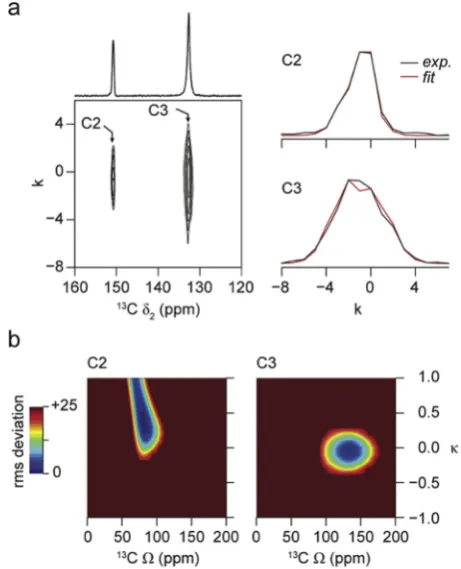
confirm the presence of rotational disorder for one of the three[49]. Furthermore,13C spectra could distinguish between the different poly-morphic forms of this material, showing changes upon an increase in temperature[49]. Additional information on a larger number of ZIFs that contain the same linker but exhibit different topologies (and/or vice versa)is required to investigate the structure sensitivity of NMR further. In addition to the isotropic chemical shift, solid-state NMR also en-ables access to the anisotropic component (parameterised byΩandκ) of this interaction. Although these are typically removed under the sample rotation used to achieve high resolution, they can be measured either from a slow MAS spectrum or from the sideband pattern in the indirect dimension of a two-dimensional experiment that recouples, or selectively reintroduces, the anisotropic shielding[26,27]. It has been shown that the anisotropic shielding, the asymmetry and the tensor orientation can provide information on the local structure including,e.g., bond distances and point symmetry [15,16,24,30]. Given the complexity of the 13C spectra of most ZIFs, slow MAS experiments result in considerable spectral overlap, and CSA-amplified PASS experiments have been used to reintroduce and measure the anisotropic shielding. Fig. 3a shows an example of a13C two-dimensional CSA-amplified PASS spectrum (for ZIF-65), along with the extracted sideband manifolds for the two C sites in the nIm linker. A scaling factor, NT, of 5 was used, resulting in an apparent
MAS rate in the indirect dimension of 2.5 kHz.Fig. 3b shows the contour plots of the rms deviation found for analyticalfitting of each sideband manifold (arbitrarily limited to 25). Values ofΩandκextracted from similar spectra for all single-linker ZIFs are given inTable 2. The low rms
[image:5.595.39.557.73.331.2]deviations observed for ZIF-7, ZIF-8 and ZIF-11 indicate that accurate values can be extracted, although for ZIF-8 it is not possible to measure
Table 2
Experimental13C (14.1 T) NMR parameters (δ
iso,Ω,κ) and rms errors, along with the assignment of resonances observed for a range of single-linker ZIFs.
δiso(ppm) Ω(ppm) κ rms error Assignment
ZIF-7 151.3±0.1 134.7±7.1 0.0±0.1 1.38 N–C(H)–N C2
149.7±0.1 134.6±7.3 0.1±0.1 4.07 N–C(H)–N C2
141.6±0.1 148.9±6.8 0.5±0.1 0.57 N–C––C–N C3
141.1±0.1 149.0±6.3 0.5±0.1 1.53 N–C––C–N C3
122.9±0.1 185.8±9.8 0.2±0.1 1.38 N–C–CH––CH C4
121.9±0.1 185.6±10.1 0.1±0.1 2.30 N–C–CH––CH C4
121.0±0.1 189.0±9.4 0.2±0.1 1.32 N–C–CH––CH C4
116.7±0.1 162.4±7.7 0.3±0.1 0.99 N–C–CH––CH C5
115.7±0.1 158.4±7.8 0.3±0.1 0.57 N–C–CH––CH C5
ZIF-8 150.9±0.1 139.2±6.7 0.1±0.1 0.80 N–C(CH3)–N C2
125.6±0.1 139.0±7.0 0.2±0.1 0.58 N–CH––CH–N C3
14.9±0.1 –CH3a C4
ZIF-11 150.5±0.1 122.2±7.7 0.0±0.1 6.02 N–C(H)–N C2
148.3±0.1 122.5±7.9 0.0±0.1 4.32 N–C(H)–N C2
141.9±0.1 145.7±6.0 0.5±0.1 0.52 N–C––C–N C3
141.5±0.1 144.4±6.1 0.6±0.1 0.26 N–C––C–N C3
140.8±0.1 140.9±5.9 0.5±0.1 0.22 N–C––C–N C3
122.6±0.1 173.8±10.3 0.1±0.1 0.81 N–C–CH––CH C4
116.1±0.1 160.2±9.2 0.3±0.1 17.14 N–C–CH––CH C5
115.0±0.1 162.3±8.6 0.2±0.1 0.89 N–C–CH––CH C5
114.4±0.1 158.1±7.9 0.2±0.1 0.20 N–C–CH––CH C5
ZIF-20 158.4±0.5 159.7±7.7 0.4±0.1 8.88 N–C(¼C–N)–N C9/4
155.7±0.5 163.3±7.3 0.4±0.1 1.37 N–C(H)–N C2/5/7
152.0±0.5 150.9±8.4 0.2±0.1 7.20 C–CH––N–CH C2/5/7
144.5±0.5 155.9±7.3 0.5±0.1 7.25 N¼CH–N C2/5/7
131.4±0.5 141.8±7.1 0.2±0.1 6.71 N–C––C–N C9/4
ZIF-65 150.6±0.1 82.1±5.1 0.4±0.1 0.52 N–C(NO2)–N C2
132.6±0.1 133.8±8.0 0.0±0.1 2.53 N–CH––CH–N C3
aInsufficient number of data points to determine the CSA parameters accurately.
[image:5.595.315.546.399.684.2]the CSA parameters for C4 of the mIm linker (δiso¼14.9 ppm), owing to
the very small magnitude of this interaction (and the insufficient number of sidebands produced even when the CSA is amplified). For ZIF-20, the higher rms error suggests greater uncertainties in the values determined, probably as a result of the greater overlap of the spectral resonances arising from the similarity in the C environments of the pur linker. The proposed crystal structure for ZIF-20 does suggest some disorder in the exact orientation of the linker molecules, with the positions of C and N in the six-membered ring not defined[35].
Table 2shows that in many cases the range ofΩfor a type of C species is greater than that of the correspondingδisovalues, potentially enabling
better distinction between ZIFs with similar linkers but different topol-ogies, and a greater sensitivity to small structural changes upon loading or post-synthetic modification. For example, for the C2 (N–C(X)–N) sites in ZIF-7 (X ¼H), ZIF-8 (X ¼CH3), ZIF-11 (X¼ H) and ZIF-65
(X ¼NO2)δisolies between 148 and 151 ppm (despite the different
chemical nature of these species), but Ωvaries from 82 to 139 ppm. Although it can be difficult to relate the anisotropic shielding parameters directly to the symmetry and arrangement of the coordinating atoms in anything other than the simplest molecular systems, it is noticeable that a much lowerΩ(82 ppm) is observed for ZIF-65, perhaps reflecting more symmetrical arrangement of the atoms bonded to C2 in this linker (i.e., three N atoms, rather than two N and one C). There is also a more sig-nificant change inκfor this species. Furthermore, for the two ZIFs with bIm linkers (ZIF-7 and ZIF-11)δisofor the N–C–CH––CH species (C4) is
very similar (i.e., within 1 ppm), while there is a ~15 ppm difference inΩ.
In ZIFs the N atoms on the imidazolate ring coordinate to the metal centre (in this case Zn2þ). In principle, N NMR could provide information on the linker(s) present, but may also be more sensitive to the Zn coor-dination environment (and also, therefore, to the framework topology). Furthermore, as there are typically fewer crystallographically-unique nitrogen species in ZIFs, it should be more straightforward to assign N NMR spectra than the corresponding13C NMR spectra. There are two NMR-active isotopes of N, 14N and15N. Although 14N has the much higher natural abundance (~99.7%), it also has spin quantum number I¼ 1, resulting in lineshapes that are broadened by the quadrupolar interaction. However, although having I¼1/2, the low natural abun-dance and low γ of 15N, results in very poor sensitivity (receptivity relative to1H is 1.04103), resulting in long experimental times unless efficient CP can be achieved[15,16].Fig. 4shows15N CP MAS NMR spectra of the single-linker ZIFs, ZIF-7, ZIF-8, ZIF-11, ZIF-20 and ZIF-65. (Peaks due to the spinning sidebands are indicated by *). Sharp15N spectral resonances can be observed for ZIF-7, ZIF-8 and ZIF-65, reflecting the ordered nature of these materials. For ZIF-20, broader spectral lines are observed, resulting from the disorder in the material discussed previously. Proposed assignments of the15N spectra are given inTable 3.
For ZIF-7, two15N resonances are observed in a 3:1 ratio, attributed to the four chemically-similar but crystallographically-distinct nitrogen sites in the proposed crystal structure, suggesting that three of the nitrogen species have very similar (although not identical) NMR parameters. The same bIm linker is present in ZIF-11 and, while similar chemical shifts are observed, the15N CP MAS spectrum is different in detail from that of ZIF-7, with ~4 distinct N shifts present (although each has a different in-tensity). Although there are four formally crystallographically-distinct N species predicted in the crystal structure (as with ZIF-7), there is also some uncertainty over the exact orientation of the bIm linker[1], leading to the additional splittings and line broadening observed in the spectrum. The
15N chemical shifts for the ZIFs containing mIm and nIm differ from those
with bIm (with downfield shifts of between 15 and 20 ppm observed for the linker N). For ZIF-65, a second N resonance is seen at27 ppm, corresponding to–NO2(N4). The two broad resonances for the pur linker
(at 121 and 192 ppm) can be assigned as N6/N8 and N1/N3, respectively, by comparison to the other ZIFs. Although, in general, the chemical shifts observed reflect the chemical nature of the linker, the15N
CP MAS spectra do appear to be more sensitive to changes in topology or variation in the local geometry around the metal centre than the corre-sponding 13C spectra, enabling different ZIFs to be more easily distinguished.
15N CSA-amplified PASS experiments were also performed for the
single-linker ZIFs, and the values of Ωand κextracted are given in
Table 3. Owing to the broad resonances observed for ZIF-20, it was not possible to obtain the CSA parameters for this material on a reasonable timescale. In general,Ωfor the imidazolate N species varies between 167 and 308 ppm, with the largest value (for theN–C(NO2)–N
environ-ment) seen for ZIF-65. The relatively low rms values (Table 3) reveal the good confidence in thefits, suggesting that the15NΩis less dependent purely on the chemical nature of the linker (and more sensitive to the three-dimensional structure). For example, for the four imidazolate N species in ZIF-11 (all chemically equivalent) a spread of nearly 50 ppm in
Ωis observed. AlthoughΩfor the–NO2group in the nIm linker in ZIF-65
[image:6.595.316.544.56.506.2]is one of the largest measured, and larger than mostΩvalues for the heterocyclic N species, it is similar to the imidazolate N for this ZIF.
As described above, it is difficult to performfirst-principles calcula-tions to aid in spectral assignment or to provide support for the experi-mental values measured, owing to the large size of many ZIF systems and the disorder/dynamics of the solvent molecules present (many of which are not placed accurately in models derived from diffraction). It is, however, possible to calculate NMR parameters for ZIFs where all sol-vent/water molecules have been removed from the pores. While this is not necessarily a true reflection of the as-prepared materials under experimental study here, it can potentially provide a better representa-tion of the chemical nature of the system (i.e., Im linkers attached to Zn2þ cations in a three-dimensional extended structure) and, therefore, more accurate NMR parameters can be obtained, than solution-state NMR spectra of the neutral linker molecules. Periodic DFT calculations were carried out for ZIF-7, ZIF-8 and ZIF-65 (i.e., single-linker ZIFs with no framework disorder). Any atoms from solvent molecules present in the diffraction-based structural models were removed from the pores (see the
Supporting Information for further discussion). Table 4 shows the calculated13C and15N NMR parameters for these three ZIFs. Although the exact values ofδisoandΩdo not match experiment exactly, as perhaps
expected owing to the changes made to the structure, they are in good
qualitative agreement and can be used to confirm the spectral assign-ments given inTables 2 and 3This can be seen clearly in the plots of calculated against experimental NMR parameters shown in the Sup-porting Information. In particular, for ZIF-7 (and, by analogy, ZIF-11) they suggest thatδisovalues for C4 and C5 (on the benzene ring of the
bIm linker) are 121–123 and 116–117 ppm, respectively, (rather than
vice versa), in contrast to the assignment that would be indicated if solution-state NMR shifts only were used. It is noticeable for ZIF-8 that the agreement between experiment and calculation forΩis poorer than for the other ZIFs, perhaps reflecting the dynamics of the–CH3group.
More complex ZIFs can be synthesised using more than one type of linker, producing materials that not only have different pore shapes and sizes, but that can also have different chemical properties (e.g., hydro-phobic or hydrophilic pores)[2,4–9]. The13C CP MAS NMR spectra of ZIF-68, ZIF-70 and ZIF-78 are shown in Fig. 2, with expansions also shown inFig. 5. These dual-linker ZIFs all possess the same topology (GME)[3,11], and all contain the nIm linker. However, the second linker for each ZIF varies, as shown inTable 1, with bIm for 68, Im for ZIF-70 and nbIm for ZIF-78. The increased complexity of the spectra (particularly in the crowded aromatic region) make assignment more challenging, and DFT calculations are not feasible (owing to the size of the unit cell and potential disorder of the linkers). Some progress can be made by comparing spectra to those for the single-linker ZIFs (Fig. 5and
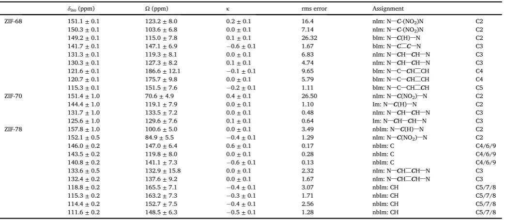
Section S3 in the Supporting Information), and by using dipolar dephasing experiments (also in the Supporting Information). Further-more,13C 2D CSA-amplified PASS experiments enable measurement of the anisotropic shielding parameters, and provide further support for the spectral assignment. The proposed assignments of the resonances are given inTable 5.
For ZIF-68, the spectrum contains resonances in very similar positions to those in ZIF-7/ZIF-11 (bIm linkers) and to those in ZIF-65 (nIm linker), enabling the type of C species to be easily assigned, as shown inFig. 5. Likewise, for ZIF-70 and ZIF-78 peaks are also observed in very similar positions to those for the nIm linker in ZIF-65. For ZIF-70 very broad spectral lines are seen, reflecting the considerable disorder of the posi-tions of the Im and nIm linkers in the structure. Although the resonances for ZIF-78 appear much narrower, many different resonances are present, reflecting the loss of the symmetry in the bIm linker through the intro-duction of the NO2group at the 6 position (seeFig. 1). Furthermore, the
crystal structure suggests some uncertainty over the exact orientation of a portion of the nIm linkers, and also in the position of the NO2group on
[image:7.595.36.555.73.185.2]the nbIm ring, although the linkers do appear to be ordered in terms of their position within the structure. Although it is possible to assign a number of C species for this material, it is not possible to distinguish easily between the three quaternary C species in nbIm (C4, C6 and C9) or between the three nbIm aromatic CH species (C5, C7 and C8), as shown inTable 2(although the quaternary and CH carbons are readily distin-guished from each other by dipolar dephasing experiments–see Sup-porting Information). In general, it is clear that the observation made earlier, that13Cδisovalues depend primarily on the chemical nature of Table 3
Experimental15N (9.4 T) NMR parameters (δ
iso,Ω,κ) and rms errors, along with the assignment of resonances observed for a range of single-linker ZIFs.
δiso(ppm) Ω(ppm) κ rms error Assignment
ZIF-7 192.6±0.1 233.5±17.2 0.7±0.2 3.03 N–C(H)–N N1
194.2±0.1 250.9±12.0 0.3±0.2 2.75 N–C(H)–N N1
ZIF-8 175.5±0.1 241.0±12.2 0.9±0.1 0.51 N–C(CH3)–N N1
ZIF-11 188.6±0.1 215.4±12.3 0.5±0.2 3.96 N–C(H)–N N1
190.3±0.1 175.1±16.2 0.8±0.1 5.81 N–C(H)–N N1
191.3±0.1 172.0±11.8 1.0±0.1 1.35 N–C(H)–N N1
193.4±0.1 167.1±12.1 0.8±0.1 1.93 N–C(H)–N N1
ZIF-20a 192.1±0.5 N1þN3
120.8±0.5 N6þN8
ZIF-65 27.2±0.1 290.9±15.4 0.3±0.1 0.54 C(NO2) N4
169.2±0.1 308.6±15.8 0.6±0.1 0.51 N–C(NO2)–N N1
aSignal intensity too low to extract CSA parameters accurately.
Table 4
Calculated (using CASTEP),13C and15N NMR parameters (δ
iso,Ω,κ) for ZIF-7, ZIF-8 and ZIF-65.
δiso(ppm) Ω(ppm) κ Assignment
ZIF-7 13C 153.35 149.10 0.08 N–C(H)–N C2
151.58 148.63 0.01 N–C(H)–N C2
147.84 166.14 0.57 N–C––C–N C3
147.76 166.84 0.57 N–C––C–N C3
147.69 169.17 0.61 N–C––C–N C3
147.53 169.77 0.61 N–C––C–N C3
129.33 224.11 0.18 N–C–CH––CH C4
128.91 223.69 0.15 N–C–CH––CH C4
127.59 223.01 0.15 N–C–CH––CH C4
127.23 223.69 0.15 N–C–CH––CH C4
121.90 198.40 0.29 N–C–CH––CH C5
121.89 197.89 0.29 N–C–CH––CH C5
120.73 193.27 0.30 N–C–CH––CH C5
120.72 192.14 0.28 N–C–CH––CH C5
15N 195.02 255.02 0.66 N–C(H)–N N1
193.36 258.80 0.69 N–C(H)–N N1
194.77 253.63 0.67 N–C(H)–N N1
192.38 261.04 0.69 N–C(H)–N N1
ZIF-8 13C 155.01 135.22 0.23 N–C(CH
3)–N C2
130.94 155.55 0.15 N–CH––CH–N C3
129.56 152.58 0.14 N–CH––CH–N C3
14.90 22.75 0.91 N–C(CH3)–N C4
15N 175.50 281.88 0.69 N–C(CH
3)–N N1
173.81 271.67 0.74 N–C(CH3)–N N1
ZIF-65 13C 157.5 78.90 0.03 N–C(NO
2)–N C2
139.40 157.95 0.08 N–CH––CH–N C3
139.40 157.95 0.08 N–CH––CH–N C3
15N 170.71 322.32 0.64 N–C(NO
2)–N N1
170.71 322.32 0.64 N–C(NO2)–N N1
[image:7.595.39.289.466.742.2]the linker and only to a lesser extent on longer-range structure and to-pology, proves to be extremely useful. When complex multi-linker ZIF structures are produced (and complex NMR spectra result), the linkers present can be identified (and spectra partially or wholly assigned) by comparison to simpler materials. In general,13CΩalso appears to be broadly similar to the values observed for single-linker ZIFs, but displays a greater variation thanδiso. For example, C2 of the nIm linker varies
between ~124 and 104 ppm (in ZIF-68), ~71 ppm (in ZIF-70), ~85 ppm (in ZIF-78), compared to ~82 ppm in the single-linker ZIF-65.
As with the single-linker ZIFs, the15N CP MAS spectra (shown in
Fig. 4, with expansions given in theSupporting Information) of the dual-linker materials contain fewer resonances than the corresponding 13C spectra. The values of15Nδisoand ofΩandκwere extracted from
CSA-amplified PASS experiments (where sufficient sensitivity was obtained) and are given inTable 6. The nIm–NO2groups are straightforward to
assign (by comparison to ZIF-65), with resonances in very similar posi-tions in all three ZIFs (26.9,27.5,27.1 ppm). For ZIF-78, the–NO2
group on the nbIm linker also appears with a very high chemical shift (10.0 ppm). For all three dual-linker ZIFs, the nIm N species have very similar shifts to those found in ZIF-65, and the bIm N species in ZIF-68 can be assigned by comparison to ZIF-7. This leaves the peaks atca. 185 ppm in ZIF-78 as the nbIm aromatic N species. For ZIF-70, it would appear that the Im N species overlap with those from nIm, producing a
very broad resonance atca.176 ppm.
Although the exact ratio of the two (or, in some cases, more) linkers that are used in an initial synthesis is known exactly, these may not be incorporated into thefinal product in the same ratios. A related problem is encountered for aluminophosphate frameworks, where multiple structure-directing agents can be used in the synthesis, but are incorpo-rated to a lesser extent (or in some cases not at all) into thefinal product
[50]. Determining the relative proportion of linkers present is important for understanding the structure and chemical properties of a material (and for reproducing it accurately), but can pose a challenge for diffraction when very similar linkers (e.g., those that differ only one functional group) are used, or where there is considerable disorder in the position of the linkers within the material. For example, in the original work, ZIF-70 was prepared using a 1:1 ratio of Im and nIm, but thefinal product exhibits a ratio of 1.3:1 (as determined from elemental analysis)
[3]. One approach to investigate linker ratios is to use acid hydrolysis to decompose the framework into individual molecules and employ solution-state NMR spectroscopy. Clearly, this has the significant disad-vantage of being a destructive technique that will be incompatible with some functional groups. In principle, the non-destructive approach of solid-state NMR spectroscopy should be ideally suited to investigate the proportions of linker present in thefinal material directly. However, as discussed above, CP is inherently non quantitative and, particularly for materials with many closely spaced or overlapped spectral lines, this can be a problem[15,16]. Although some advances towards quantitative CP measurements have been made recently using a multiple-contact method
[51], this does rely and strongly-coupled1H spin baths and so has been suggested not to be of use in the more sparse1H distributions found in MOFs. Therefore, we have focussed only on the comparison of resonances from chemically-similar species in conventional CP spectra to investigate the linker ratios.
For ZIF-68, it is difficult to determine the linker ratio unambiguously, as the13C spectrum contains both sharp and broader resonances, and it is not clear if there is additional unreacted linker present within the pores. When considering the sharper peaks only, an integral ratio of ~1:1 would be obtained for the C3 (nIm) and C4 or C5 (bIm), suggesting that the linker ratio is also close to 1:1. The relative intensities of the imidazole N in the15N spectra are closer to 1:2 (nIm:bIm), but it should be noted that the N species differ in the number (and types) of surrounding H. As described in the Supporting Information, the nIm:bIm linker ratio determined from1H solution-state NMR of the digested ZIF was 1:2, in better agreement with the 15N data. However, this value would be affected by the presence of any unreacted linker within the pores, as indicated by the13C solid-state NMR spectra. ZIF-70 also poses a chal-lenge owing to the broad peaks resulting from the linker disorder. However, decomposition of the C3 peaks in the13C NMR spectrum, gives an approximate ratio of 1.5:1 for Im:nIm. It is not possible to resolve the two chemically-similar N1 species in the15N NMR spectrum. A solution-state1H NMR spectrum after digestion gave an Im:nIm ratio of 1.9:1, higher than the 1.3:1 observed in the original preparation of ZIF-70[3]. It is not possible to resolve the two chemically-similar N1 species in the
15N NMR spectrum. For ZIF-78, although the13C spectra cannot be fully
assigned, the relative intensities of the C3 (nIm) and C5þC7þC8 (nbIm) signals are ~1:1.5 (or 2:3), as expected for a 1:1 ratio of the linkers. In the
15N NMR spectrum, the relative intensity ratio of N1 (nIm) and N1þN3
(nbIm) is ~1:1.2 and, although the exact chemical environment of the imidazole N is different, the relatively low number of proximate H in each case probably supports the conclusion from the13C NMR spectrum. As expected, the relative ratios of the signals from the–NO2groups are
not in agreement (~1.8:1 for N4:N10), reflecting the greater number of nearby1H in the nbIm linker. The solution-state1H NMR spectrum of the digested ZIF confirmed a 1:1.1 (nIm:nbIm), in very good agreement with the solid-state NMR data.
Fig. 6a shows a plot of the experimental13CδisoagainstΩfor each
[image:8.595.51.276.55.451.2]resonance in the single- and dual-linker ZIFs (with the exception of ZIF-20). Symbols denote the chemical type of species/linker and these are
coloured according to the ZIF from which they result. The points clearly fall into separate regions, with N–C(X)–N species (shown by crosses) having highδiso(between 145 and 155 ppm). There is a variation inΩ
(between 80 and 140 ppm), with the smallestΩobserved when R¼NO2, i.e., when C2 is bonded to three N. HighestΩare found for R¼H. The N–CH––CH–N species in the Im, mIm and nIm linkers (denoted by the
filled circles) have very similarδiso(125–132 ppm) andΩ(~130 ppm).
For bIm-based linkers, the related, but now unprotonated, N–C––C–N species (denoted byfilled and open squares) generally have higherδiso
(140–145 ppm), but similarΩ. The C species in the benzene rings have the lowest δisoand the highestΩ, with the introduction of the–NO2
group for ZIF-78 resulting in a decrease in δiso and Ω. A plot of the
experimental15NδisoagainstΩis shown inFig. 6b. The N species can be
clearly differentiated by theirδisovalues, with–NO2at much higher
shifts (25 to 0 ppm) and Im N species between150 and200 ppm. Unlike13C, a fairly wide range ofΩvalues are observed for the Im N, with little clear correlation withδisoor with the chemical nature of the linker.
Owing to low sensitivity, it was not possible to measureΩfor all–NO2
groups, so it is not currently possible to comment more generally on the typicalΩfor these species.
4. Conclusions
In this work we have investigated the13C and15N NMR parameters of a series of ZIFs, prepared using one or more functionalised imidazolate
and/or benzimidazolate linkers. In most cases, the resonances in the solid-state NMR spectra can be tentatively assigned by combining a basic knowledge of the chemical shifts observed for linker molecules in solu-tion with dipolar dephasing experiments to identify quaternary C species. Owing to the chemical complexity of the systems studied,i.e., the size of the unit cells, the presence of linker disorder, disorder in the orientation of functionalised linkers and the presence and dynamics of solvent/water molecules within the pores of the materials, it is difficult to assign res-onances to specific crystallographic sites, or to use periodic DFT calcu-lations to predict exact shifts. However, calcucalcu-lations performed for ordered model systems were able to aid and confirm the spectral assignment for some of the simpler ZIFs. Measurement of the anisotropic component of the shielding, specificallyΩ, using amplified PASS exper-iments provides additional support for the spectral assignment.
The plots inFig. 6indicate that the13C and15N NMR parameters are sufficiently sensitive to detect small differences in chemical environ-ments,e.g., changes to topology, linker functionalisation or the presence of molecules within the pores, when comparing different ZIFs or different forms of the same ZIF. However, the magnitude ofδisoandΩobserved
[image:9.595.39.561.71.298.2]relate primarily to the chemical nature of the linker, and so measurement of both parameters, particularly for13C, provides strong support for determining the type(s) of linkers present. For example, spectra of the dual-linker ZIFs contain resonances with very similar shifts to those of ZIFs composed of just one linker, and so the chemical nature of the C species can be determined despite considerable spectral overlap in some
Table 5
Experimental13C (14.1 T) NMR parameters (δ
iso,Ω,κ) and rms errors, along with the assignment of resonances observed for a range of dual-linker ZIFs.
δiso(ppm) Ω(ppm) κ rms error Assignment
ZIF-68 151.1±0.1 123.2±8.0 0.2±0.1 16.4 nIm: N–C-(NO2)N C2
150.3±0.1 103.6±6.8 0.0±0.1 7.14 nIm: N–C-(NO2)N C2
149.2±0.1 115.0±7.8 0.1±0.1 26.32 bIm: N–C(H)–N C2
141.7±0.1 147.1±6.9 0.6±0.1 1.67 bIm: N–C––C–N C3
131.3±0.1 119.3±8.1 0.0±0.1 6.83 nIm: N–CH–CH–N C3
130.3±0.1 127.3±8.2 0.1±0.1 4.74 nIm: N–CH–CH–N C3
121.6±0.1 186.6±12.1 0.1±0.1 9.65 bIm: N–C–CH––CH C4
120.7±0.1 175.7±9.8 0.0±0.1 5.79 bIm: N–C–CH––CH C4
115.3±0.1 151.5±7.6 0.2±0.1 1.11 bIm: N–C–CH––CH C5
ZIF-70 151.4±1.0 70.6±4.9 0.4±0.1 26.50 nIm: N–C(NO2)–N C2
144.4±1.0 119.1±7.9 0.0±0.1 1.10 Im: N–C(H)–N C2
131.7±1.0 133.5±7.2 0.0±0.1 0.48 nIm: N–CH–CH–N C3
125.6±1.0 129.6±7.6 0.1±0.1 0.64 Im: N–CH–CH–N C3
ZIF-78 157.8±1.0 100.6±5.0 0.0±0.1 3.49 nbIm: N–C(H)–N C2
152.1±0.5 84.9±5.5 0.4±0.1 1.29 nIm: N–C(NO2)–N C2
146.0±0.2 147.0±6.4 0.6±0.1 0.17 nbIm: C C4/6/9
143.5±0.2 119.8±8.0 0.0±0.1 0.28 nbIm: C C4/6/9
140.8±0.2 141.1±7.3 0.6±0.1 0.13 nbIm: C C4/6/9
133.6±0.5 132.9±15.8 0.0±0.1 2.32 nIm: N–CH––CH–N C3
132.4±0.2 137.6±9.2 0.0±0.1 1.67 nIm: N–CH––CH–N C3
118.8±0.2 165.5±7.1 0.4±0.1 3.07 nbIm: CH C5/7/8
115.3±0.2 163.2±7.3 0.3±0.1 1.71 nbIm: CH C5/7/8
114.4±0.2 152.7±7.5 0.4±0.1 2.56 nbIm: CH C5/7/8
111.6±0.2 148.5±6.3 0.5±0.1 1.28 nbIm: CH C5/7/8
Table 6
Experimental15N (9.4 T) NMR parameters (δ
iso,Ω,κ) and rms errors, along with the assignment of resonances observed for a range of dual-linker ZIFs.
δiso(ppm) Ω(ppm) κ rms error Assignment
ZIF-68 26.9±0.1a nIm: C(NO
2) N4
170.9±0.1 231.8±9.6 1.0±0.1 3.46 nIm:N–C(NO2)–N N1
172.9±0.1 201.3±11.2 1.0±0.1 129.68 nIm:N–C(NO2)–N N1
191.6±0.1 189.5±21.8 0.6±0.3 70.39 bIm:N–C(H)–N N1
ZIF-70 27.5±0.2a nIm: C(NO
2) N4
175.0±0.2 219.1±17.4 0.5±0.2 6.12 nIm/Im:N N1
ZIF-78 10.0±0.1 220.0±11.0 0.2±0.2 21.87 nbIm: C(NO2) N10
27.1±0.1 210.0±11.0 0.0±0.2 8.93 nIm: C(NO2) N4
173.4±0.1 237.3±11.2 0.5±0.2 4.50 nIm:N–C(NO2)–N N1
184.6±0.1 205.6±10.5 0.4±0.2 20.32 nbIm:N–C(H)–N N1
188.4±0.1 200.9±18.0 0.6±0.2 3.59 nbIm:N–C(H)–N N1
a
[image:9.595.37.563.336.450.2]cases, demonstrating the value of this type of linker“library”. For these materials, some indication of the relative proportion of each linker pre-sent can be obtained from the relative intensities of resonances from chemically-similar C species that are well-resolved in the13C CP MAS NMR spectrum, although care must be taken to ensure that the distri-butions of nearby H are sufficiently similar. Some progress may be made in this respect in the future by investigating the applicability of the multiple-contact CP approach,[51] to ZIFs (and MOFs more generally). This work has initiated the more general task of creating a library of NMR parameters of single- and dual-linker ZIFs that can be used not only by the solid-state NMR community, but also the wider materials chemistry community to help to understand and assign NMR spectra. The results will also be useful to help solve the structures of new (and particularly complex and disordered) ZIFs and to characterise those subjected to post-synthetic modification.
Acknowledgements
We are grateful to EPSRC computational support through the Collaborative Computational Project on NMR Crystallography (CCP-NC), via EP/M022501/1, and for other support through EP/G062129/1 (JK) and EP/M506631/1 (SS). AFO acknowledges funding from the European Community Seventh Framework Program (FP7/2007–2013 [grant agreement number 608490], Project M4CO2). SEA would also like to thank the Royal Society and Wolfson Foundation for a merit award. AFO would also like to acknowledge the SCI for a scholarship for her PhD
studies. Some of the calculations were performed on the ARCHER UK National Supercomputing Service, and were supported by CCP-NC. The research data (and/or materials) supporting this publication can be accessed at http://dx.doi.org/10.17630/7959a81e-161d-4ada-9914-08d3d235ce88.
Appendix A. Supplementary data
Supplementary data related to this article can be found athttp://dx. doi.org/10.1016/j.ssnmr.2017.09.001.
References
[1] K.S. Park, Z. Ni, A.P. C^ote, J.Y. Choi, R. Huang, F.J. Uribe-Romo, H.K. Chae, M. O'Keeffe, O.M. Yaghi, Exceptional chemical and thermal stability of zeolitic imidazolate frameworks, Proc. Nat. Acad. Sci. U.S.A. 103 (2006) 10186–10191. [2] A. Phan, C.J. Doonan, F.J. Uribe-Romo, C.B. Knobler, M. O'Keeffe, O.M. Yaghi,
Synthesis, structure, and carbon dioxide capture properties of zeolitic imidazolate frameworks, Acc. Chem. Res. 43 (2010) 58–67.
[3] R. Banerjee, A. Phan, B. Wang, C. Knobler, H. Furukawa, M.O.' Keeffe, O.M. Yaghi, High-throughput synthesis of zeolitic imidazolate frameworks and application to CO2 capture, Science 319 (2008) 939–943.
[4] B. Chen, Z. Yang, Y. Zhu, Y. Xia, Zeolitic imidazolate framework materials: recent progress in synthesis and applications, J. Mater. Chem. A 2 (2014) 16811–16831. [5] B.R. Pimentel, A. Parulkar, E.-K. Zhou, N.A. Brunelli, R.P. Lively, Zeolitic
imidazolate frameworks: next-generation materials for energy-efficient gas separations, ChemSusChem 7 (2014) 3202–3240.
[6] B. Wang, A.P. Cot^e, H. Furukawa, M. O'Keeffe, O.M. Yaghi, Colossal cages in zeolitic imidazolate frameworks as selective carbon dioxide reservoirs, Nature 453 (2008) 207–211.
[7] Y.-Q. Tian, Y.-M. Zhao, Z.-X. Chen, G.-N. Zhang, L.-H. Weng, D.-Y. Zhao, Design and generation of extended zeolitic metal–organic frameworks (ZMOFs): synthesis and crystal structures of zinc(II) imidazolate polymers with zeolitic topologies, Chem. Eur. J. 13 (2007) 4146–4154.
[8] P. Horcajada, T. Chalati, C. Serre, B. Gillet, C. Sebrie, T. Baati, J.F. Eubank, D. Heurtaux, P. Clayette, C. Kreuz, J.-S. Chang, Y.K. Hwang, V. Marsaud, P.-N. Bories, L. Cynober, S. Gil, G. Ferey, P. Couvreur, R. Gref, Porous metal– organic-framework nanoscale carriers as a potential platform for drug delivery and imaging, Nat. Mater 9 (2009) 172–178.
[9] J. An, S.J. Geib, N.L. Rosi, Cation-triggered drug release from a porous ZincAdeninate MetalOrganic framework, J. Am. Chem. Soc. 131 (2009) 8376–8377.
[10] W. Morris, C.J. Doonan, H. Furukawa, R. Banerjee, O.M. Yaghi, Crystals as molecules: postsynthesis covalent functionalization of zeolitic imidazolate frameworks, J. Am. Chem. Soc. 130 (2008) 12626–12627.
[11] R. Banerjee, H. Furukawa, D. Britt, C. Knobler, M. O'Keeffe, O.M. Yaghi, Control of pore size and functionality in isoreticular zeolitic imidazolate frameworks and their carbon dioxide selective capture properties, J. Am. Chem. Soc. 131 (2009) 3875–3877.
[12] Y. Ban, Y. Li, Y. Peng, H. Jin, W. Jiao, X. Liu, W. Yang, Metal-substituted zeolitic imidazolate framework ZIF-108: gas-sorption and membrane-separation properties, Chem. Eur. J. 20 (2014) 11402–11409.
[13] H. Fei, J.F. Cahill, K.A. Prather, S.M. Cohen, Tandem postsynthetic metal Ion and ligand exchange In zeolitic imidazolate frameworks, Inorg. Chem. 52 (2013) 4011–4016.
[14] M. Kim, J.F. Cahill, H. Fei, K.A. Prather, S.M. Cohen, Postsynthetic ligand and cation exchange in robust metal–organic frameworks, J. Am. Chem. Soc. 134 (2012) 18082–18088.
[15] S.E. Ashbrook, D.M. Dawson, J.M. Griffin, Local Structural Characterisation, John Wiley&Sons Ltd, Chichester, UK, 2014.
[16] K.J.D. MacKenzie, M.E. Smith, Multinuclear Solid-state NMR of Inorganic Materials, Pergamon Press, Oxford, UK, 2002.
[17] S.E. Ashbrook, D.M. Dawson, V.R. Seymour, Recent developments in solid-state NMR spectroscopy of crystalline microporous materials, Phys. Chem. Chem. Phys. 16 (2014) 8223–8242.
[18] A. Sutrisno, Y. Huang, Solid-state NMR: a powerful tool for characterization of metal–organic frameworks, Solid State Nucl. Magn. Reson 49–50 (2013) 1–11. [19] A. Sutrisno, L. Liu, J. Xu, Y. Huang, Natural abundance solid-state67Zn NMR
characterization of microporous zinc phosphites and zinc phosphates at ultrahigh magneticfield, Phys. Chem. Chem. Phys. 13 (2011) 16606–16617.
[20] A. Sutrisno, V.V. Terskikh, Q. Shi, Z. Song, J. Dong, S.Y. Ding, W. Wang, B.R. Provost, T.D. Daff, T.K. Woo, Y. Huang, Characterization of Zn-Containing metal–organic frameworks by solid-state67Zn NMR spectroscopy and computational modeling, Chem. Eur. J. 18 (2012) 12251–12259.
[21] P. He, B.E.G. Lucier, V.V. Terskikh, Q. Shi, J. Dong, Y. Chu, A. Zheng, A. Sutrisno, Y. Huang, Spies within metal-organic frameworks: investigating metal centers using solid-state NMR, J. Phys. Chem. C 118 (2014) 23728–23744.
[22] S.E. Ashbrook, S. Sneddon, New methods and applications in solid-state NMR spectroscopy of quadrupolar nuclei, J. Am. Chem. Soc. 136 (2014) 15440–15456. [23] R.F. Moran, D.M. Dawson, S.E. Ashbrook, Exploiting NMR spectroscopy for the
study of disorder in solids, Int. Rev. Phys. Chem. 36 (2017) 39–115. [24] C. Bonhomme, C. Gervais, F. Babonneau, C. Coelho, F. Pourpoint, T. Azaïs,
[image:10.595.48.275.58.426.2]S.E. Ashbrook, J.M. Griffin, J.R. Yates, F. Mauri, C.J. Pickard, First-principles Fig. 6.Plot of (a)13C and (b)15NΩagainstδ
calculation of NMR parameters using the gauge including projector augmented wave method: a Chemist's point of view, Chem. Rev. 112 (2012) 5733–5779. [25] S.E. Ashbrook, D. McKay, Combining solid-state NMR spectroscopy withfirst
principles calculations–a guide to NMR Crystallography, Chem. Commun. 52 (2016) 7186–7204.
[26] O.N. Antzutkin, Sideband manipulation in magic-angle-spinning nuclear magnetic resonance, Prog. Nucl. Magn. Reson. Spectrosc. 35 (1999) 203–266.
[27] L. Shao, J.J. Titman, Chemical shift anisotropy amplification, Prog. Nucl. Magn. Reson. Spectrosc. 51 (2007) 103–137.
[28] R.M. Orr, M.J. Duer, Applications of the CSA-amplified PASS experiment, Solid State Nucl. Magn. Reson 30 (2006) 1–8.
[29] R.M. Orr, M.J. Duer, S.E. Ashbrook, Correlating fast and slow chemical shift spinning sideband patterns in solid-state NMR, J. Magn. Reson 174 (2005) 301–309. [30] M.R. Mitchell, D. Carnevale, R. Orr, K.R. Whittle, S.E. Ashbrook, Exploiting the
chemical shielding anisotropy to probe structure and disorder in ceramics:89Y MAS NMR andfirst-principles calculations, J. Phys. Chem. C 116 (2012) 4273–4286. [31] D. Carnevale, S.E. Ashbrook, G. Bodenhausen, Solid-state NMR measurements and
DFT calculations of the magnetic shielding tensors of protons of water trapped in barium chlorate monohydrate, RSC Adv. 4 (2014) 56248–56258.
[32] S.E. Ashbrook, M.R. Mitchell, S. Sneddon, R.F. Moran, M. de los Reyes, G.R. Lumpkin, K.R. Whittle, New insights into phase distribution, phase composition and disorder in Y2(Zr,Sn)2O7ceramics from NMR spectroscopy, Phys. Chem. Chem. Phys. 17 (2015) 9049–9059.
[33] S. Sneddon, University of St Andrews, PhD Thesis, 2016.
[34] E.F. Baxter, T.D. Bennett, C. Mellot-Draznieks, C. Gervais, F. Blanc, A.K. Cheetham, Combined experimental and computational NMR study of crystalline and amorphous zeolitic imidazolate frameworks, Phys. Chem. Chem. Phys. 17 (2005) 25191–25196.
[35] X. Huang, J. Zhang, X. Chen, [Zn(bim)2]⋅(H2O)1.67: a metal-organic open-framework with sodalite topology, Chin. Sci. Bull. 48 (2003) 1531–1534. [36] H. Hayashi, A.P. C^ote, H. Furukawa, M. O'Keeffe, O.M. Yaghi, Zeolite a imidazolate
frameworks, Nat. Mater 6 (2007) 501–506.
[37] A.E. Bennett, C.M. Rienstra, M. Auger, K.V. Lakshmi, R.G. Griffin, Heteronuclear decoupling in rotating solids, J. Chem. Phys. 103 (1995) 6951–6958. [38] D. Sandstr€om, M.H. Levitt, Structure, Molecular, Ordering of a nematic liquid
crystal studied by natural-abundance double-quantum13C NMR, J. Am. Chem. Soc. 118 (1996) 6966–6974.
[39] M.H. Levitt, P.K. Madhu, C.E. Hughes, Cogwheel phase cycling, J. Magn. Reson 155 (2002) 300–306.
[40] M. Bak, J.T. Rasmussen, N.C. Nielsen, SIMPSON: a general simulation Program for solid-state NMR spectroscopy, J. Magn. Reson 147 (2000) 296–330.
[41] J. Herzfeld, A.E. Berger, Sideband intensities in NMR spectra of samples spinning at the magic angle, J. Chem. Phys. 73 (1980) 6021–6030.
[42] R.K. Harris, E.D. Becker, S.M.C. de Menezes, P. Granger, R.E. Hoffman, K.W. Zilm, Further conventions for NMR shielding and chemical shifts (IUPAC
recommendations 2008), Pure Appl. Chem. 80 (2008) 59–84.
[43] M.D. Segall, P.J.D. Lindan, M.J. Probert, C.J. Pickard, P.J. Hasnip, S.J. Clark, M.C. Payne, First-principles simulation: ideas, illustrations and the CASTEP code, J. Phys. Condens. Matter 14 (2002) 2717–2744.
[44] C.J. Pickard, F. Mauri, All-electron magnetic response with pseudopotentials: NMR chemical shifts, Phys. Rev. B 63 (2001) 245101.
[45] J.P. Perdew, K. Burke, M. Ernzerhof, Generalized gradient approximation made simple, Phys. Rev. Lett. 77 (1996) 3865–3868.
[46] J.R. Yates, C.J. Pickard, F. Mauri, Calculation of NMR chemical shifts for extended systems using ultrasoft pseudopotentials, Phys. Rev. B 76 (2007) 024401. [47] E.R. McNellis, J. Meyer, K. Reuter, Azobenzene at Coinage Metal Surfaces: role of
Dispersive van der Waals Interactions, Phys. Rev. B 80 (2009) 205414. [48] A. Tkatchenko, M. Scheffler, Accurate Molecular van der Waals Interactions from
Ground-State Electron Density and Free-Atom Reference Data, Phys. Rev. Lett. 102 (2009) 073005.
[49] A. Orsi, D.J. Price, J. Kahr, R.S. Pillai, S. Sneddon, S. Cao, V. Benoit, M.M.Łozinska, D.B. Cordes, A.M.Z. Slawin, P.L. Llewellyn, I. Casely, S.E. Ashbrook, G. Maurin, P.A. Wright, Porous zinc and cobalt 2-nitroimidazolate frameworks with six-membered ring windows and a layered cobalt 2-nitroimidazolate polymorph, CrystEngComm 19 (2017) 1377–1388.
[50] Z. Han, A.L. Picone, A.M.Z. Slawin, V.R. Seymour, S.E. Ashbrook, W. Zhou, S.P. Thompson, J.E. Parker, P.A. Wright, Novel large-pore aluminophosphate molecular sieve STA-15 prepared using the tetrapropylammonium cation as a structure directing agent, Chem. Mater 22 (2010) 338–346.