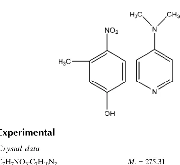
3-Methyl-4-nitrophenol–4-dimethyl-aminopyridine (1/1)
Srinivasan Muralidharan,aPerumal Nagapandiselvi,a Thothadri Srinivasan,bRengasamy Gopalakrishnanaand Devadasan Velmuruganb*
aDepartment of Physics, Anna University, Chennai 600 025, India, andbCentre of
Advanced Study in Crystallography and Biophysics, University of Madras, Guindy Campus, Chennai 600 025, India
Correspondence e-mail: [email protected]
Received 20 September 2012; accepted 5 October 2012
Key indicators: single-crystal X-ray study;T= 293 K; mean(C–C) = 0.002 A˚; Rfactor = 0.056;wRfactor = 0.185; data-to-parameter ratio = 19.0.
In the title adduct, C7H7NO3 .
C7H10N2, the dihedral angle
betwen the benzene ring and pyridine rings is 9.60 (8)while
the nitro group attached to the benzene ring makes a dihedral angle of 21.76 (13). The hydroxyl O atom deviates by 0.0247 (15) A˚ from the plane of the benzene ring. The crystal packing features O—H N hydrogen bonds.
Related literature
For a related structure, see: Dong & Cheng (2012).
Experimental
Crystal data
C7H7NO3C7H10N2 Mr= 275.31
a= 11.4923 (9) A˚ b= 9.8362 (8) A˚ c= 12.7781 (10) A˚
= 103.870 (5)
V= 1402.3 (2) A˚3
MoKradiation
= 0.09 mm1 T= 293 K
0.350.300.30 mm
Data collection
Bruker SMART APEXII area-detector diffractometer Absorption correction: multi-scan
(SADABS; Bruker, 2008) Tmin= 0.968,Tmax= 0.973
13307 measured reflections 3498 independent reflections 2469 reflections withI> 2(I) Rint= 0.028
Refinement
R[F2> 2(F2)] = 0.056 wR(F2) = 0.185 S= 1.03 3498 reflections
184 parameters
H-atom parameters constrained max= 0.35 e A˚
3 min=0.27 e A˚
[image:1.610.48.234.460.630.2]3
Table 1
Hydrogen-bond geometry (A˚ ,).
D—H A D—H H A D A D—H A
O3—H3 N3i 0.82 1.79 2.594 (2) 168
Symmetry code: (i)x;y1 2;zþ
3 2.
Data collection:APEX2(Bruker, 2008); cell refinement:SAINT (Bruker, 2008); data reduction:SAINT; program(s) used to solve structure:SHELXS97(Sheldrick, 2008); program(s) used to refine structure: SHELXL97 (Sheldrick, 2008); molecular graphics: ORTEP-3(Farrugia, 1997); software used to prepare material for publication:SHELXL97andPLATON(Spek, 2009).
The authors thank the TBI X-ray facility, CAS in Crystal-lography and Biophysics, University of Madras, India, for the data collection. TS thanks the DST for an Inspire fellowship.
Supplementary data and figures for this paper are available from the IUCr electronic archives (Reference: PV2590).
References
Bruker (2008).APEX2, SAINTandSADABS. Bruker AXS Inc., Madison, Wisconsin, U. S. A.
Dong, S.-L. & Cheng, X. (2012).Acta Cryst.E68, o518. Farrugia, L. J. (1997).J. Appl. Cryst.30, 565.
Sheldrick, G. M. (2008).Acta Cryst.A64, 112–122. Spek, A. L. (2009).Acta Cryst.D65, 148–155. Structure Reports
Online
supporting information
Acta Cryst. (2012). E68, o3106 [doi:10.1107/S1600536812041670]
3-Methyl-4-nitrophenol
–
4-dimethylaminopyridine (1/1)
Srinivasan Muralidharan, Perumal Nagapandiselvi, Thothadri Srinivasan, Rengasamy
Gopalakrishnan and Devadasan Velmurugan
S1. Comment
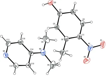
In the molecule (fig.1),the pyridine ring (N3/C10/C11/C12/C13/C14) makes a dihedral angle of 9.60 (8)° with the phenyl
ring (C1/C2/C3/C4/C5/C6) system. The oxygen atom O3 deviates by -0.0247 (15)Å from the plane of the phenyl ring.
The carbon atom C7 deviates by deviates by 0.0677 (21)Å from the plane of the phenyl ring.
The nitrogen atom N1 deviates by -0.0285 (18)Å from the plane of the phenyl ring. The nitrogen atom N2 devaites by
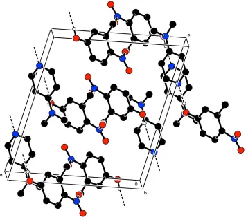
-0.0292 (18)Å from the plane of the pyridine ring. The crystal packing is stabilized by intermolecular O—H···N hydrogen
bonds
S2. Experimental
4-Dimethylaminopyridine and 3-methyl-4-nitrophenol were taken in equimolar (1:1) ratio using acetone as solvent. The
solution was filtered in a clean beaker and optimally closed. The prepared solution was kept at room temperature for two
days after which crystals suitable for X-ray diffraction were obtained.
S3. Refinement
The hydrogen atoms were placed in calculated positions with C—H = 0.93 Å to 1.08 Å refined in the riding model with
Figure 1
The molecular structure of the title compound, showing displacement ellipsoids drawn at the 30% probability level. H
Figure 2
The crystal packing of the title compound viewed down c axis. H-atoms not involved in H-bonds have been excluded for
clarity.
3-Methyl-4-nitrophenol–4-dimethylaminopyridine (1/1)
Crystal data
C7H7NO3·C7H10N2 Mr = 275.31
Monoclinic, P21/c Hall symbol: -P 2ybc
a = 11.4923 (9) Å
b = 9.8362 (8) Å
c = 12.7781 (10) Å
β = 103.870 (5)°
V = 1402.3 (2) Å3 Z = 4
F(000) = 584
Dx = 1.304 Mg m−3
Mo Kα radiation, λ = 0.71073 Å Cell parameters from 3498 reflections
θ = 1.8–28.4°
µ = 0.09 mm−1 T = 293 K Block, colourless 0.35 × 0.30 × 0.30 mm
Data collection
Bruker SMART APEXII area-detector diffractometer
Radiation source: fine-focus sealed tube Graphite monochromator
ω and φ scans
Absorption correction: multi-scan (SADABS; Bruker, 2008)
13307 measured reflections 3498 independent reflections 2469 reflections with I > 2σ(I)
Rint = 0.028
θmax = 28.4°, θmin = 1.8°
h = −15→12
Refinement on F2 Least-squares matrix: full
R[F2 > 2σ(F2)] = 0.056 wR(F2) = 0.185 S = 1.03 3498 reflections 184 parameters 0 restraints
Primary atom site location: structure-invariant direct methods
Secondary atom site location: difference Fourier map
Hydrogen site location: inferred from neighbouring sites
H-atom parameters constrained
w = 1/[σ2(F
o2) + (0.0939P)2 + 0.361P] where P = (Fo2 + 2Fc2)/3
(Δ/σ)max = 0.001 Δρmax = 0.35 e Å−3 Δρmin = −0.27 e Å−3
Special details
Geometry. All esds (except the esd in the dihedral angle between two l.s. planes) are estimated using the full covariance matrix. The cell esds are taken into account individually in the estimation of esds in distances, angles and torsion angles; correlations between esds in cell parameters are only used when they are defined by crystal symmetry. An approximate (isotropic) treatment of cell esds is used for estimating esds involving l.s. planes.
Refinement. Refinement of F2 against ALL reflections. The weighted R-factor wR and goodness of fit S are based on F2, conventional R-factors R are based on F, with F set to zero for negative F2. The threshold expression of F2 > 2sigma(F2) is used only for calculating R-factors(gt) etc. and is not relevant to the choice of reflections for refinement. R-factors based on F2 are statistically about twice as large as those based on F, and R- factors based on ALL data will be even larger.
Fractional atomic coordinates and isotropic or equivalent isotropic displacement parameters (Å2)
x y z Uiso*/Ueq
O3 0.15952 (12) −0.10464 (16) 0.99019 (11) 0.0722 (4)
H3 0.1231 −0.0878 0.9281 0.108*
N3 −0.03318 (13) 0.41197 (16) 0.70694 (12) 0.0580 (4)
N1 0.55651 (14) 0.22673 (19) 1.12638 (14) 0.0661 (4)
O1 0.63106 (15) 0.1883 (2) 1.20437 (15) 0.0964 (6)
N2 0.17044 (15) 0.41211 (18) 1.02568 (13) 0.0637 (4)
C14 0.10524 (14) 0.41079 (16) 0.92229 (13) 0.0474 (4)
C6 0.38178 (14) 0.15782 (17) 0.97841 (13) 0.0475 (4)
C1 0.45173 (14) 0.14230 (17) 1.08398 (13) 0.0498 (4)
C13 0.13746 (15) 0.48787 (18) 0.84064 (15) 0.0524 (4)
H13 0.2061 0.5415 0.8567 0.063*
C3 0.32858 (16) −0.0365 (2) 1.12182 (14) 0.0568 (4)
H3A 0.3113 −0.1011 1.1691 0.068*
C10 0.00038 (16) 0.33308 (18) 0.88898 (16) 0.0569 (4)
H10 −0.0253 0.2778 0.9381 0.068*
C5 0.28341 (14) 0.07288 (18) 0.94833 (13) 0.0499 (4)
H5 0.2347 0.0806 0.8791 0.060*
C4 0.25448 (14) −0.02386 (17) 1.01789 (13) 0.0507 (4)
C11 −0.06391 (16) 0.33887 (19) 0.78414 (17) 0.0615 (5)
H11 −0.1342 0.2882 0.7652 0.074*
C2 0.42568 (15) 0.0457 (2) 1.15360 (13) 0.0558 (4)
H2 0.4749 0.0369 1.2226 0.067*
O2 0.56459 (19) 0.3367 (2) 1.08688 (19) 0.1206 (8)
C12 0.06747 (16) 0.48361 (18) 0.73744 (15) 0.0560 (4)
C7 0.40796 (19) 0.2548 (2) 0.89618 (16) 0.0650 (5)
H7A 0.3587 0.2327 0.8264 0.098*
H7B 0.4909 0.2478 0.8948 0.098*
H7C 0.3910 0.3460 0.9148 0.098*
C9 0.1410 (3) 0.3213 (3) 1.10553 (18) 0.0842 (7)
H9A 0.0657 0.3478 1.1194 0.126*
H9B 0.2026 0.3264 1.1711 0.126*
H9C 0.1353 0.2297 1.0787 0.126*
C8 0.26892 (19) 0.5044 (3) 1.06203 (19) 0.0842 (7)
H8A 0.3380 0.4714 1.0400 0.126*
H8B 0.2870 0.5109 1.1392 0.126*
H8C 0.2475 0.5926 1.0311 0.126*
Atomic displacement parameters (Å2)
U11 U22 U33 U12 U13 U23
O3 0.0643 (8) 0.0827 (10) 0.0624 (8) −0.0234 (7) 0.0014 (6) 0.0170 (7)
N3 0.0535 (8) 0.0548 (8) 0.0598 (9) 0.0094 (7) 0.0020 (6) −0.0021 (7)
N1 0.0526 (9) 0.0713 (11) 0.0682 (10) −0.0036 (8) 0.0023 (7) −0.0149 (8)
O1 0.0690 (10) 0.1098 (13) 0.0881 (11) −0.0055 (9) −0.0249 (8) −0.0143 (10)
N2 0.0660 (10) 0.0714 (10) 0.0511 (8) 0.0051 (8) 0.0087 (7) −0.0005 (7)
C14 0.0456 (8) 0.0435 (8) 0.0533 (9) 0.0073 (6) 0.0119 (6) −0.0005 (7)
C6 0.0458 (8) 0.0496 (9) 0.0482 (8) 0.0043 (6) 0.0132 (6) −0.0018 (6)
C1 0.0406 (8) 0.0544 (9) 0.0515 (9) 0.0044 (7) 0.0056 (6) −0.0099 (7)
C13 0.0444 (8) 0.0520 (9) 0.0602 (10) −0.0030 (7) 0.0112 (7) 0.0025 (7)
C3 0.0553 (9) 0.0649 (11) 0.0485 (9) 0.0058 (8) 0.0093 (7) 0.0111 (8)
C10 0.0567 (10) 0.0480 (9) 0.0698 (11) −0.0029 (7) 0.0224 (8) 0.0054 (8)
C5 0.0484 (8) 0.0584 (9) 0.0399 (8) −0.0007 (7) 0.0047 (6) 0.0034 (7)
C4 0.0459 (8) 0.0555 (9) 0.0488 (9) −0.0016 (7) 0.0078 (6) 0.0023 (7)
C11 0.0453 (9) 0.0537 (10) 0.0813 (13) −0.0029 (7) 0.0069 (8) −0.0095 (9)
C2 0.0508 (9) 0.0687 (11) 0.0422 (8) 0.0103 (8) 0.0001 (7) −0.0002 (7)
O2 0.1045 (15) 0.1048 (15) 0.1303 (17) −0.0483 (12) −0.0155 (12) 0.0154 (13)
C12 0.0585 (10) 0.0538 (9) 0.0563 (10) 0.0064 (8) 0.0150 (8) 0.0068 (7)
C7 0.0666 (11) 0.0673 (12) 0.0613 (11) −0.0061 (9) 0.0157 (9) 0.0084 (9)
C9 0.1090 (18) 0.0927 (16) 0.0549 (11) 0.0277 (14) 0.0273 (11) 0.0153 (11)
C8 0.0637 (12) 0.115 (2) 0.0667 (13) 0.0000 (12) 0.0009 (10) −0.0245 (13)
Geometric parameters (Å, º)
O3—C4 1.328 (2) C3—C4 1.401 (2)
O3—H3 0.8200 C3—H3A 0.9300
N3—C12 1.331 (2) C10—C11 1.367 (3)
N3—C11 1.335 (3) C10—H10 0.9300
N1—O2 1.207 (3) C5—C4 1.396 (2)
N1—O1 1.209 (2) C5—H5 0.9300
N1—C1 1.456 (2) C11—H11 0.9300
N2—C14 1.354 (2) C2—H2 0.9300
C14—C10 1.404 (2) C7—H7B 0.9600
C14—C13 1.409 (2) C7—H7C 0.9600
C6—C5 1.384 (2) C9—H9A 0.9600
C6—C1 1.403 (2) C9—H9B 0.9600
C6—C7 1.502 (3) C9—H9C 0.9600
C1—C2 1.383 (3) C8—H8A 0.9600
C13—C12 1.371 (3) C8—H8B 0.9600
C13—H13 0.9300 C8—H8C 0.9600
C3—C2 1.359 (2)
C4—O3—H3 109.5 O3—C4—C3 118.28 (15)
C12—N3—C11 115.74 (15) C5—C4—C3 118.74 (15)
O2—N1—O1 121.00 (19) N3—C11—C10 124.75 (16)
O2—N1—C1 119.70 (17) N3—C11—H11 117.6
O1—N1—C1 119.19 (19) C10—C11—H11 117.6
C14—N2—C8 121.99 (18) C3—C2—C1 120.37 (15)
C14—N2—C9 120.72 (18) C3—C2—H2 119.8
C8—N2—C9 117.27 (18) C1—C2—H2 119.8
N2—C14—C10 122.35 (17) N3—C12—C13 124.50 (17)
N2—C14—C13 122.36 (16) N3—C12—H12 117.8
C10—C14—C13 115.29 (15) C13—C12—H12 117.8
C5—C6—C1 116.28 (15) C6—C7—H7A 109.5
C5—C6—C7 118.45 (15) C6—C7—H7B 109.5
C1—C6—C7 125.23 (16) H7A—C7—H7B 109.5
C2—C1—C6 122.06 (15) C6—C7—H7C 109.5
C2—C1—N1 116.10 (15) H7A—C7—H7C 109.5
C6—C1—N1 121.85 (17) H7B—C7—H7C 109.5
C12—C13—C14 119.88 (16) N2—C9—H9A 109.5
C12—C13—H13 120.1 N2—C9—H9B 109.5
C14—C13—H13 120.1 H9A—C9—H9B 109.5
C2—C3—C4 119.98 (16) N2—C9—H9C 109.5
C2—C3—H3A 120.0 H9A—C9—H9C 109.5
C4—C3—H3A 120.0 H9B—C9—H9C 109.5
C11—C10—C14 119.82 (17) N2—C8—H8A 109.5
C11—C10—H10 120.1 N2—C8—H8B 109.5
C14—C10—H10 120.1 H8A—C8—H8B 109.5
C6—C5—C4 122.56 (14) N2—C8—H8C 109.5
C6—C5—H5 118.7 H8A—C8—H8C 109.5
C4—C5—H5 118.7 H8B—C8—H8C 109.5
O3—C4—C5 122.98 (15)
C8—N2—C14—C10 −172.07 (18) C13—C14—C10—C11 −1.7 (2)
C9—N2—C14—C10 6.4 (3) C1—C6—C5—C4 −0.3 (2)
C8—N2—C14—C13 7.6 (3) C7—C6—C5—C4 177.63 (17)
C9—N2—C14—C13 −174.00 (17) C6—C5—C4—O3 179.17 (16)
C5—C6—C1—C2 1.3 (2) C6—C5—C4—C3 −0.7 (3)
C5—C6—C1—N1 −178.98 (15) C2—C3—C4—C5 0.7 (3)
C7—C6—C1—N1 3.2 (3) C12—N3—C11—C10 −0.4 (3)
O2—N1—C1—C2 −156.9 (2) C14—C10—C11—N3 1.8 (3)
O1—N1—C1—C2 19.3 (3) C4—C3—C2—C1 0.2 (3)
O2—N1—C1—C6 23.4 (3) C6—C1—C2—C3 −1.3 (3)
O1—N1—C1—C6 −160.43 (18) N1—C1—C2—C3 178.99 (16)
N2—C14—C13—C12 −179.41 (16) C11—N3—C12—C13 −1.2 (3)
C10—C14—C13—C12 0.3 (2) C14—C13—C12—N3 1.2 (3)
N2—C14—C10—C11 178.01 (17)
Hydrogen-bond geometry (Å, º)
D—H···A D—H H···A D···A D—H···A
O3—H3···N3i 0.82 1.79 2.594 (2) 168