
organic papers
Acta Cryst.(2006). E62, o1513–o1515 doi:10.1107/S1600536806009639 Luet al. C
15H15NS
o1513
Acta Crystallographica Section E Structure Reports
Online
ISSN 1600-5368
N-[(R)-1-Phenylethyl]thiobenzamide
Zhong-Lin Lu,aHoong-Kun Fun,b* Suchada Chantra-prommac* and R. Stan Browna
aDepartment of Chemistry, Queen’s University,
Kingston, Ontario, Canada K7L 3N6,bX-ray Crystallography Unit, School of Physics, Universiti Sains Malaysia, 11800 USM, Penang, Malaysia, andcDepartment of Chemistry, Faculty of Science, Prince of Songkla University, Hat-Yai, Songkhla 90112, Thailand
Correspondence e-mail: [email protected], [email protected]
Key indicators
Single-crystal X-ray study T= 100 K
Mean(C–C) = 0.003 A˚ Rfactor = 0.047 wRfactor = 0.117
Data-to-parameter ratio = 21.4
For details of how these key indicators were automatically derived from the article, see http://journals.iucr.org/e.
Received 13 March 2006 Accepted 14 March 2006
#2006 International Union of Crystallography All rights reserved
The title compound, C15H15NS, crystallizes with two
crystal-lographically independent and conformationally almost iden-tical molecules in the asymmetric unit. Both molecules are non-planar, the dihedral angle between the two phenyl planes in each molecule being 76.41 (11) and 86.65 (10). An anti
conformation of the amide and thio groups is observed in the thioamide fragment. In the solid state, the structure is stabilized by weak C—H S intramolecular interactions, intermolecular N—H S hydrogen bonds and C—H
interactions.
Comment
Chiral ligands containing N and S donor atoms have received considerable attention in asymmetric catalysis (e.g.Begumet al., 2006; Brunneret al., 1996). Thioamides especially can act as anionic monodentate or bidentate, chelating or bridging ligands after deprotonation, and they can also provide a potential source of axial chirality owing to their non-planarity (Achet al., 2002; Artis & Lipton, 1998; Hossainet al., 2003;). In this paper, we report the synthesis and crystal structure ofN -[(R)-1-phenylethyl]thiobenzamide (I), with an aim to subse-quently investigate the axial chirality of its metal complexes.
The title compound crystallizes with two crystal-lographically independent molecules,A andB, in the asym-metric unit (Fig. 1). Both molecules are non-planar, the dihedral angles between the two phenyl planes being 76.41 (11)(in moleculeA) and 86.65 (10) (in moleculeB).
These values are comparable to the analogous dihedral angle of 71.7 (2) in N-benzyl-2-methylfuran-3-thiocarboxanilide
(Pavlovic´ et al., 2004). The angles between the two planes defined by the thioamide groups (S1/C7/N1) and the attached phenyl rings are 29.89 (11)(in moleculeA) and 34.43 (9)(in
moleculeB). These values are larger than that of 19.1 (2)in N-benzyl-2-methylfuran-3-thiocarboxanilide (Pavlovic´ et al., 2004), but much smaller than that of 83.0 (1) in N ,2-dimethylthionaphthamide (Achet al., 2002).
thioamide compounds [C—S = 1.676 (3) A˚ in N -benzyl-2-methylfuran-3-thiocarboxanilide (Pavlovic´ et al., 2004), 1.667 (3) A˚ in N,2-dimethyl-3-thiofuramide (Popovic´ et al., 2001) and 1.688 (3) A˚ in N,2-dimethylthionaphthamide (Ach
et al., 2002); C—N = 1.327 (4) A˚ inN -benzyl-2-methylfuran-3-thiocarboxanilide, 1.335 (3) A˚ inN,2-dimethyl-3-thiofuramide and 1.334 (3) A˚ inN,2-dimethylthionaphthamide]. The short N1—C7 bond distance indicates significant double-bond character, in contrast to the N1—C8 bond distance (Table 1). However, the N1—C7 bond in (I) is still longer than the corresponding C N distance [1.254 (9) A˚ ] found for (I) acting as a deprotonated ligand in the complex bis{(1,5-cyclooctadiene)[m-N-(R )-1-phenylethyl)thiobenzamidato]-rhodium} (Brunneret al., 1996).
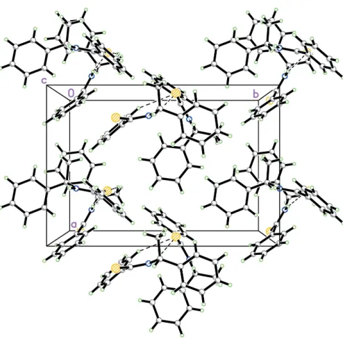
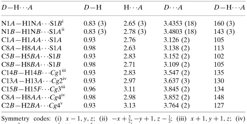
In the crystal packing, molecules are connected into infinite chains along thea axis (Fig. 2) by N—H S intermolecular hydrogen bonds between the amide group and the thioamide S atom (Table 2). The crystal structure is further stabilized by weak intramolecular C—H S interactions and C—H
interactions.
Experimental
A solution containing O-4-nitrobenzyl benzothioate (0.260 g, 1.0 mmol) and N-(R)-methylbenzylamine (0.122 g, 1.0 mmol) in dichloromethane (20 ml) was stirred overnight. The productN-[(R )-1-phenylethyl]thiobenzamide was separated by chromatography on silica gel (yield 0.18 g, 75%). Yellow needle-like single crystals of (I) of X-ray diffraction quality were obtained by slow evaporation of a dichloromethane/n-hexane (1:3) solution (m.p. 338–339 K).
Crystal data
C15H15NS
Mr= 241.35
Orthorhombic,P212121
a= 10.4149 (3) A˚
b= 15.0106 (5) A˚
c= 16.3110 (5) A˚
V= 2549.96 (14) A˚3
Z= 8
Dx= 1.257 Mg m 3
MoKradiation Cell parameters from 6772
reflections = 1.8–29.0 = 0.23 mm1
T= 100.0 (1) K Needle, yellow 0.500.240.24 mm
Data collection
Bruker SMART APEX2 CCD area-detector diffractometer !scans
Absorption correction: multi-scan (SADABS; Bruker, 2005)
Tmin= 0.936,Tmax= 0.946 50018 measured reflections
6772 independent reflections 6150 reflections withI> 2(I)
Rint= 0.056
max= 29.0
h=12!14
k=20!20
l=22!22
Refinement
Refinement onF2
R[F2> 2(F2)] = 0.047
wR(F2) = 0.117
S= 1.13 6772 reflections 317 parameters
H atoms treated by a mixture of independent and constrained refinement
w= 1/[2(F
o2) + (0.0579P)2 + 0.9357P]
whereP= (Fo2+ 2Fc2)/3 (/)max= 0.001
max= 0.43 e A˚
3
min=0.35 e A˚
3
Absolute structure: Flack (1983), 2982 Friedel pairs
Flack parameter: 0.02 (6)
Table 1
Selected geometric parameters (A˚ ,).
S1A—C7A 1.688 (2) N1A—C7A 1.321 (3) N1A—C8A 1.473 (3)
S1B—C7B 1.681 (2) N1B—C7B 1.322 (3) N1B—C8B 1.476 (3) C7A—N1A—C8A 126.46 (18)
N1A—C7A—C6A 115.71 (18) N1A—C7A—S1A 123.48 (16) C6A—C7A—S1A 120.81 (16)
C7B—N1B—C8B 125.49 (17) N1B—C7B—C6B 116.18 (17) N1B—C7B—S1B 123.30 (15) C6B—C7B—S1B 120.51 (15) C1A—C6A—C7A—N1A 149.5 (2)
C5A—C6A—C7A—N1A 30.0 (3)
C1B—C6B—C7B—N1B 34.8 (3) C5B—C6B—C7B—N1B 145.5 (2)
organic papers
o1514
Luet al. C [image:2.610.49.295.71.242.2]15H15NS Acta Cryst.(2006). E62, o1513–o1515
Figure 1
[image:2.610.47.294.294.538.2]The asymmetric unit of (I), with the atom-labelling scheme; displacement ellipsoids are drawn at the 60% probability level.
Figure 2
Table 2
Hydrogen-bond geometry (A˚ ,).
Cg1,Cg2,Cg3 andCg4 are the centroids of the phenyl rings C1A–C6A, C1B– C6B, C9A–C14Aand C9B–C14B, respectively.
D—H A D—H H A D A D—H A
N1A—H1NA S1Bi
0.83 (3) 2.65 (3) 3.4353 (18) 160 (3) N1B—H1NB S1Aii
0.83 (3) 2.78 (3) 3.4803 (18) 143 (3) C1A—H1AA S1A 0.93 2.76 3.126 (2) 105 C8A—H8AA S1A 0.98 2.63 3.138 (2) 113 C5B—H5BA S1B 0.93 2.83 3.152 (2) 102 C8B—H8BA S1B 0.98 2.71 3.109 (2) 105 C14B—H14B Cg1iii
0.93 2.83 3.547 (2) 135 C13A—H13A Cg2iv 0.93 2.97 3.637 (3) 130 C15B—H15F Cg3iii
0.96 3.11 3.845 (2) 134 C8A—H8AA Cg4iv
0.98 2.98 3.852 (2) 148 C2B—H2BA Cg4v 0.93 3.13 3.764 (2) 127
Symmetry codes: (i) x1;y;z; (ii) xþ3
2;yþ1;z 1
2; (iii) xþ1;yþ1;z; (iv)
xþ3
2;yþ1;zþ12; (v)xþ52;yþ1;z12.
H atoms attached to N were located in a difference map and refined isotropically. The remaining H atoms were placed in calcu-lated positions, with C—H distances in the range 0.93–0.98 A˚ ; the
Uiso(H) values were constrained to be 1.2Ueqof the carrier atom.
Data collection:APEX2(Bruker, 2005); cell refinement:APEX2; data reduction: SAINT (Bruker, 2005); program(s) used to solve structure: SHELXTL (Sheldrick, 1998); program(s) used to refine structure:SHELXTL; molecular graphics:SHELXTL; software used to prepare material for publication:SHELXTLandPLATON(Spek, 2003).
The authors thank the Malaysian Government and the Universiti Sains Malaysia for Scientific Advancement Grant Allocation (SAGA) grant No. 304/PFIZIK/653003/A118, and the National Science and Engineering Research Council of Canada and Queen’s University for support of this work.
References
Ach, D., Reboul, V. & Metzner, P. (2002).Eur. J. Org. Chem.pp. 2573–2586. Allen, F. H., Kennard, O., Watson, D. G., Brammer, L., Orpen, A. G. & Taylor,
R. (1987).J. Chem. Soc. Perkin Trans. 2, pp. S1–19.
Artis, D. R. & Lipton, M. A. (1998).J. Am. Chem. Soc.120, 12200–12206. Begum, R. A., Powell, D. & Bowman-James, K. (2006).Inorg. Chem.45, 964–
966.
Bruker (2005).APEX2(Version 1.27),SAINT(Version 7.12A) andSADABS
(Version 2004/1) . Bruker AXS Inc., Madison, Wisconsin, USA.
Brunner, H., Buegler, J. & Nuber, B. (1996).Tetrahedron Asymmetry,7, 3095– 3098.
Flack, H. D. (1983).Acta Cryst.A39, 876–881.
Hossain, M. A., Llinares, J. M., Powell, D. & Bowman-James, K. (2003).Inorg. Chem.42, 5043–5045.
Pavlovic´, G., Tralic-Kulenovic, V. & Popovic, Z. (2004).Acta Cryst.E60, o637– o639.
Popovic´, J., Mrvos-Sermek, D. & Tralic-Kulenovic, V. (2001).Acta Cryst.E57, o893–o894.
Sheldrick, G. M. (1998).SHELXTL.Version 5.10. Bruker AXS Inc., Madison, Wisconsin, USA.
Spek, A. L. (2003).J. Appl. Cryst.36, 7–13.
organic papers
Acta Cryst.(2006). E62, o1513–o1515 Luet al. C
supporting information
sup-1 Acta Cryst. (2006). E62, o1513–o1515
supporting information
Acta Cryst. (2006). E62, o1513–o1515 [https://doi.org/10.1107/S1600536806009639]
N
-[(
R
)-1-Phenylethyl]thiobenzamide
Zhong-Lin Lu, Hoong-Kun Fun, Suchada Chantrapromma and R. Stan Brown
N-[(R)-1-Phenylethyl]thiobenzamide
Crystal data
C15H15NS Mr = 241.35
Orthorhombic, P212121
Hall symbol: P 2ac 2ab
a = 10.4149 (3) Å
b = 15.0106 (5) Å
c = 16.3110 (5) Å
V = 2549.96 (14) Å3 Z = 8
F(000) = 1024
Dx = 1.257 Mg m−3
Melting point = 338–339 K Mo Kα radiation, λ = 0.71073 Å Cell parameters from 6772 reflections
θ = 1.8–29.0°
µ = 0.23 mm−1 T = 100 K Needle, yellow 0.50 × 0.24 × 0.24 mm
Data collection
Bruker SMART APEX2 CCD area-detector diffractometer
Radiation source: fine-focus sealed tube Graphite monochromator
Detector resolution: 8.33 pixels mm-1 ω scans
Absorption correction: multi-scan (SADABS; Bruker, 2005)
Tmin = 0.936, Tmax = 0.946
50018 measured reflections 6772 independent reflections 6150 reflections with I > 2σ(I)
Rint = 0.056
θmax = 29.0°, θmin = 1.8° h = −12→14
k = −20→20
l = −22→22
Refinement
Refinement on F2
Least-squares matrix: full
R[F2 > 2σ(F2)] = 0.047 wR(F2) = 0.117 S = 1.13 6772 reflections 317 parameters 0 restraints
Primary atom site location: structure-invariant direct methods
Secondary atom site location: difference Fourier map
Hydrogen site location: inferred from neighbouring sites
H atoms treated by a mixture of independent and constrained refinement
w = 1/[σ2(F
o2) + (0.0579P)2 + 0.9357P]
where P = (Fo2 + 2Fc2)/3
(Δ/σ)max = 0.001
Δρmax = 0.43 e Å−3
Δρmin = −0.35 e Å−3
Absolute structure: Flack (1983), 2982 Friedel pairs
supporting information
sup-2 Acta Cryst. (2006). E62, o1513–o1515
Special details
Experimental. The low-temparture data were collected with the Oxford Cyrosystem Cobra low-temperature attachment.
Geometry. All e.s.d.'s (except the e.s.d. in the dihedral angle between two l.s. planes) are estimated using the full covariance matrix. The cell e.s.d.'s are taken into account individually in the estimation of e.s.d.'s in distances, angles and torsion angles; correlations between e.s.d.'s in cell parameters are only used when they are defined by crystal symmetry. An approximate (isotropic) treatment of cell e.s.d.'s is used for estimating e.s.d.'s involving l.s. planes.
Refinement. Refinement of F2 against ALL reflections. The weighted R-factor wR and goodness of fit S are based on F2,
conventional R-factors R are based on F, with F set to zero for negative F2. The threshold expression of F2 > σ(F2) is used
only for calculating R-factors(gt) etc. and is not relevant to the choice of reflections for refinement. R-factors based on F2
are statistically about twice as large as those based on F, and R- factors based on ALL data will be even larger.
Fractional atomic coordinates and isotropic or equivalent isotropic displacement parameters (Å2)
x y z Uiso*/Ueq
supporting information
sup-3 Acta Cryst. (2006). E62, o1513–o1515
H1BA 1.0998 0.3779 0.4694 0.025* C2B 1.1882 (2) 0.29161 (15) 0.39296 (13) 0.0234 (4) H2BA 1.1489 0.3113 0.3451 0.028* C3B 1.2763 (2) 0.22240 (16) 0.39018 (13) 0.0252 (5) H3BA 1.2960 0.1955 0.3404 0.030* C4B 1.3349 (2) 0.19329 (15) 0.46175 (14) 0.0254 (5) H4BA 1.3943 0.1471 0.4596 0.031* C5B 1.3058 (2) 0.23241 (14) 0.53607 (13) 0.0235 (4) H5BA 1.3451 0.2122 0.5838 0.028* C6B 1.2174 (2) 0.30230 (14) 0.53970 (12) 0.0188 (4) C7B 1.1866 (2) 0.34414 (14) 0.62053 (12) 0.0186 (4) C8B 1.1217 (2) 0.48434 (14) 0.69057 (12) 0.0199 (4) H8BA 1.1621 0.4586 0.7394 0.024* C9B 1.1718 (2) 0.57873 (14) 0.68147 (11) 0.0187 (4) C10B 1.0941 (2) 0.65238 (15) 0.69479 (13) 0.0225 (4) H10B 1.0088 0.6443 0.7101 0.027* C11B 1.1424 (3) 0.73789 (16) 0.68549 (13) 0.0272 (5) H11B 1.0889 0.7867 0.6937 0.033* C12B 1.2696 (3) 0.75125 (16) 0.66406 (13) 0.0272 (5) H12B 1.3015 0.8087 0.6578 0.033* C13B 1.3486 (2) 0.67860 (16) 0.65210 (14) 0.0264 (5) H13B 1.4343 0.6871 0.6382 0.032* C14B 1.3005 (2) 0.59282 (15) 0.66078 (13) 0.0225 (4) H14B 1.3544 0.5442 0.6527 0.027* C15B 0.9769 (2) 0.47600 (16) 0.70140 (14) 0.0261 (5) H15D 0.9533 0.4142 0.7014 0.039* H15E 0.9342 0.5060 0.6571 0.039* H15F 0.9521 0.5026 0.7525 0.039* H1NB 1.158 (3) 0.4566 (18) 0.5744 (18) 0.030 (7)* H1NA 0.304 (3) 0.364 (2) 0.827 (2) 0.048 (9)*
Atomic displacement parameters (Å2)
U11 U22 U33 U12 U13 U23
supporting information
sup-4 Acta Cryst. (2006). E62, o1513–o1515
C13A 0.0378 (13) 0.0247 (12) 0.0270 (11) −0.0003 (11) −0.0006 (10) −0.0044 (9) C14A 0.0290 (12) 0.0257 (12) 0.0212 (10) −0.0064 (9) 0.0015 (9) −0.0034 (9) C15A 0.0324 (12) 0.0254 (11) 0.0206 (10) 0.0001 (10) 0.0005 (9) 0.0037 (9) S1B 0.0360 (3) 0.0208 (2) 0.0140 (2) −0.0022 (2) −0.0013 (2) 0.00253 (19) N1B 0.0280 (10) 0.0206 (9) 0.0108 (7) 0.0003 (8) 0.0002 (7) 0.0010 (6) C1B 0.0254 (11) 0.0207 (10) 0.0161 (9) 0.0025 (9) 0.0006 (8) −0.0005 (8) C2B 0.0293 (11) 0.0263 (11) 0.0146 (9) −0.0017 (10) −0.0004 (8) −0.0012 (8) C3B 0.0300 (12) 0.0268 (11) 0.0188 (10) −0.0036 (10) 0.0041 (9) −0.0056 (9) C4B 0.0271 (12) 0.0233 (11) 0.0259 (11) 0.0047 (9) 0.0029 (9) −0.0019 (9) C5B 0.0277 (11) 0.0225 (11) 0.0204 (10) 0.0002 (9) −0.0019 (8) −0.0008 (8) C6B 0.0229 (10) 0.0188 (10) 0.0147 (9) −0.0030 (8) 0.0010 (7) −0.0015 (7) C7B 0.0216 (10) 0.0199 (10) 0.0144 (8) −0.0020 (9) −0.0021 (8) −0.0012 (7) C8B 0.0269 (11) 0.0204 (10) 0.0124 (9) −0.0002 (9) 0.0008 (8) −0.0013 (7) C9B 0.0248 (11) 0.0206 (10) 0.0108 (8) −0.0019 (8) −0.0026 (7) −0.0002 (7) C10B 0.0266 (11) 0.0262 (11) 0.0148 (9) 0.0023 (9) −0.0010 (8) −0.0033 (8) C11B 0.0391 (14) 0.0232 (12) 0.0192 (10) 0.0067 (10) −0.0054 (9) −0.0035 (8) C12B 0.0415 (14) 0.0217 (11) 0.0184 (10) −0.0084 (10) −0.0059 (9) 0.0005 (8) C13B 0.0257 (12) 0.0328 (13) 0.0208 (10) −0.0055 (10) −0.0019 (9) 0.0002 (9) C14B 0.0229 (11) 0.0269 (11) 0.0176 (9) 0.0023 (9) −0.0009 (8) −0.0007 (8) C15B 0.0282 (12) 0.0290 (11) 0.0211 (10) −0.0046 (9) 0.0040 (9) −0.0032 (9)
Geometric parameters (Å, º)
supporting information
sup-5 Acta Cryst. (2006). E62, o1513–o1515
C12A—C13A 1.391 (4) C12B—C13B 1.380 (4) C12A—H12A 0.9300 C12B—H12B 0.9300 C13A—C14A 1.381 (4) C13B—C14B 1.389 (3) C13A—H13A 0.9300 C13B—H13B 0.9300 C14A—H14A 0.9300 C14B—H14B 0.9300 C15A—H15A 0.9600 C15B—H15D 0.9600 C15A—H15B 0.9600 C15B—H15E 0.9600 C15A—H15C 0.9600 C15B—H15F 0.9600
supporting information
sup-6 Acta Cryst. (2006). E62, o1513–o1515
C11A—C12A—C13A 119.0 (2) C13B—C12B—C11B 119.5 (2) C11A—C12A—H12A 120.5 C13B—C12B—H12B 120.3 C13A—C12A—H12A 120.5 C11B—C12B—H12B 120.3 C14A—C13A—C12A 120.3 (2) C12B—C13B—C14B 120.2 (2) C14A—C13A—H13A 119.8 C12B—C13B—H13B 119.9 C12A—C13A—H13A 119.8 C14B—C13B—H13B 119.9 C13A—C14A—C9A 121.3 (2) C13B—C14B—C9B 120.7 (2) C13A—C14A—H14A 119.4 C13B—C14B—H14B 119.6 C9A—C14A—H14A 119.4 C9B—C14B—H14B 119.6 C8A—C15A—H15A 109.5 C8B—C15B—H15D 109.5 C8A—C15A—H15B 109.5 C8B—C15B—H15E 109.5 H15A—C15A—H15B 109.5 H15D—C15B—H15E 109.5 C8A—C15A—H15C 109.5 C8B—C15B—H15F 109.5 H15A—C15A—H15C 109.5 H15D—C15B—H15F 109.5 H15B—C15A—H15C 109.5 H15E—C15B—H15F 109.5
supporting information
sup-7 Acta Cryst. (2006). E62, o1513–o1515
Hydrogen-bond geometry (Å, º)
D—H···A D—H H···A D···A D—H···A
N1A—H1NA···S1Bi 0.83 (3) 2.65 (3) 3.4353 (18) 160 (3)
N1B—H1NB···S1Aii 0.83 (3) 2.78 (3) 3.4803 (18) 143 (3)
C1A—H1AA···S1A 0.93 2.76 3.126 (2) 105 C8A—H8AA···S1A 0.98 2.63 3.138 (2) 113 C5B—H5BA···S1B 0.93 2.83 3.152 (2) 102 C8B—H8BA···S1B 0.98 2.71 3.109 (2) 105 C14B—H14B···Cg1iii 0.93 2.83 3.547 (2) 135
C13A—H13A···Cg2iv 0.93 2.97 3.637 (3) 130
C15B—H15F···Cg3iii 0.96 3.11 3.845 (2) 134
C8A—H8AA···Cg4iv 0.98 2.98 3.852 (2) 148
C2B—H2BA···Cg4v 0.93 3.13 3.764 (2) 127
![2 (4 Isobutylphenyl) N′ [1 (4 nitrophenyl)ethylidene]propanohydrazide](data:image/gif;base64,R0lGODlhAQABAIAAAP///wAAACH5BAEAAAAALAAAAAABAAEAAAICRAEAOw==)