Original Article
Expression of POSTN mRNA is associated
with osteosarcoma prognosis
Chao Lu, Dianming Jiang
Department of Orthopedics, The First Affiliated Hospital of Chongqing Medical University, Chongqing, China
Received January 4, 2016; Accepted March 18, 2016; Epub October 1, 2016; Published October 15, 2016
Abstract: Aim: This study aimed to investigate the effect of POSTN mRNA expression on osteosarcoma prognosis. Methods: The carcinoma and adjacent tissues were collected from 81 primary osteosarcoma patients. The expres-sion level of POSTN mRNA in bone tissues was detected by real-time polymerase chain reaction (RT-PCR) method and the differences between two groups were compared. The relationships of POSTN mRNA expression with clinical indexes and the survival rate of osteosarcoma patients during follow-up were also analyzed. Results: The over-ex-pression level of POSTN mRNA in carcinoma tissues was significantly higher than that in adjacent tissues (P<0.05). The clinical stages, soft tissue infiltration and preoperative metastasis of osteosarcoma patients were related to
POSTN mRNA expression. Postoperative follow-up results displayed that with different POSTN mRNA expression levels, the survival time of patients was significantly different, and that the higher the POSTN mRNA over-expression was, the shorter the patients survived, with statistically significant differences (P<0.05). The clinical stages, soft tissue infiltration, preoperative metastasis and POSTN mRNA expression were proved by Cox regression analysis to be the prognosis-related factors of osteosarcoma (P<0.05). Conclusions: POSTN mRNA may be an important factor for osteosarcoma prognosis, which needs to be further investigated in terms of the protein level.
Keywords: Osteosarcoma, POSTN, RT-PCR, prognosis
Introduction
Osteosarcoma (OS), a common tumor of
ortho-pedics, ranks first in primary malignant bone
tumors [1]. Characterized by spindle neoplastic cells in osteoid tissues, osteosarcoma belongs to mesenchymal malignant tumors [2]. OS mainly occurs in adolescents aged 10~20 with metaphysis of limb bones as the primary posi-tion, and its annual incidence is 4~5 per 1 mil-lion people [3]. With high malignancy, strong invasion and high local recurrence rate as well as rapid progression, osteosarcoma severely hazards physical health of adolescents [4, 5]. Surgery assisted with neoadjuvant chemother-apy is the main clinical treatment of osteosar-coma. However, the 5-year survival rate of patients developing tumor metastasis after limb salvage surgery is only 20% or so.
New techniques for prognosis and treatment of osteosarcoma are continually improved along with the development of science and the deep-ening study on the pathogenesis of
osteosar-coma. Gene prognosis and molecular targeted therapy based on the development of modern tumor molecular biology and molecular genet-ics have opened up a new way for prognosis and treatment of osteosarcoma [6-8]. Previous studies have indicated that the occurrence and development of osteosarcoma are closely cor-related with the abnormal activation of protoon-cogenes or the abnormal inactivation of antion-cogenes [9-11]. Therefore, it is very important for the prognosis of osteosarcoma to explore new marker genes.
In recent years, the expression of POSTN has been found by numerous studies to have sig-
nificant differences between normal tissues
and tumor tissues, which suggests that POSTN
may be closely associated with the occurrence, development and prognosis of malignant tum- ors [12-14]. And for this reason, the present study detected the role of POSTN gene in
diag-nosis and identification of patients with
Materials and methods
Objects of the study
81 osteosarcoma patients were selected into
the case group from The First Affiliated Hospital
of Chongqing Medical University during July, 2009 and July, 2012. They all diagnosed with primary osteosarcoma by pathological diagno-sis, X ray and CT scan. The cases were aged 16~40 with an average age of 28.6±7.8, and did not get radiotherapy or chemotherapy before operation. The osteosarcoma tissue
samples without inflammation or necrosis were
collected from the primary origin during sur-gery, put into liquid nitrogen for quick-freeze and preserved at -80°C in ultra cold storage freezer.
The control group contained 12 benign osteo-chondroma patients from the same hospital during the same period. Specimen collection and operation were the same as the case group. The ethics committee of The First
Affiliated Hospital of Chongqing Medical
University gave permission to this study. The tissue collection was performed under the informed consent of all the subjects and com-plied with the provisions of ethics.
POSTN mRNA expression tested with RT-PCR Total RNA extraction: appropriate-quality bone tissues, added with pro rata Trizol (Invitrogen company), was made into homogenate using homogenizer (OV5 type, Sino instrument Co., Ltd.). Then chloroform, isopropanol and ethanol were added into the homogenate following steps to extract the total RNA. RNA was exam-ined its concentration and purity by ultraviolet spectroscopy method and its integrity by elec-trophoresis in 1% agarose gel (containing EB). Synthesis of cDNA by reverse transcription
5 μg of total RNA was added with 0.5 μg Oligo
(dT), undergone degeneration at 75°C for 5 min
The primers and Taq Man probes were design- ed and synthesized by Shanghai Sangon Biotech Co., Ltd.. The forward and reverse prim-er sequences of POSTN gene were 5’-AATGG- AAGGAATGAAAGGCTG-3’ and 5’-CCTCGATeTc- eTeecTeAGT-3’ respectively. The TaqMan probe was 5’-(FAM)-AGCAGTTTTGCCCATTGACCATGTT (TAMRA)-3’. Glyceraldehyde 3 phosphate dehy-drogenase (GAPDH) was the reference gene, and its forward and reverse primer sequences were 5’-GAAGGTGAAGGTCGGAGTC-3’ and 5’- GAAGATGGTGATGGGATTTC-3’ respectively, and its TaqMan probe was 5’-(FAM)-CAAGCTTCCC- GTTCTCAGCC(TAMRA)-3’.
The RT-PCR solution contained 5.0 μL cDNA, 0.75 μL forward primer, 0.75 μL reverse primer, 12.5 μL SYBGREEN Master Mix (Roche Com-pany) and 6.0 μL DEPC water. The PCR condi -tions were: initial denaturation at 95°C for 10 min; followed by 45 cycles of degeneration at 95°C for 45 s, annealing at 65°C for 1 min and extension at 72°C for 1 min; at last extension at 72°C for 10 min. Represented by the expres-sion level ratio of POSTN to corresponding GAPDH, the relative expression levels of POSTN
mRNA from 0 to the maximum were averagely divided into three equal parts, namely negative (-), weak positive (+) and strong positive (++). Weak and strong positive were collectively referred to as positive. Positive rate = (Weak positive cases + Positive cases)/Total cases ×100%.
Statistical methods
All statistical tests were performed by SPSS- 18.0 software. Data comparison between gro-
ups was conducted by χ2 test. Survival data
were analyzed by Kaplan-Meier method. The Cox regression model was used for analysis of
[image:2.612.90.320.97.176.2]prognosis-related factors. Statistical signifi -cance existed when P<0.05.
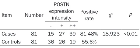
Table 1. Expression levels of POSTN in case group and control group
Item Number
POSTN expression
intensity Positive rate χ2 P
- + ++
Cases 81 15 27 39 81.48% 18.923 <0.01 Controls 81 36 26 19 55.6%
and added to 12.5 μl with DEPC water. Then
the operation was as follows: ice-bath for 30 s, adding 40 U RNA Inhibitor, 10 nmol dNTP Mix and First-Strand Buffer, incuba-tion at 42°C for 5 min, adding 200 U SuperScriptTMII and reaction for 50 min.
Finally the reaction was finished after
Results
Comparison of POSTN mRNA expression levels
The POSTN mRNA expression levels of the two groups are shown in Table 1. There were 15 negative cases, 27 weak positive cases and 39 strong positive cases in 81 osteosarco- ma cases with positive rate of 81.48%. The control group had 36 negative cases, 26 weak
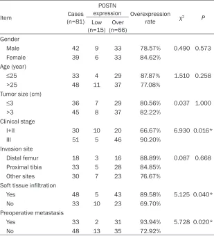
According to the malignant degree, osteosar-coma was divided into 3 levels in this study: Level I and II were in the low-grade osteosarco-ma group while Level III in the high-grade group. The POSTN expression positive rate of the above two groups was 66.67% and 90.20% respectively and this difference was
statistical-ly significant (P<0.05). The soft tissue infiltra -tion and preoperative metastasis analyses of osteosarcoma patients with different POSTN
mRNA expression levels proved that the posi-tive rate of POSTN mRNA expression was
sig-nificantly higher in osteosarcoma patients with soft tissue infiltration than those without infil -tration (89.58% vs. 69.70%) and statistical
sig-nificance existed in the difference (P<0.05); the positive rate in patients with preoperative metastasis (93.94%) was obviously higher than that in patients without metastasis (72.92%),
and the difference was statistically significant
[image:3.612.92.401.96.436.2](P<0.05). The results are listed in Table 2.
Table 2. Correlation of POSTN mRNA expression levels of osteosarcoma patients with clinical indexes
Item (n=81)Cases
POSTN
expression Overexpression
rate χ2 P
Low
(n=15) (n=66)Over Gender
Male 42 9 33 78.57% 0.490 0.573
Female 39 6 33 84.62%
Age (year)
≤25 33 4 29 87.87% 1.510 0.258
>25 48 11 37 77.08%
Tumor size (cm)
≤3 36 7 29 80.56% 0.037 1.000
>3 45 8 37 82.22%
Clinical stage
I+II 30 10 20 66.67% 6.930 0.016*
III 51 5 46 90.20%
Invasion site
Distal femur 18 3 16 88.89% 0.087 0.668
Proximal tibia 33 5 28 84.85%
Other sites 30 7 23 76.67%
Soft tissue infiltration
Yes 48 5 43 89.58% 5.125 0.040*
No 33 10 23 69.70%
Preoperative metastasis
Yes 33 2 31 93.94% 5.728 0.020*
No 48 13 35 72.92%
*stands for statistical significance.
positive cases and 19 strong positive cases, and its positive rate was 55.6%. The result showed that the POSTN
expression levels and positive rate in case group were higher than those in control group, and that the differences between two groups
had statistical signifi
-cance (χ2 = 18.923, P<
0.01).
Correlation of POSTN mRNA expression lev -els with clinical indexes of osteosarcoma
Further analysis of the relationship of POSTN
mRNA expression levels with the clinical indexes of the cases manifested that the differences of
POSTN expression lev-els in age, gender, tu- mor size and invasion site of cases had no
statistical significance
(P>0.05).
Table 3. Association of POSTN mRNA expression with survival time of osteosarcoma patients Duration of
follow-up/month casesInitial during follow-upDeath cases in follow-upCases lost
0~12 81 14 1
13~24 66 9 1
25~36 56 8 2
37~48 46 8 3
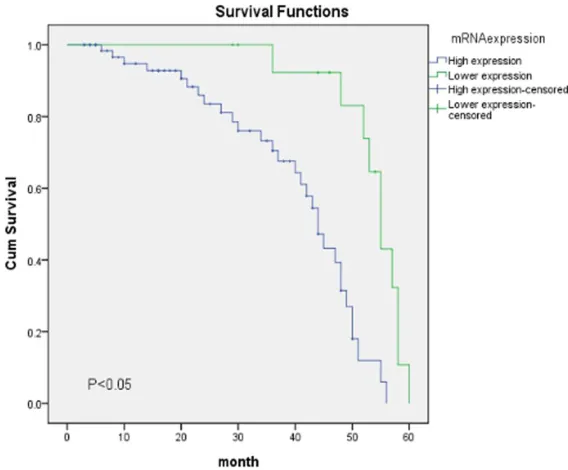
[image:3.612.91.302.492.583.2]Relationship between POSTN mRNA
expres-sion levels and survival time of patients
Table 3 shows the relationship between POSTN
mRNA expression levels and the follow-up results. In the 60-month follow-up, there were 41 death cases and 11 missing cases among a total of 81 osteosarcoma subjects. During the follow-up, the survival time of patients with low
POSTN mRNA expression levels was remark-ably longer than that of patients with high
lev-els and the statistically significant difference in
survival time was observed (P<0.05) (Figure 1). Cox regression analysis
Cox regression model was used to investigate osteosarcoma-related factors, such as gender, age, tumor size and position, clinical stages, soft tissue invasion and preoperative metasta-sis. We found that the prognosis of
[image:4.612.90.374.72.307.2]osteosar-and type I collagen to maintain the stabil-ity of the extracellular matrix [17]. Moreover, periostin takes part in the recruitment, adhesion and migration of cells, and regulates cell proliferation, dif-ferentiation and apoptosis through multi-ple signal transduction pathways, which is closely correlated with the occurrence of many tumors [18, 19].
Figure 1. It showed the relationship between POSTN mRNA expression levels and survival time of osteosarcoma patients.
coma was related to clinical
stages, soft tissue infiltration,
preoperative metastasis and
[image:4.612.90.324.380.449.2]POSTN mRNA expressions. The results can be found in
Table 4.
Discussion
POSTN gene located on human chromosome 13q13.3 is about 36 kb long and encodes periostin (PN) of 836 amino acids [15]. As a mem-ber of fasciclin FASI super-family, POSTN is also known
as osteoblast-specific factor 2 (OSF-2) [16]. Periostin, specifi -cally secreted by interstitial cells, mainly distributes in extracellular matrix. It works together with many extracel-lular matrix proteins such as
type W collagen, fibronectin
Table 4. Cox regression multivariate analysis of prog-nosis of 81 osteosarcoma patients
Factor P OR (95% CI)
Clinical stages 0.034 2.103 (1.060-4.175) Soft tissue infiltration 0.042 2.224 (1.029-4.808) Preoperative metastasis 0.010 3.040 (1.307-7.072)
POSTN mRNA overexpression 0.000 6.478 (2.463-17.039)
POSTN gene and its encoded periostin have been proved by multiple researches to be closely related to the occurrence, development and metastasis of a variety of malignant tumors in clinic [20, 21]. Sasaki H pointed out that the serum periostin levels of patients of non-small cell lung cancer and thymic carcinoma were
sig-nificantly high and had much to do with the
development and prognosis of tumors. In addi-tion, the prognosis was worse in patients with high POSTN mRNA expression levels than those with low expression levels [22, 23]. Shao et al.
confirmed the positive expression of periostins
expres-sion and osteosarcoma, therefore, the present study chose to discuss this association.
The present study found that the expression level of POSTN mRNA was low in control group yet high in case group. The overexpression rate of POSTN mRNA in case group was obviously
higher than that in control group (χ2 = 18.923, P<0.01). The clinical data of osteosarcoma patients, such as age, gender, tumor size and
invasion site, had no significant correlation
with POSTN mRNA expression levels. As for tumor grade, the positive rate of POSTN mRNA expression was remarkably higher in high-grade osteosarcoma group than in low-high-grade group (89.58% vs. 69.7%). Additionally, among
patients with soft tissue infiltration or preoper -ative metastasis, the positive expression rate of POSTN mRNA was comparatively higher, and
the differences had statistical significance
(P<0.05). The follow-up results demonstrated the survival rate was higher in patients with low expression levels than those with high levels. With different POSTN mRNA expression levels, patients had notably different survival times. Moreover, the higher the POSTN mRNA overex-pression was, the shorter the patients survived,
and the differences were statistically signifi -cant (P<0.05). In addition to POSTN mRNA expression levels, the clinical stages, soft
tis-sue infiltration and preoperative metastasis
were also served as the indicators of osteosar-coma prognosis shown by Cox regression multi-variate analysis.
To sum up, POSTN gene may have great influ -ence on the occurr-ence, development and prognosis of osteosarcoma. However, function-al proteins play a direct role in living organisms, and genes are likely to change during the transcription process. Thus, the association between POSTN gene and osteosarcoma needs to be further investigated in terms of the protein level.
Disclosure of conflict of interest
None.
Address correspondence to: Chao Lu, Department of Orthopedics, The First Affiliated Hospital of Chongqing Medical University, Chongqing 400016, China. E-mail: [email protected]
References
[1] Song JG. [Current treatment of primary and metastatic osteosarcoma]. Zhonghua Zhong
[2] Papagelopoulos PJ, Galanis EC, Vlastou C, Nikiforidis PA, Vlamis JA, Boscainos PJ, Fragiadakis EG, Stamos KG, Pantazopoulos T and Sim FH. Current concepts in the eva- luation and treatment of osteosarcoma. Orthopedics 2000; 23: 858-867; quiz 868-859.
[3] Qiu Z and Liao Q. [Progress of osteosarcoma therapy]. Zhongguo Xiu Fu Chong Jian Wai Ke Za Zhi 2010; 24: 1469-1475.
[4] Zhang FY, Tang W, Zhang ZZ, Huang JC, Zhang SX and Zhao XC. Systematic review of high-dose and standard-high-dose chemotherapies in the treatment of primary well-differentiated osteosarcoma. Tumour Biol 2014; 35: 10419-10427.
[5] Geetha N, Kumar A, Ramachandran K, Abraham E and Joseph F. Osteosarcoma of the sella. Australas Radiol 1999; 43: 517-519. [6] Herold T, Metzeler KH, Vosberg S, Hartmann L,
Rollig C, Stolzel F, Schneider S, Hubmann M, Zellmeier E, Ksienzyk B, Jurinovic V, Pasalic Z, Kakadia PM, Dufour A, Graf A, Krebs S, Blum H, Sauerland MC, Buchner T, Berdel WE, Woermann BJ, Bornhauser M, Ehninger G, Mansmann U, Hiddemann W, Bohlander SK, Spiekermann K and Greif PA. Isolated trisomy 13 defines a homogeneous AML subgroup with high frequency of mutations in spliceo-some genes and poor prognosis. Blood 2014; 124: 1304-1311.
[7] Vidula N and Rugo HS. Translating the molecu-lar message of triple-negative breast cancer into targeted therapy. Clin Cancer Res 2015; 21: 1511-1513.
[8] Kim KH and Park KK. Small RNA- and DNA-based gene therapy for the treatment of liver cirrhosis, where we are? World J Gastroenterol 2014; 20: 14696-14705.
[9] Somers GR, Ho M, Zielenska M, Squire JA and Thorner PS. HER2 amplification and overex-pression is not present in pediatric osteosar-coma: a tissue microarray study. Pediatr Dev Pathol 2005; 8: 525-532.
[10] Yi C, Li X, Xu W and Chen A. Relationship be-tween the expression of MTA-1 gene and the metastasis and invasion in human osteosar-coma. J Huazhong Univ Sci Technolog Med Sci 2005; 25: 445-447.
[11] Martin JW, Chilton-MacNeill S, Koti M, van Wijnen AJ, Squire JA and Zielenska M. Digital expression profiling identifies RUNX2, CDC5L, MDM2, RECQL4, and CDK4 as potential pre-dictive biomarkers for neo-adjuvant chemo-therapy response in paediatric osteosarcoma. PLoS One 2014; 9: e95843.
signature predicts poor prognosis in serous epithelial ovarian cancer. Gynecol Oncol 2014; 132: 334-342.
[13] Forsti A, Jin Q, Altieri A, Johansson R, Wagner K, Enquist K, Grzybowska E, Pamula J, Pekala W, Hallmans G, Lenner P and Hemminki K. Polymorphisms in the KDR and POSTN genes: association with breast cancer susceptibility and prognosis. Breast Cancer Res Treat 2007; 101: 83-93.
[14] Tian B, Zhang Y and Zhang J. Periostin is a new potential prognostic biomarker for glioma. Tumour Biol 2014; 35: 5877-5883.
[15] Hong LZ, Wei XW, Chen JF and Shi Y. Over- expression of periostin predicts poor prognosis in non-small cell lung cancer. Oncol Lett 2013; 6: 1595-1603.
[16] Nuzzo PV, Buzzatti G, Ricci F, Rubagotti A, Argellati F, Zinoli L and Boccardo F. Periostin: a novel prognostic and therapeutic target for genitourinary cancer? Clin Genitourin Cancer 2014; 12: 301-311.
[17] Snider P, Hinton RB, Moreno-Rodriguez RA, Wang J, Rogers R, Lindsley A, Li F, Ingram DA, Menick D, Field L, Firulli AB, Molkentin JD, Markwald R and Conway SJ. Periostin is re-quired for maturation and extracellular matrix stabilization of noncardiomyocyte lineages of the heart. Circ Res 2008; 102: 752-760. [18] Wong GS, Habibollahi P, Heidari P, Lee JS,
Klein-Szanto AJ, Waldron TJ, Gimotty P, Nakagawa H, Taylor PR, Wang TC, Mahmood U and Rustgi AK. Optical imaging of periostin en-ables early endoscopic detection and charac-terization of esophageal cancer in mice. Gastroenterology 2013; 144: 294-297.
[19] Lv Y, Wang W, Jia WD, Sun QK, Huang M, Zhou HC, Xia HH, Liu WB, Chen H, Sun SN and Xu GL. High preoparative levels of serum periostin are associated with poor prognosis in patients with hepatocellular carcinoma after hepatectomy. Eur J Surg Oncol 2013; 39: 1129-1135.
[20] Li M, Li C, Li D, Xie Y, Shi J, Li G, Guan Y, Zhang P, Peng F, Xiao Z and Chen Z. Periostin, a stro-ma-associated protein, correlates with tumor invasiveness and progression in nasopharyn-geal carcinoma. Clin Exp Metastasis 2012; 29: 865-877.
[21] Wang Z and Ouyang G. Periostin: a bridge be-tween cancer stem cells and their metastatic niche. Cell Stem Cell 2012; 10: 111-112. [22] Sasaki H, Dai M, Auclair D, Fukai I, Kiriyama M,
Yamakawa Y, Fujii Y and Chen LB. Serum level of the periostin, a homologue of an insect cell adhesion molecule, as a prognostic marker in nonsmall cell lung carcinomas. Cancer 2001; 92: 843-848.
[23] Sasaki H, Dai M, Auclair D, Kaji M, Fukai I, Kiriyama M, Yamakawa Y, Fujii Y and Chen LB. Serum level of the periostin, a homologue of an insect cell adhesion molecule, in thymoma patients. Cancer Lett 2001; 172: 37-42. [24] Shao R, Bao S, Bai X, Blanchette C, Anderson
RM, Dang T, Gishizky ML, Marks JR and Wang XF. Acquired expression of periostin by human breast cancers promotes tumor angiogenesis through up-regulation of vascular endothelial growth factor receptor 2 expression. Mol Cell Biol 2004; 24: 3992-4003.
[25] Kudo Y, Ogawa I, Kitajima S, Kitagawa M, Kawai H, Gaffney PM, Miyauchi M and Takata T. Periostin promotes invasion and anchorage-independent growth in the metastatic process of head and neck cancer. Cancer Res 2006; 66: 6928-6935.