Original Article
Expression of interleukin-5 in different
pathologic types of nasal polyp tissues
Zongfeng Wang1*, Yuanyuan Wang1*, Zhili Zhang2, Xiyin Sun3
Departments of 1Otolaryngology & Head and Neck Surgery, 2Orthodoutics, 3Pathology, Dongying People’s
Hospital, Dongying, China. *Equal contributors.
Received September 4, 2016; Accepted November 18, 2016; Epub March 1, 2017; Published March 15, 2017
Abstract: Interleukin-5 (IL-5) exhibits aberrantly high expression in nasal polyps (NP) tissues, which is closely associ-ated with the formation and development of NP. Antagonizing IL-5 is recognized as a promising strategy for NP pa-tients. However, the expression of IL-5 in different pathologic types of NP has not yet been completely understood. In order to explore immunohistochemical IL-5 expression in NP, compare the results in different NP types, 52 cases of NP tissues and 20 cases of inferior turbinate mucosa tissues as healthy control were collected. Hematoxylin-eosin
staining was adopted to detect four NP types: inflammatory infiltrated (15.38%), adenocystic (11.54%), edematous (57.69%) and fibrous polyp (15.38%). Immunohistochemical analysis confirmed that the expression of IL-5 in NP patients (73.08%) were significantly increased compared with controls (χ2 = 19.871, p<0.001). IL-5 expression
exhibited obvious difference in different pathological types of NP (χ2 = 47.333, p<0.001). We did not detect IL-5
expression in adenocystic polyp tissues. Among patients with IL-5 positive expression, the levels of IL-5 in inflamma
-tory infiltrated, edematous and fibrous polyp were weak (+), moderate (+ for 2, ++ for 13, and +++ for 15 patients) and strong (+++) respectively. Collectively, IL-5 was overexpressed in NP tissues in general, but varied notably in dif -ferent NP types, the stronger expression of IL-5 indicating the more severe NP condition. Our data support IL-5 as a potential target for the treatment of NP, but different types of polyps should be dependent on different approaches.
Keywords: Nasal polyps, interleukin-5, inflammatory infiltrated, adenocystic, edematous, fibrous
Introduction
Nasal polyps (NP) accounts for 1-4% in the adult population and derives from the abnor-mal mucosal protrusions on nasal mucosa or paranasal sinuses along with mucosal epitheli-al hyperplasia, infiltration of inflammatory cells, neo-vascularization, and remarkable edema [1, 2]. The etiology and pathophysiology of NP are still controversial. 80-90% of the NPs are char-acterized by prominent eosinophilia (especially the activated eosinophils) in the middle and inferior turbinate bones [3]. The recruitment of mucosal eosinophils facilitated by cytokines and chemokines reflects a more extensive dis-ease and a decrdis-eased surgical success rate [4, 5]. Besides, the released cytokines and chemo-kines also contribute to the accumulation of other inflammatory cells, such as B cells, plas-ma cells, plas-macrophages, and neutrophils and induce the development of NP [6, 7]. Currently, intranasal glucocorticoids is the most common
strategy for NP, and endoscopic sinus surgery is reserved for cases refractory to medical treat-ment [8, 9], but still remains 10-40% recur-rence rate [10]. Hence, finding the key mole-cules involved in NP is crucial for revealing the pathological progression and developing novel approaches for NP.
asthma and atopic diseases [13], helminth infections [14] and drug hypersensitivity [15]. In NP, the expression of IL-5 in polyp tissues was increased significantly at both mRNA and pro-tein levels, which was closely related to the for-mation of edema and development of NP [16, 17]. Apart from that, Xu et al. reported that the expression of IL-5 in eosinophils was remark-ably stronger during the progression of NP com-pared with the healthy turbinate mucosa [18]. Neutralizing IL-5 with humanized anti-IL-5 monoclonal antibodies suppressed the inflam-matory reaction and reduced the size of NP in half of the patients with NP [19-22]. However, limited attention has been directed towards the expression of IL-5 in patients with different types of NP.
In the present study, clinical research on tis-sues from 52 NP patients and 20 healthy con-trol, hematoxylin-eosin (HE) staining, immuno-histochemistry, quantitative real time RT-PCR and Western blotting assay were performed to explore the types of NP and the expression of IL-5 in different NP types. We found that the expression of IL-5 in NP patients were notably increased compared with controls, IL-5 expres-sion varied in different NP types, and the higher expression of IL-5 pointed to the more severe NP condition.
Materials and methods
Patients
We studied NP tissues from 52 patients who underwent endoscopic sinus surgery between January 1999 and March 2009 at the Ear-Nose-Throat (E.N.T) department of Dongying People’s Hospital, Shandong, China, and a total of 20 healthy nasal mucosa tissue samples (15 cases received correction of deviated nasal septum surgery and 5 cases received dacryo-cystorhinostomy) were taken as healthy con-trol. All tissue specimens were obtained with permission from the Medical Ethics Committee of Dongying People’s Hospital. The median age of all patients was 35.6 years (range, 18-69 years); 29 cases were male, and 23 cases were female. There were 14 cases patients experi-enced the NP course for 5 months to 2 years, 15 cases for 2 years to 4 years and 23 cases for over 4 years. NP diagnosis was based on a CT examination, endoscopic sinus surgery and postoperative pathological examination, which
was complied with new diagnostic criteria of nasal polyps formulated by E.N.T textbook. None of the patients suffered from cystic fibro-sis or received any related drugs before sur-gery. Patients and controls with asthma, aspirin specific reactive bronchial dilation, chronic obstructive pulmonary and autoimmune dis-eases were excluded. All participants were without acute upper respiratory tract infection or steroid drug history before 4 weeks of sur-gery, and agreed to participate in our study and signed an informed consent.
Specimens
Fresh tissues removed from the operating room were immediately sectioned. One part of them was fixed in 10% formaldehyde at room tem-perature for 4-12 h and washing with phos-phate buffered saline (PBS, pH 7.2-7.4). The other part of tissues was preserved in liquid nitrogen tank. The fixed tissues were then dehy-drated through graded ethanol solutions (70% for 2 h, 80% overnight, 90% for 2 h and 100% for 2 h), followed by post-fixed into dimethylben-zene for 30 min and embedded in paraffin. 5 µm thick sections were cut subsequently and placed onto slides and dried in a 70°C chamber for 40 min. Thereafter, sections were dewaxed by ethanol and soaked into PBS for further HE staining and immunohistochemical analysis.
Hematoxylin-eosin (HE) staining
Sections were soaked in sequence with hema-toxylin (Solarbio, Beijing, China) for 5-8 min, ddH2O for 5 min and 1% hydrochloric ethanol for 3 s. After rinsing with ddH2O for 20 min, sec-tions were stained with 1% eosin for 5 min and dehydrated by a graded ethanol series (75%, 95%, 95%, and 100%) for 5 min respectively, followed by permeabilizing twice with dimethyl-benzene for 10 min. Then sections were mount-ed with neutral gum and observmount-ed using a microscope (Olympus, Tokyo, Japan).
Immunohistochemical analysis
block-ing with normal goat serum. In brief, sample sections were incubated with mouse anti-IL-5 monoclonal antibody (1:50 diluted) (Abcam, Cambridge, MA, USA) overnight at 4°C and stained with UltraSensitiveTM SP (Mouse) IHC Kit (Maixin Biotech, Fuzhou, China) strictly fol-lowing the manufacturer’s instruction. Dia- minobenzidine (DAB) was applied as the chro-mogen, and nuclei were stained by hema- toxylin.
Result determination
The results were evaluated separately by two pathologists who were blinded to the clinical parameters. For each section, 5 high-power fields that contained at least 100 cells for each field were selected randomly. Results were determined by the percentage of IL-5 positive staining cells (0% = -, <30% = +, 31-80% = ++, ≥81% = +++).
Quantitative real time RT-PCR (qRT-PCR)
Total RNA was extracted from each group using Trizol reagent (TaKaRa) according to manufac-turers’ protocols. Real-Time PCR Kit (DBI) was used to carry out reverse transcription to obtain the cDNA; Expression of IL-5 was examined using SYBR® Premix Ex TaqTM II (TaKaRa) and GAPDH was served as internal reference; IL-5 upstream primer sequence: 5’-ACCTTGGC- ACTGCTTTCTACTC-3’, downstream primer se- qu-ence: 5’-GGTTTACTCTCCGTCTTTCTTCTC-3’; GAPDH upstream primer sequence: 5’-ATGGC- ATGGACTGTGGTCAT-3’, downstream primer se- qu-ence: 5’-GGTTTACTCTCCGTCTTTCTTCTC-3’; The PCR conditions were 94°C for 2 min, and then 40 cycles of 94°C for 20 sec, 58°C for 20 sec, and 72°C for 20 sec, and a final extension at 72°C for 10 min. All experiments were performed in duplicate. Results are repre-sented as fold induction using the 2-ΔΔCt method.
Western blotting assay
Total protein was extracted from 40-100 mg
tis-membrane was incubated with HRP-conjugated secondary antibody at room temperature for 1 h; proteins were detected using an enhanced chemiluminescence (ECL) kit (Pierce). The pro-tein expression were adjusted to correspond internal reference expression (IOD value of tar-get protein versus IOD of correspond internal reference).
Statistical analysis
Experimental results were expressed as mean ± SD. Statistical analysis was performed with SPSS 19.0 software. One-way ANOVA was used for the analysis of the differences between the groups. The categorical data was analyzed by the chi-square test. Differences between IL-5 expressions in positive NP pathologic types were assessed by Friedman rank sum test.
p<0.05 or p<0.01 was considered to be statisti-cally significant difference.
Results
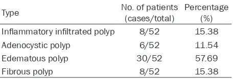
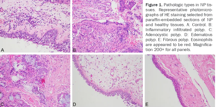
Pathologic types in NP tissues
[image:3.612.91.319.85.163.2]We employed HE staining to identify the patho-logic types among 52 NP tissues. The results were shown in Table 1 and Figure 1, four diff- erent histopathologic types were detected according to the observed structural charactre-istics. 8 patients (15.38%) were detected as inflammatory infiltrated type of NP, 6 patients (11.54%) were showed as adenocystic type of NP, 30 patients (57.69%) were demonstrated as edematous type of NP, and 8 patients (15.38%) were proved to be fibrous type of NP. As shown in Figure 1, for inflammatory infiltrat-ed type of NP, there were a large number of inflammatory cells densely infiltrated in the lamina of mucous membrane, which was domi-nated by eosinophils, but without fibroblast pro-liferation (Figure 1B). Mucous gland hyperpla-sia with gland expansion and mucus retention, slightly inflammatory cell infiltration in stroma without fibroblast proliferation, and squamous Table 1. Pathologic types of NP tissues
Type No. of patients(cases/total) Percentage(%)
Inflammatory infiltrated polyp 8/52 15.38
Adenocystic polyp 6/52 11.54
Edematous polyp 30/52 57.69
Fibrous polyp 8/52 15.38
(Figure 1C). The edematous polyp is morpho-logically characterized by pseudostratified cili-ated columnar epithelium, thickening of the basement membrane, edema even present- ing the edema pool, scattered cells with numer-ous leukocytes, predominantly eosinophils, tiny amounts of fibroblast proliferation (Figure 1D). The last histological type is fibrous polyp char-acterized by local or total fiber tissue prolifera-tion, elevated fibroblast cells, and collagen fiber deposition (Figure 1E).
Enhanced expression of IL-5 in NP tissues
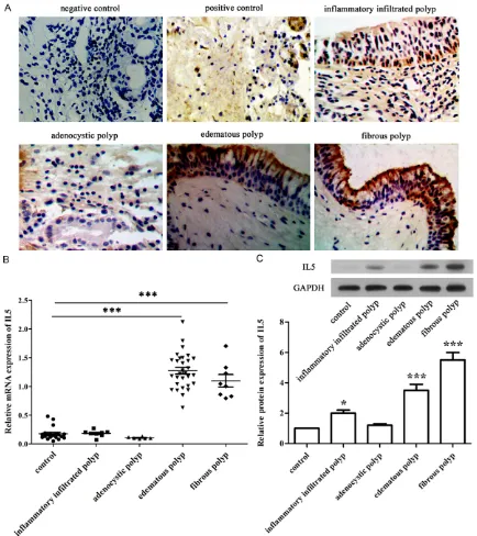
Immunohistochemistry was carried out to anal-ysis the expression of IL-5 in NP tissues. We observed a slightly expression of IL-5 in control tissues, which accounted for 15% (3/20 cases),
trated polyp tissues, only one case (12.5%) showed positive IL-5 expression. All adenocys-tic polyp tissues exhibited negative IL-5 expres-sion. All edematous and fibrous polyp tissues were 100% IL-5 positive rate. Chi-square test demonstrated that the expression of IL-5 in dif-ferent pathological types differed from each other (χ2 = 47.333, P<0.001). We further veri-fied these findings by immunohistochemical analysis, quantitative real time RT-PCR and western blotting assay. As shown in Figure 2A, IL-5 positive cells which stained yellow or tan can be observed in most nasal polyps tissues and a few control tissues. And the IL-5 positive staining part was mainly distributed in fibro-blasts cytoplasmic. In different histopathologic types, IL-5 was highly expressed in the
edema-Figure 1. Pathologic types in NP tis-sues. Representative photomicro-graphs of HE staining selected from
paraffin-embedded sections of NP
and healthy tissues. A: Control. B:
Inflammatory infiltrated polyp. C:
Adenocystic polyp. D: Edematous polyp. E: Fibrous polyp. Eosinophils
[image:4.612.90.524.72.287.2]are appeared to be red. Magnifica -tion 200× for all panels.
Table 2. Expression of IL-5 in NP tissues
Type n PositiveIL-5 expressionNegative Percentage (%)
Control 20 3 17 15.00
NP 52 38 14 73.08
Table 3. IL-5 expression in different types of NP tissues
Type n IL-5 expression Positive rate (%) Positive Negative
Inflammatory infiltrated polyp 8 1 7 12.50
Adenocystic polyp 6 0 6 0
Edematous polyp 30 30 0 100
Fibrous polyp 8 8 0 100
and a dispersed expression of IL-5 in NP stroma, which accounted for 73.08% (38/52 cases) (Table 2). Chi-square test revealed a statistically significant differ-ence between NP tissues and control tis-sues (χ2 = 19.871, p<0.001), which indi-cating that the expression of IL-5 in NP tissues was increased significantly com-pared with control tissues.
Expression of IL-5 in different NP patho-logic types
[image:4.612.88.330.323.376.2] [image:4.612.91.329.413.493.2]infil-tous polyp and fibrous polyp tissues compared with control tissues (P<0.01). The expression of IL-5 in inflammatory infiltrated polyp and ade-nocystic polyp tissues was also higher than control tissues (P<0.05). The quantitative real time RT-PCR results demonstrated the ralative
[image:5.612.89.523.71.559.2]mRNA expression of IL-5 in edematous polyp, fibrous polyp tissues were significantly incre- ased compared with control (P<0.01). Mean- while, The relative protein expression of IL-5 in edematous polyp, fibrous polyp tissues were significantly increased compared with control Figure 2. Expression of IL-5 in different NP pathologic types. A: Immunohistochemical detection on NP and healthy tissues stained with antibody to IL-5, and representative examples of images are shown. Nuclei stained by
hematox-ylin were observed to be blue or purple. The yellow, clay bank particles were target proteins. (Original magnification
and inflammatory infiltrated polyp (P<0.01, P<0.05). The above results suggested that IL-5 expression was associated with the pathologi-cal types in NP tissues.
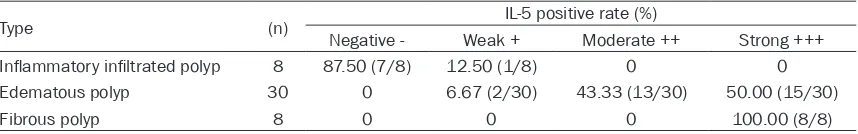
Immunohistochemical intensity of IL-5 in posi-tive NP pathologic types
We invited two pathologists to investigate the staining intensity of IL-5 in positive NP patho-logic types. As shown in Table 4, an IL-5 score of + was found in 1 of 8 inflammatory infiltrated polyps (12.5%). Of the 30 edematous polyp tis-sues, IL-5 staining was graded as + in 2 tissues (6.67%), ++ in 13 tissues (43.33%) and +++ in 15 tissues (50%). All adenocystic polyp and fibrous polyp tissues were scored as +++ (6/6 cases, 100%, 8/8 cases, 100%). Friedman rank sum test confirmed that IL-5 expression levels varied notably in different NP types (χ2 = 36.772, P<0.001), and the stronger expression of IL-5 indicating the more serious NP.
Discussion
NP is a chronic inflammatory disease charac-terized histologically by the infiltration of inflam-matory cells like eosinophils or neutrophils; simultaneously, IL-5 is overexpressed and pro-motes the progression of NP. However, less is known the expression of IL-5 in different patho-logic types of NP. This study revealed the appar-ently elevated IL-5 expression in NP patients through examining 52 cases of NP and 20 cases of healthy nasal mucosa tissues. But the expressions of IL-5 were different in detected four NP types, and the higher expression of IL-5 suggested the more serious NP condition. Li et al. proved that IL-5 synthesis is upregulat-ed in human NP tissue accompaniupregulat-ed by the elevated number of eosinophils [23]. Here, we detected the increased IL-5 in NP tissues com-pared with healthy nasal mucosa, which was consistent with previous studies.
In this study, we found that IL-5 was positively stained in inflammatory infiltrated, edematous and fibrous polyps but negatively stained in adenocystic polyp, suggesting that the expres-sion of IL-5 was associated with the pathologic type of NP. Bachert et al. revealed that IL-5 was secreted by antigen activated T cells at the ini-tial stage of NP, then the activated eosinophils replaced T cells as the main source of IL-5 with the development of NP [17]. So one possible explanation of our result is that there were eosinophilia and eosinophil infiltration in the three positive NP types, but with less eosino-phils sequenced in adenocystic polyp which may lead to the decreased distribution of IL-5. On the other side, Zhang et al. and Cao et al. both point out that NPs in Chinese patients are clinically indistinguishable from polyps of their white counterparts, but part of them lack IL-5 and eotaxin expression in NP tissue, indicating different pathologic processes in them [24, 25], which is similar with the adenocystic polyp in this study. Furthermore, except for eosino-phils infiltration, mast cells and neutroeosino-phils infiltration also play an important role in the progression of NP. We therefore speculate that adenocystic polyp may exhibit a pathologic mechanism different from other three NP types. Our experiment also indicates that differ-ent types of polyps require differdiffer-ent treatmdiffer-ents based on the respective pathophysiology. Prior studies pointed out that eosinophil-fibro-blast interactions facilitated the development of eosinophil-associated diseases, eosinophils express at least 2 potent mediators (IL-1β and TGF-β) to induce a fibrogenic fibroblast pheno-type in asthma and allergen-induced subepi-thelial and peribronchial fibrosis [26, 27]. Rudack et al. proved that fibroblasts secreted GM-CSF prolonged eosinophil survival in vitro
[image:6.612.93.522.84.151.2]combined with IL-3 and IL-5 produced by eo- sinophils in NP development [28]. Apart from that fibroblasts act as important immune regu-latory cells via their ability to cross-talk with T cells, it can promote Th2 polarization in Th-cell Table 4. Immunohistochemical intensity of IL-5 in positive NP types
Type (n) Negative - Weak +IL-5 positive rate (%)Moderate ++ Strong +++
Inflammatory infiltrated polyp 8 87.50 (7/8) 12.50 (1/8) 0 0
Edematous polyp 30 0 6.67 (2/30) 43.33 (13/30) 50.00 (15/30)
responses [29]. So fibroblast proliferation is related to a serious degree of NP, moreover, both Th2 and eosinophils are the main source of IL-5. The present study observed a weakly stained IL-5 in inflammatory infiltrated polyp, moderate stained IL-5 (positive were range from weak to strong) in edematous polyp, and high stained IL-5 in all fibrous polyp, indicating that the pathological type of NP was gradually become serious along with the increased expression of IL5.
In summary, our present study demonstrated that the increased expression of IL-5 was var-ied in different NP types, and the higher expres-sion of IL-5 indicated the more serious NP type except for the non-IL-5 expressed adenocystic polyp. Our data support anti-IL-5 as a promising candidate for the treatment of NP, but different types of polyps should dependent on different approaches.
Disclosure of conflict of interest
None.
Address correspondence to: Yuanyuan Wang, De- partment of Otolaryngology & Head and Neck Surgery, Dongying People’s Hospital, Dongying
257091, Shandong, China. Tel: +86-15990981437;
E-mail: [email protected]
References
[1] Cheng J, Ouyang H, Du J. Expression and regu-lation of interleukin-37 in pathogenesis of na-sal polyps. Indian J Otolaryngol Head Neck Surg 2014; 66: 401-406.
[2] Salaria N, Sharma N, Garg U, Saluja SK,
Agarwal R. Inflammatory septal nasal polyp.
Iranian J Otorhinolaryngol 2015; 27: 319-323. [3] Stoop AE, van der Heijden HA, Biewenga J,
van der Baan S. Eosinophils in nasal polyps and nasal mucosa: an immunohistochemical study. J Allergy Clin Immunol 1993; 91: 616-622.
[4] Newman LJ, Platts-Mills TA, Phillips CD, Hazen KC, Gross CW. Chronic sinusitis. Relationship
of computed tomographic findings to allergy,
asthma, and eosinophilia. JAMA 1994; 271: 363-367.
[5] Marks SC, Shamsa F. Evaluation of prognostic factors in endoscopic sinus surgery. Am J Rhi-nol 1997; 11: 187-191.
[6] Mygind N, Dahl R, Bachert C. Nasal polyposis,
eosinophil dominated inflammation, and aller -gy. Thorax 2000; 55 Suppl 2: S79-83.
[7] Bachert C, Patou J, Van Cauwenberge P. The role of sinus disease in asthma. Curr Opin Allergy Clin Immunol 2006; 6: 29-36.
[8] Sharma R, Lakhani R, Rimmer J, Hopkins C. Surgical interventions for chronic rhinosinus-itis with nasal polyps. Cochrane Database Syst Rev 2014; 11: CD006990.
[9] Rajguru R. Nasal polyposis: Current trends. Indian J Otolaryngol Head Neck Surg 2014; 66 Suppl 1: 16-21.
[10] Wynn R, Har-El G. Recurrence rates after endo-scopic sinus surgery for massive sinus polypo-sis. Laryngoscope 2004; 114: 811-813. [11] Gevaert P, Hellman C, Lundblad L, Lundahl J,
Holtappels G, van Cauwenberge P, Tavernier J, Bachert C. Differential expression of the inter-leukin 5 receptor alpha isoforms in blood and tissue eosinophils of nasal polyp patients. Allergy 2009; 64: 725-732.
[12] Takatsu K. Interleukin-5 and IL-5 receptor in health and diseases. Proc Jpn Acad Ser B Phys Biol Sci 2011; 87: 463-485.
[13] Corren J. Anti-interleukin-5 antibody therapy in asthma and allergies. Curr Opin Allergy Clin Immunol 2011; 11: 565-570.
[14] Tagboto SK. Interleukin-5, eosinophils and the control of helminth infections in man and labo-ratory animals. J Helminthol 1995; 69: 271-278.
[15] Mikami C, Ochiai K, Kagami M, Tomioka H, Tanabe E. In vitro interleukin-5 (il-5) production by peripheral blood mononuclear cells from patients with drug hypersensitivity. J Dermatol 1996; 23: 379-381.
[16] Zhao A, Wang H, Wu H, Yang Y, Xu H, Wang D. [Quantitative analysis of interleukin-5 mrna and protein in nasal polyps]. Lin Chuang Er Bi Yan Hou Tou Jing Wai Ke Za Zhi 2014; 28: 1053-1056.
[17] Bachert C, Wagenmann M, Hauser U, Rudack C. Il-5 synthesis is upregulated in human nasal polyp tissue. J Allergy Clin Immunol 1997; 99: 837-842.
[18] Xu R, Li Y, Xie M, Xu G, Zhang G, Wang S. [The concentration and expression of il-5 in human nasal polyp tissue]. Zhonghua Er Bi Yan Hou Ke Za Zhi 2000; 35: 251-254.
[19] Gevaert P, Van Bruaene N, Cattaert T, Van Steen K, Van Zele T, Acke F, De Ruyck N, Blomme K, Sousa AR, Marshall RP, Bachert C. Mepolizumab, a humanized anti-il-5 mab, as a treatment option for severe nasal polyposis. J Allergy Clin Immunol 2011; 128: 989-99, e981-988.
nasal polyps. J Allergy Clin Immunol 2006; 118: 1133-1141.
[21] Guo J, Feng J, Lin L, Zhao X, Wu H. [Effect of
specific immunotherapy on gm-csf and il-5 in
the tissues of recurrent nasal polyps]. Lin Chuang Er Bi Yan Hou Tou Jing Wai Ke Za Zhi 2015; 29: 2023-2025.
[22] Langier S, Landsberg R, Sade K, Kivity S. Anti-il-5 immunomodulates the effect of staphylo-coccus aureus enterotoxin on t cell response in nasal polyps. Rhinology 2011; 49: 570-576. [23] Li TY, Xia LP, Wang ZF, Chen MY. [Study on the
association between interleukin-5 and eosino-phil in nasal polyp]. Lin Chuang Er Bi Yan Hou Ke Za Zhi 2000; 14: 488-490.
[24] Cao PP, Li HB, Wang BF, Wang SB, You XJ, Cui YH, Wang DY, Desrosiers M, Liu Z. Distinct im-munopathologic characteristics of various ty- pes of chronic rhinosinusitis in adult chinese. J Allergy Clin Immunol 2009; 124: 478-484, 484, e471-472.
[25] Zhang N, Holtappels G, Claeys C, Huang G, van
Cauwenberge P, Bachert C. Pattern of inflam -mation and impact of staphylococcus aureus enterotoxins in nasal polyps from southern china. Am J Rhinol 2006; 20: 445-450.
[26] Gomes I, Mathur SK, Espenshade BM, Mori Y,
Varga J, Ackerman SJ. Eosinophil-fibroblast in
-teractions induce fibroblast il-6 secretion and
extracellular matrix gene expression: Implica-
tions in fibrogenesis. J Allergy Clin Immunol
2005; 116: 796-804.
[27] Tanaka H, Komai M, Nagao K, Ishizaki M, Kajiwara D, Takatsu K, Delespesse G, Nagai H. Role of interleukin-5 and eosinophils in aller-gen-induced airway remodeling in mice. Am J Respir Cell Mol Biol 2004; 31: 62-68.
[28] Rudack C, Hauser U, Stoll W. [Effect of
cyto-kines and fibroblasts on eosinophilic granulo -cyte survival in polyposis nasi]. Laryngorhin- ootologie 1999; 78: 378-381.