Original Article
Effects of EGCG on LPS-induced elevation of
inflammatory factors in human gingival
fibroblasts and functional mechanisms
Fei Huang
1,2*, Jie Ding
3*, Li Miao
4*, Yingxuan Zhao
1, Mingye Zhang
1, Caiyun Chen
1, Gaoyi Wu
5, Songshan
Lin
1, Feng Wang
11Department of Stomatology, PLA Navy General Hospital, Beijing, China; 2Department of Medical Genetics and Development Biology, Fourth Military Medical University, Xi’an, Shaanxi, China; 3Department of Dermatology, PLA 307 Hospital, Beijing, China; 4Department of Stomatology, PLA General Hospital of Beijing, Beijing, China; 5Department of Stomatology, The General Hospital of Jinan Military Commend, Jinan, China. *Equal contributors.
Received February 21, 2017; Accepted March 21, 2017; Epub May 1, 2017; Published May 15, 2017
Abstract: Periodontal disease is one bacterial infectious disorder featured with elevated inflammatory factor level. Epigallocatechin-3-gallate (EGCG) is one factor derived from green tea with anti-inflammation effects. The role of
EGCG in bacterial periodontal disease is unclear, this study aimed to illustrate the role of green teat extract EGCG
in LPS-induced inflammatory factors of human gingival cells along with related mechanism. In vitro cultured human
gingival fibroblasts were stimulated by LPS. Inflammatory cytokine IL-8 and IL-6 secretion level was measured after treatment using serial concentrations of EGCG (20-100 μM). Western blotting and p65-DNA analyzed the effect of EGCG on NF-κB pathway. Phosphorylation of Akt, p38, ERK and JNK was measured by Western blotting. EGCG treat
-ment significantly suppressed LPS-induced expression of inflammatory cytokines IL-8 (P<0.01 at 100 μM, P<0.05 at 20 μM) and IL-6 (P<0.01 at 100 μM, P<0.05 at 20 μM) in human gingival fibroblast in a dose-dependent manner. By mechanism analysis, we found that EGCG treatment remarkably suppressed LPS induced p65 and IκB phosphoryla
-tion in human gingival cells, and affected p65 nuclear transloca-tion. Meanwhile, EGCG effectively suppressed Akt phosphorylation, and p38, ERK or JNK phosphorylation in MAPK pathway. EGCG can alleviate LPS-induced inflam
-matory response in human gingival fibroblast by modulating NF-κB, PI3K/Akt signal pathway and MAPK pathway. Keywords: EGCG, human gingival fibroblast, inflammation, signal pathway
Introduction
Periodontal disease is one bacterial infectious
disease, and is featured with gingival inflamma
-tion and destruc-tion of supporting tissues.
Body immunity and inflammation response can
facilitate progression of periodontal disease
[1]. Porphyromonas gingivalis is the major
bac-terial strain in periodontal disease [2], with LPS
as the major toxicity factor [3]. As one LPS
response cell surface receptor, Toll like recep
-tor and related signal pathway participate in
recognition of microbes and production of
pro-inflammatory factors [4]. Human gingival fibro
-blast is the major subtype of periodontal cells.
Previous studies showed the surface
expres-sion of Toll like receptor 4 in gingival fibroblast
and its participation in LPS induced inflamma
-tory factor production [5-7]. Gingival fibroblast
induced inflammatory response is believed to
participate in reconstruction of gingival tissues
[7]. Therefore, the decrease of inflammatory
factor level could reduce cell oxidative stress
response and inflammatory response, thus
ef-fectively managing progression of periodontal
disease.
pro-cess via mediating PI3K-Akt signal pathway
[13]. Recent study revealed that EGCG could
inhibit inflammatory cytokine and chemokine
production in human cells [14]. The effect of
EGCG on LPS-induced elevation of inflammato
-ry factors of human gingival fibroblast has not
been reported.
In this study, we generated an
in vitro
system of
LPS-stimulated human gingival fibroblast to
confirm the protective effect of green tea
ex-tract EGCG on human gingival cells, along with
related mechanism. Our results revealed that
EGCG could reduce LPS-induced human
gingi-val fibroblast inflammation by mediating NF-κB,
PI3K/Akt and MAPK pathways.
Materials and methods
Materials
Green tea active extracts EGCG were
pur-chased from Sigma-Aldrich (US). DMEM cell
cul-ture medium, fetal bovine serum (FBS) and
reagent/disposable materials for cell culture
were purchased from Gibco (US). P-p65, p-IκB,
p-Akt, p-Erk, p-JNK, p-p38 and GAPDH were
purchased from Cell signaling Technology (US).
LPS derived from Porphyromonas gingivalis
was purchased from InvivoGen (US). NF-κB p65
transcriptional factor analysis kit was pur
-chased from Abcam (US). ELISA kits for IL-6 and
IL-8 were purchased from R&D (US).
Cell culture
Human gingival fibroblast separation and cul
-ture were performed as previously described in
literatures [15]. Transplant was obtained from
patient impacted tooth. After PBS rinsing, tis
-sues were cut into small pieces and were
cul-tured in DMEM medium containing 10% FBS
and 1% streptomycin-penicillin at 37°C with 5%
CO
2. After 3 days culture, cells grew from
tis-sues. After reaching 70% confluence, cells were
digested for passage culture.
ELISA
Human gingival fibroblast was inoculated into
24-well plate at 2×10
5cells per well density for
24 h to reach attached growth. Cells were then
incubated with EGCG (20-100 μM) for 1 h, fol
-lowed by LPS (1 μg/mL) treatment for 1 h.
Supernatant was collected from measuring IL-6
and IL-8 levels by ELISA method, suing test kit
purchased from R&D (US), following manual
instruction.
Western blotting
Human gingival fibroblasts were inoculated into
6-well plate at 1×10
6cells per well density for
[image:2.612.91.520.70.233.2]24 h to make attached growth. Cells were then
incubated with EGCG (20-100 μM) for 1 h, fol
-lowed by LPS (1 μg/mL) treatment for 1 h. Cells
were collected for twice rinsing in PBS. Cells
Figure 1. EGCG treatment decreased LPS induced inflammatory cytokine in human gingival fibroblast. Human gin
-gival fibroblast was incubated with EGCG (20-100 μM) for 1 h, followed by LPS (1 μg/mL) treatment for 1 h. Cell culture supernatant was quantified for IL-6 (A) and IL-8 (B) by ELISA. (A) IL-6 concentrations in control, LPS and EGCG groups were 66.67±12.82, 638.46±53.85, 458.97±48.72, 320.51±48.72 and 120.51±38.46 pg/mL. (B) IL-8con -centrations in control, LPS and EGCG groups were 100±31.11, 622.22±75.56, 488.89±57.78, 293.33±35.56 and
were then lysed in RIPA buffer containing
phos-phatase inhibitor. BCA method was used to
qu-antify proteins. Equal volume of protein was se-
parated by 12% SDS-PAGE, followed by
trans-ferring to PVDF membrane, which was blocked
by TBST buffer containing 5% defatted milk
po-wder. Rabbit anti-human monoclonal antibody
(Cell Signaling Technology, 1:1000 dilution) was
used for 4°C overnight incubation. Goat
anti-rabbit secondary antibody (Vector Laboratory,
1:5000) was added for incubation. After
wash-ing, ECL was used for development. Image J
software was used to measure optical density.
Statistical analysis
All statistical analysis was performed by SPSS
19.0 software. Measurement data were prese-
nted by mean ± standard deviation (SD). Com-
parison among multiple groups was performed
by analysis of variance (ANOVA). A statistical
significance was identified when P<0.05.
Results
EGCG treatment decreased LPS induced
in-flammatory cytokines in human gingival fibro
-blast
Human gingival fibroblast was incubated with
EGCG (20-100 μM) for 1 h, followed by LPS (1
μg/mL) treatment for 1 h. Cell culture superna
-tant was quantified for IL-6 and IL-8 by ELISA.
Our results (
Figure 1
) showed that EGCG
treat-ment significantly depressed LPS induced
hu-man gingival fibroblast inflammatory factor IL-6
and IL-8 elevation in a dose dependent man-
ner.
EGCG treatment suppressed LPS induced p65
or IκB phosphorylation in human gingival fibro
-blast
Protein samples were collected and quantified
for p65 and IκB phosphorylation in human gin
-gival fibroblast by Western blot. Our results
showed that LPS treatment induced elevated
p65 and IκB phosphorylation level in human
gingival fibroblast, whilst EGCG treatment sig
-nificantly suppressed p65 and IκB phosphory
-lation (
Figure 2
).
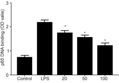
EGCG treatment decreased p65 nuclear
trans-location in LPS stimulated human gingival
fibroblast
ELISA was used to measure binding affinity
between NF-κB transcriptional and DNA. Our
results showed that LPS treatment induced
elevated binding affinity between p65 and DNA
Figure 2. EGCG treatment decreased LPS induced
p65 or IκB phosphorylation in human gingival fibro
-blast. (A) Human gingival fibroblast was incubated with EGCG (20-100 μM) for 1 h, followed by LPS (1 μg/mL) treatment for 1 h. Protein samples were col
-lected for measuring p65 and IκB phosphorylation by Western blot. (B) LPS treatment induced elevated p65 and IκB phosphorylation in human gingival fibro
[image:3.612.93.296.70.367.2]in human gingival fibroblast, indicating
enhan-ced nuclear translocation, whilst EGCG
treat-ment significantly depressed binding affinity
between p65 and DNA (
Figure 3
).
EGCG treatment depressed PI3K-Akt or MAPK
signal pathway activity in LPS stimulated
hu-man gingival fibroblast
Protein samples were collected and measured
for AKt, p38, Erk1/2 and JNK phosphorylation
level in human gingival fibroblast by Western
blot. Our results showed that LPS treatment
elevated phosphorylated level of Akt, p38,
Erk1/2 and JNK in human gingival fibroblast,
whilst EGCG treatment significantly depressed
phosphorylation level of Akt, p38, Erk1/2 and
JNK (
Figure 4
).
Discussion
Periodontal disease derives from inflammatory
response of gingival tissues [16]. Gingival fibro
-blast is the major cell component of gingival
mesenchymal tissues, and plays important
roles in periodontal disease. LPS component of
Porphyromonas gingivalis can stimulate
expres-sion of inflammatory cytokines in human gingi
-val fibroblast [3]. Therefore, LPS is one impor
-tant pathogenic factor of Porphyromonas
gingi-valis. In this study, we generated an
in vitro
the protective effect of green tea extracts
EGCG on human gingival fibroblast along with
the investigation of related mechanism. Our
results showed that EGCG treatment signifi
-cantly suppressed LPS induced expression of
inflammatory factors IL-8 and IL-6 in human gin
-gival fibroblast in a dosage dependent manner.
The analysis of mechanism revealed that EGCG
treatment remarkably suppressed LPS induced
p65 or IκB phosphorylation in LPS induced
human gingival fibroblast, and affected p65
nuclear translocation. Meanwhile, EGCG
effec-tively down-regulated Akt phosphorylation and
phosphorylation of p38, Erk and JNK in MAPK
pathway. It is the first time that the role of EGCG
in human gingival fibroblast inflammatory
res-ponse was reported.
Previous study showed that various cells could
express IL-6 and IL-8, both of which are believed
to participate in tissue injury related with
infla-mmation or neutrophil [17]. These ILs have
str-ong anti-inflammatory effects, and are believed
to be involved in pathological process of
peri-odontal disease. Our results showed that EGCG
significantly decreased levels of these two
in-flammatory cytokines.
[image:4.612.91.287.71.207.2]Moreover, some study showed that LPS induced
IL-6 and IL-8 production in human gingival fibro
-blast mainly via affecting NF-κB signal pathway
activation [18]. As one transcriptional factor,
NF-κB can enhance expression of multiple
ge-nes related with inflammatory response. P65
subunit and p50 subunit form heterodimer as
one major activated form to interact with DNA
binding site. Under normal circumstance, such
heterodimer can bind with IκB protein in cyto
-plasm. Under LPS stimulation, IκB is undergone
phosphorylation to induce the release of p65
and p50. These subunits then translocate into
nucleus. After binding with DNA binding sites,
transcription of target gene is initiated. Previous
study showed that EGCG could inhibit NF-κB
activation in human head-neck cancer H891
cell line and breast cancer MDA-MB-231 cells
[19]. Other study showed that EGCG treatment
could affect intracellular IκB level in dosage-
and time-dependent manner, and inhibit NF-κB
nuclear translocation [20]. Under UV irradiation
induced NF-κB activation process of normal
human epithelial keratocytes, EGCG could
in-hibit NF-κB activation and nuclear transloca
-tion [20]. We observed EGCG could affect p65
Figure 3. EGCG treatment suppressed LPS induced
p65 nuclear translocation in human gingival fibro
-blast. Human gingival fibroblast was incubated with EGCG (20-100 μM) for 1 h, followed by LPS (1 μg/
mL) treatment for 1 h. ELISA was performed to test
the binding affinity between NF-κB transcriptional
and DNA. LPS treatment induced elevated binding
affinity between p65 and DNA in human gingival fi -broblast, indicating enhanced nuclear translocation,
whilst EGCG treatment significantly depressed bind
Figure 4. EGCG treatment depressed PI3K-Akt or MAPK signal pathway activity in LPS stimulated human gingival fibroblast. Human gingival fibroblast was incubated with EGCG (20-100 μM) for 1 h, followed by LPS (1 μg/mL) treatment for 1 h. Protein samples were collected and measured for AKt, p38, Erk1/2 and JNK phos
ylation level of p65 and IκB.
PI3K/AKT signal pathway is one important reg
-ulatory pathway for NF-κB activation, and is
involved in cell migration, cell growth
facilita-tion and blockade of cell apoptosis [21].
Pre-vious study showed the implication of PI3K/
AKT pathway related molecules in treating peri
-odontal diseases [22]. Therefore, this study
observed the effect of EGCG on PI3K/AKT sig
-nal pathway. Results showed that in
LPS-induced inflammatory model of human gingival
fibroblast, EGCG significantly depressed PI3K/
AKT signal pathway activity.
Besides PI3K/AKT signal pathway, MAPK signal
pathway can also enhance expression of inflam
-matory cytokines in various immune cells. It is
also involved in LPS induced inflammatory
res-ponse of human gingival fibroblast [23]. Our
study showed that EGCG treatment also
affect-ed phosphorylation of MAPK signal pathway
related molecules p38, Erk1/2 and JNK, as
consistent with previous study regarding the
participation of EGCG in the suppression of
MAPK signal pathway in mouse epithelium [24]
and NHEK cells [25].
In summary, we confirmed that EGCG could
ex-ert anti-inflammatory effects via mediating
phosphorylation level of p65 and IκB, as well as
p65 nuclear translocation. Moreover, PI3K-Akt
signal pathway and MAPK signal pathway was
also found to be involved in anti-inflammatory
effects of EGCG.
Acknowledgements
This work was supported by National Natural
Science Foundation of China (31301127); Post-
doctoral Science Foundation of China (2012M-
521869); Cultivating Innovation Foundation of
PLA General Navy Hospital (CXPY201519).
Disclosure of conflict of interest
None.
Address correspondence to: Dr. Gaoyi Wu, Depart-
ment of Stomatology, The General Hospital of Jinan
Military Commend, 25 Shi Fan Road, Jinan, China.
Tel: +86-531-51666423; Fax: +86-531-51666423;
E-mail: [email protected]; Drs. Songshan Lin and Feng Wang, Department of Stomatology, PLA
Navy General Hospital, Beijing, China. Tel:
+86-10-(FW)
References
[1] Ji S, Choi YS and Choi Y. Bacterial invasion and
persistence: critical events in the pathogene-sis of periodontitis? J Periodontal Res 2015; 50: 570-85.
[2] Darveau RP. The oral microbial consortium’s
interaction with the periodontal innate defense
system. DNA Cell Biol 2009; 28: 389-95.
[3] Yucel-Lindberg T and Bage T. Inflammatory me -diators in the pathogenesis of periodontitis. Expert Rev Mol Med 2013; 15: e7.
[4] Holden JA, Attard TJ, Laughton KM, Mansell A, O’Brien-Simpson NM and Reynolds EC. Porphy-romonas gingivalis lipopolysaccharide weakly activates M1 and M2 polarized mouse macro
-phages but induces inflammatory cytokines.
Infect Immun 2014; 82: 4190-203.
[5] Jian CX, Li MZ, Zheng WY, He Y, Ren Y, Wu ZM,
Fan QS, Hu YH and Li CJ. Tormentic acid inhib
-its LPS-induced inflammatory response in hu
-man gingival fibroblasts via inhibition of TLR4-mediated NF-kappaB and MAPK signalling pathway. Arch Oral Biol 2015; 60: 1327-32.
[6] Wang QB, Sun LY, Gong ZD and Du Y. Veratric
acid inhibits LPS-induced IL-6 and IL-8
produc-tion in human gingival fibroblasts. Inflammaproduc-tion
2016; 39: 237-42.
[7] Scheres N and Crielaard W. Gingival fibroblast
responsiveness is differentially affected by Porphyromonas gingivalis: implications for the pathogenesis of periodontitis. Mol Oral Micro- biol 2013; 28: 204-18.
[8] Singh BN, Shankar S and Srivastava RK. Green
tea catechin, epigallocatechin-3-gallate (EG- CG): mechanisms, perspectives and clinical
applications. Biochem Pharmacol 2011; 82:
1807-21.
[9] Yang CS, Liao J, Yang GY and Lu G. Inhibition of lung tumorigenesis by tea. Exp Lung Res 2005; 31: 135-44.
[10] Doss MX, Potta SP, Hescheler J and Sachinidis
A. Trapping of growth factors by catechins: a
possible therapeutical target for prevention of
proliferative diseases. J Nutr Biochem 2005;
16: 259-66.
[11] Navarro-Peran E, Cabezas-Herrera J, Sanchez-Del-Campo L, Garcia-Canovas F and Rodriguez-Lopez JN. The anti-inflammatory and anti-can -cer properties of epigallocatechin-3-gallate are mediated by folate cycle disruption, adenosine
release and NF-kappaB suppression. Inflamm
Res 2008; 57: 472-8.
[12] Yang J, Han Y, Chen C, Sun H, He D, Guo J,
high glucose-induced endothelial cell inflam
-mation by suppression of PKC and NF-kappaB
signaling in human umbilical vein endothelial cells. Life Sci 2013; 92: 589-97.
[13] Li S, Wu L, Feng J, Li J, Liu T, Zhang R, Xu S, Cheng K, Zhou Y, Zhou S, Kong R, Chen K,
Wang F, Xia Y, Lu J, Zhou Y, Dai W and Guo C. In vitro and in vivo study of epigallocatechin-3-gallate-induced apoptosis in aerobic glyco-lytic hepatocellular carcinoma cells involving
inhibition of phosphofructokinase activity. Sci
Rep 2016; 6: 28479.
[14] Mukherjee S, Siddiqui MA, Dayal S, Ayoub YZ and Malathi K. Epigallocatechin-3-gallate sup
-presses proinflammatory cytokines and che
-mokines induced by Toll-like receptor 9 ago
-nists in prostate cancer cells. J Inflamm Res
2014; 7: 89-101.
[15] Ogata Y, Niisato N, Sakurai T, Furuyama S and
Sugiya H. Comparison of the characteristics of
human gingival fibroblasts and periodontal lig -ament cells. J Periodontol 1995; 66: 1025-31. [16] Bengtsson T, Khalaf A and Khalaf H. Secreted
gingipains from Porphyromonas gingivalis colo-nies exert potent immunomodulatory effects
on human gingival fibroblasts. Microbiol Res
2015; 178: 18-26.
[17] Erdemir EO, Hendek MK, Keceli HG and Apan TZ. Crevicular fluid levels of interleukin-8, inter
-leukin-17 and soluble intercellular adhesion
molecule-1 after regenerative periodontal the- rapy. Eur J Dent 2015; 9: 60-5.
[18] Huang Y, Guo W, Zeng J, Chen G, Sun W, Zhang
X and Tian W. Prediabetes enhances periodon
-tal inflammation consistent with activation of toll-like receptor-mediated nuclear factor-kap
-paB pathway in rats. J Periodontol 2016; 87:
e64-74.
[19] Masuda M, Suzui M, Lim JT, Deguchi A, Soh JW and Weinstein IB. Epigallocatechin-3-gallate decreases VEGF production in head and neck
and breast carcinoma cells by inhibiting EGFR-related pathways of signal transduction. J Exp
Ther Oncol 2002; 2: 350-9.
[20] Aggarwal BB and Shishodia S. Molecular tar -gets of dietary agents for prevention and
ther-apy of cancer. Biochem Pharmacol 2006; 71:
1397-421.
[21] Lim W, Yang C, Bazer FW and Song G.
Chry-sophanol induces apoptosis of
choriocarcino-ma through regulation of ROS and the AKT and ERK1/2 pathways. J Cell Physiol 2017; 232:
331-339.
[22] Atomura R, Sanui T, Fukuda T, Tanaka U, Toyo-da K, Taketomi T, Yamamichi K, Akiyama H and Nishimura F. Inhibition of Sprouty2 polarizes
macrophages toward an M2 phenotype by sti- mulation with interferon gamma and Porp- hyromonas gingivalis lipopolysaccharide. Im-
mun Inflamm Dis 2016; 4: 98-110.
[23] Park GJ, Kim YS, Kang KL, Bae SJ, Baek HS, Auh QS, Chun YH, Park BH and Kim EC. Effects
of sirtuin 1 activation on nicotine and
lipopoly-saccharide-induced cytotoxicity and inflamma
-tory cytokine production in human gingival fi -broblasts. J Periodontal Res 2013; 48: 483-92.
[24] Kim H, Ramirez CN, Su ZY and Kong AN. Epi-genetic modifications of triterpenoid ursolic acid in activating Nrf2 and blocking cellular
transformation of mouse epidermal cells. J
Nutr Biochem 2016; 33: 54-62.
[25] Konger RL, Derr-Yellin E, Hojati D, Lutz C and
Sundberg JP. Comparison of the acute ultravio-let photoresponse in congenic albino hairless