R E S E A R C H A R T I C L E
Open Access
A study of cochlear and auditory pathways
in patients with tension-type headache
Hang Shen
1, Wenyang Hao
2, Libo Li
1, Daofeng Ni
2, Liying Cui
1and Yingying Shang
2*Abstract
Background:The purpose of this study was to systematically evaluate the function of cochlear and auditory pathways in patients suffering from tension-type headache (TTH) using various audiological methods. Methods:Twenty-three TTH patients (46 ears) and 26 healthy controls (52 ears) were included, and routine diagnostic audiometry, extended high-frequency audiometry, acoustic reflex (ASR), transient evoked otoacoustic emissions (TEOAEs), distortion product otoacoustic emissions (DPOAEs) and suppression TEOAEs were tested. Results:The TTH group showed higher thresholds (P< 0.05) for both pure tone and extended high-frequency audiometry at all frequencies except for 9, 14 and 16 kHz. All ASR thresholds were significantly higher (P< 0.05) in the TTH group compared with the controls, except for the ipsilateral reflex at 1 kHz, but the threshold differences between the ASR and the corresponding pure tone audiometry did not differ (P >0.05). For the DPOAEs, the detected rates were lower (P< 0.05) in the TTH group compared with the controls at 4 and 6 kHz, and the amplitudes and signal to noise ratio (S/N) were not significantly different between groups. No differences in the TEOAEs (P> 0.05) were observed for the detected rates, amplitudes, S/Ns or contralateral suppression, except for the S/Ns of the 0.5-1 kHz TEOAE responses, which were significantly higher (P< 0.05) in the TTH group.
Conclusions:The results of our study indicate that subclinical changes in cochlear function are associated with TTH.
Keywords:Tension-type headache; Otoacoustic emissions; Acoustic reflex; Phonophobia; Brainstem
Background
Tension-type headache (TTH) is the most prevalent type of primary headache, and its lifetime prevalence in the general population ranges from 30 to 78 % [1]. TTH is characterized by episodes of headache lasting minutes to days, with mild to moderate pain that is typically bilateral, pressing or tightening in quality. TTH patho-physiology is complex and incompletely understood. Neuron sensitization in the central nervous system is regarded as one of the major mechanisms underlying this condition [2, 3].
Phonophobia is regarded as the most common audi-tory symptom in patients with migraines. However, pho-nophobia can also occur in TTH patients at relatively high rates of 38–40.9 % [4, 5], although the frequency
and intensity are not as serious as in migraineurs. Never-theless, noise can be an important trigger and aggravat-ing factor in both TTH and migraine patients [4, 6, 7]. A study by Spierings et al. [6] showed that noise is a pre-cipitating and aggravating factor in 29 % and 65 % of TTH patients, respectively. This finding led to specula-tion that these primary headaches may be associated with dysfunction of the auditory system. Several studies [8–10] examining the auditory system using auditory brainstem responses and otoacoustic emissions (OAEs) have been conducted in migraineurs, and the results suggest that subclinical dysfunctions of the cochlear and auditory pathways are related to migraines. Because phonophobia and noise are also related to TTH as complicating symptoms and participating or aggravat-ing factors, we investigated whether these types of dys-functions also exist in TTH patients. Furthermore, studies have shown that cochlear-vestibular symptoms, such as tinnitus and vertigo, are also observed in TTH * Correspondence:[email protected]
2Department of Otorhinolaryngology, Peking Union Medical College Hospital, Chinese Academy of Medical Sciences and Peking Union Medical College, Beijing, China
Full list of author information is available at the end of the article
patients, although at lower prevalences than in mi-graine patients [11, 12]. This finding is also suggestive of potential relationships between TTH and the audi-tory system. However, to our knowledge, no specific studies exploring the auditory system have been per-formed in TTH patients.
Pure tone audiometry (PTA) is the most common hearing test used to obtain hearing threshold levels in individuals for frequencies ranging from 0.25–8 kHz, enabling the identification of the type, degree and config-uration of hearing loss. Extended high-frequency audiom-etry evaluates hearing thresholds for frequencies higher than 8 kHz and has been suggested as a method for moni-toring the effects of noise exposure, ototoxic medication, and hearing loss resulting from other causes [13–17]. High frequencies appear to be more susceptible to exter-nal factors, such as medication and noise, than the middle and low frequencies [15, 17]. Although audiometry cannot clarify the lesion level of auditory dysfunction, it remains a useful tool for evaluating the integral function of the audi-tory system.
OAEs were first reported by Kemp in 1978 [18]. OAEs are sounds that arise in the cochlea and move through the middle ear and straight into the ear canal, where they can be detected using sensitive equipment. OAEs are thought to be the byproducts of the preneural mech-anisms of the cochlear amplifier and are related to the normal function of the outer hair cells (OHCs). Transi-ent evoked otoacoustic emissions (TEOAEs) and distor-tion product otoacoustic emissions (DPOAEs) are the two most common types of evoked otoacoustic emis-sions (EOAEs). For TEOAEs, a “click” stimulus is pro-vided to the ear to evoke broad-spectrum responses of the highest intensity in the mid-frequency spectrum, 1– 4 kHz. DPOAEs are the product of an intermodulation distortion generated by the cochlea responding to the stimulation of two simultaneous pure tones. The fre-quencies of these two stimuli are close to each other, and they are described as primary tones. The response is a tonal signal that does not exist in the eliciting stimuli; therefore, it is called a “distortion product”. Because EOAE testing can detect fine changes in the OHCs that are undetectable by other methods, it permits a sensitive evaluation of cochlear function [14, 17, 19] and object-ively monitors dynamic changes in the cochlea before any functional and significant hearing loss appears [20].
OAE suppression testing is a technique that assesses the brainstem auditory reflex, which is a reduction in OAE amplitude when stimulation occurs in the contra-lateral ear [21]. The reflex arc includes the auditory nerve, cochlear nucleus, trapezoid body, superior olivary complex, olivocochlear bundle, inferior vestibular nerve, outer hair cells and inner hair cells. Because contralat-eral suppression is mediated by the efferents from the
medial superior olivary complex to the OHCs, it is a useful tool for studying the cochlear efferents in the brainstem.
The acoustic reflex (ASR) refers to the reflexive con-traction of the middle ear muscles caused by loud sound stimulation. The acoustic reflex threshold (ART) is the lowest level of a sound stimulus that can elicit an ASR response, i.e., a measurable change in acoustic emittance. The ASR arc is composed of the cochlea, auditory nerve, ventral cochlear nucleus, trapezoid body, superior olivary complex, facial nucleus and facial nerve. Previous studies have shown that ARTs are re-lated to thresholds of the uncomfortable loudness level (UCL) and can be used to estimate the UCL [22–24]. In previous studies, significantly lower thresholds were ob-served in subjects with hypersensitivity to sound caused by various diseases [25, 26].
The purpose of this study was to systematically evalu-ate the function of the cochlear and brainstem auditory pathways in patients suffering from TTH using pure tone audiometry, extended high-frequency audiometry, the ASR, TEOAEs, DPOAEs and TEOAE suppression to examine the potential relationship between TTH and the auditory system.
Methods
Subjects
The TTH group included patients recruited from the Headache Clinic of the Neurology Outpatient Depart-ment at the Peking Union Medical College Hospital. Twenty-three patients with TTH (7 females and 16 males) were involved in this study, and 46 ears were tested. Of the 23 patients, 16 had frequent episodic TTH, and 7 had chronic TTH. Patients with episodic TTH were studied during attack-free periods. The mean patient age was 34 ± 9 years (range 18–52). TTH was di-agnosed according to the criteria of the International Headache Society. The diagnosis was consistent with the International Classification of Headache Disorders-3 (beta version) codes 2.1, 2.2 and 2.3 [1]. Patients with 2.4 probable TTH were excluded. No patients had any neurotologic symptom such as tinnitus, hearing loss, dizziness or vertigo. No history of chronic otological dis-ease, ear surgery, noise exposure, ototoxicity, or any sys-temic metabolic or autoimmune disease associated with hearing loss was reported, and no history of central nervous system disease or other primary headache disor-ders, except for TTH, was reported.
otological disease, noise exposure, ototoxicity, central ner-vous system disease or any systemic, metabolic or auto-immune diseases associated with hearing loss. The study protocol was approved by the Peking Union Medical College Hospital Institutional Review Board in accord-ance with the Declaration of Helsinki. Informed con-sent was obtained from all subjects.
Audiometry
All of the subjects underwent an otoscopic examination performed by an ENT doctor, and those subjects with any type of external ear or middle ear disease were ex-cluded. Next, the subjects underwent tympanometry (Madsen Otoflex 100 and Otodiagnostic Suite immit-tance meter, GN, Denmark). Subjects with any type of tympanometric disorder were excluded. Pure tone audi-ometry was performed using a Conera audiometer (Madsen, GN, Denmark) (ISO 389). First, frequencies from 0.25 to 8 kHz were tested using TDH 39 head-phones with a step size of 5 dB (ISO 8253–1:1989). The extended high frequencies (9, 10, 11.2, 12.5, 14 and 16 kHz) were tested with Sennheiser HDA-200 head-phones using the above-described method. All equip-ment was calibrated every 12 months.
ART testing
Ipsilateral and contralateral ARTs were measured using an Otoflex 100 and Otodiagnostic Suite immittance meter (Madsen, GN, Denmark). The ASR responses to four different stimuli (0.5-, 1-, 2- and 4- kHz tones) were recorded at a probe tone of 226 Hz. An auto threshold search program was applied in a combined descending-ascending manner, and an ASR response was detected when an admittance change exceeded 0.04 mmho. To begin, a stimulus was presented at an intensity of 70 dB HL, and its intensity level was decreased or increased in 5-dB steps according to the responses. The maximum output intensity for all tones was 105 dB HL.
Otoacoustic emission testing
TEOAEs and DPOAEs were evaluated with an otoacous-tic emission analyzer (Madsen, CELESTA 503, Denmark) in a soundproof booth.
For the DPOAE testing, two pure tone stimuli were presented simultaneously to the ear canal at different frequencies, and the distortion-product component at 2f1-f2 was recorded. The DPOAE response amplitude was measured as a function of f2 frequency, with the f2/ f1 ratio fixed at 1.2 at a fixed stimulus level (L1 = 65 dB SPL, L2 = 55 dB SPL). Eight pairs of stimuli were pre-sented with an f2 frequency at 0.75, 1, 1.5, 2, 3, 4, 6 or 8 kHz. For each frequency, the DPOAE signal was con-sidered detectable if the signal to noise ratio (S/N) exceeded 3 dB at 2f1-f2.
The TEOAEs were obtained at 0.5-4 kHz with a stimu-lation of 40-μsec clicks and a linear protocol. The stimu-lus level in the external ear canal was 80 ± 3 dB SPL. The click rate was 50 Hz, and the time window for ana-lysis was 6–18 ms post-stimulus. In total, 1000 sweeps were averaged. A TEOAE response was regarded detect-able if the S/N exceeded 3 dB. If the reproducibility per-centages exceeded 70 % and the stimulus stability was higher than 80 %, the response was considered accept-able for analysis. For TEOAE suppression testing, white noise was generated by an ORBITER 922 audiometer (Madsen, Denmark) and was presented to the contralat-eral ear through a TDH 39 headphone. The intensity of white noise was fixed at a 40-dB sensation level to achieve the optimal measurement parameters [21].
Statistical analysis
All statistical analyses were performed using SPSS for Windows, version 17.0. The threshold, amplitude and S/ N results for the audiometry, the ASR, DPOAEs and TEOAEs were compared in the TTH patients and con-trols using the independent samplest-test. The detected rates of ASR, DPOAEs and TEOAEs were compared be-tween the TTH patients and controls using the χ2 test. To determine whether the TEOAE amplitude changes with and without contralateral noise, pairedt-tests were used, and differences in the extent of suppression be-tween the patients and controls were compared using the Mann–WhitneyUtest.
Results
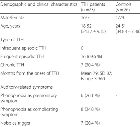
The demographic and clinical characteristics of the study groups are shown in Table 1. No differences in gender or age were detected between the TTH patients and controls.
The TTH group had higher thresholds in both pure tone and extended high-frequency audiometry (Fig. 1), with significant differences observed at all frequencies except for 9, 14 and 16 kHz.
The ipsilateral and contralateral ARTs were obtained in most subjects, and except for the ipsilateral reflex at 1 kHz, all thresholds were significantly higher in the TTH group compared with the control group (Table 2). However, the differences in threshold values between the ASR and PTA did not differ between the groups.
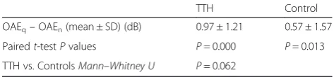
0.5 and 1 kHz (Table 4). For contralateral TEOAE sup-pression, significant suppression occurred in both groups, which was confirmed by a paired t-test to as-sess the TEOAEs in silence and with noise (Table 5). However, the Mann–WhitneyUtest showed no differ-ence in the mean suppression value between groups.
Discussion
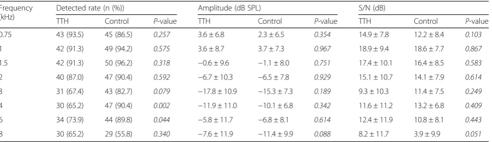
In the present study, despite the fact that subjects with complaints of hearing loss, tinnitus, vertigo or dizzi-ness were excluded from both groups to rule out the influences of complicating ear diseases, statistically sig-nificant differences were observed between the TTH patients and controls in pure tone and high-frequency hearing thresholds (Fig. 1). This finding suggests a subclinical dysfunction of the auditory system in TTH patients. Furthermore, higher cochlear dysfunction risks were indicated by the lower detected DPOAE rates at 4 and 6 kHz in the TTH patients, which could explain the audiometry results, indicating that cochlear
dysfunction may contribute to threshold elevation. The higher S/Ns in the TEOAE test at frequencies of 0.5– 1 kHz in the TTH group seem to contradict the DPOAE findings. However, the possibility that the higher S/N values may result from lower background noise, not a higher response, makes the S/N value a less than optimal parameter to evaluate cochlear function. Indeed, the S/N is more often used as a cri-terion to decide whether the response exists. In previ-ous studies, OAEs have been tested in migraineurs, and an amplitude decrease has also been implicated in subclinical cochlear dysfunction [8–10]. However, because the mechanisms of TTH and migraine are dif-ferent, the causes of cochlear dysfunction may also be different.
Contralateral suppression TEOAEs were evaluated in cochlear efferents in the brainstem, but no significant differences were observed between groups. ART is an-other test that is related to the auditory pathway at the brainstem level. The results indicate that although the ARTs were higher in the TTH group, the ART and PTA thresholds were not higher than those of the control group. Given the audiometry results, the elevation in the ARTs in the TTH group was likely a result of elevated hearing thresholds; moreover, no ART decrease related to hypersensitivity was detected in the TTH patients. However, in the present study, all hearing tests were per-formed in interictal states, except for those administered to subjects with chronic TTH, but most patients experi-enced phonophobia only during attacks. Further studies during headache attacks are required and may still dis-play positive results.
In the present study, cochlear dysfunction was ob-served in the TTH patients; however, the mechanism of cochlear dysfunction remains unclear. In addition, knowledge about the physiological mechanisms under-lying TTH is limited. At present, both peripheral and central mechanisms are thought to play important roles in TTH [2, 3]. Enhanced tenderness to palpation of pericranial myofascial tissues has been reported as the most obvious abnormality in TTH patients [27, 28],
Fig 1Comparison of pure tone and extended high-frequency hearing thresholds between the two groups (mean)
Table 1Demographic and clinical characteristics of the studied groups
Demographic and clinical characteristics TTH patients (n=23)
Controls (n= 26)
Male/female 16/7 17/9
Age, years 18-52
(34.17 ± 9.15)
24-51 (34.88 ± 7.88)
Type of TTH
-Infrequent episodic TTH 0
Frequent episodic TTH 16 (69.6 %)
Chronic TTH 7 (30.4 %)
Months from the onset of TTH Mean 79; SD 87; Range 3-360
Auditory-related symptoms
Phonophobia as premonitory symptom
6 (26.1 %)
-Phonophobia as complicating symptom
8 (34.8 %)
and impulses of nociceptive receptors in the pericranial muscles are thought to be transmitted to the brain and perceived as a headache. Myofascial tissue could also be one of the key factors in TTH [29, 30]. Central sensitization is regarded as the most likely mechanism of TTH [2, 3]. Pain sensitivity may occur at both the supraspinal level and the spinal dorsal horn/trigeminal nucleus level. More studies are required to assess whether any similar peripheral changes occur in the auditory system concurrent with the alterations of the somatosensory system.
Another possible explanation for the cochlear dysfunc-tion observed in TTH patients is the ototoxicity of pain-killers. Although their ototoxic effects are not as strong as those of other ototoxic medications, such as aminogly-cosides and chemotherapeutics, analgesics, including as-pirin, non-steroidal anti-inflammatory drugs (NSAIDs), and acetaminophen, do induce ototoxicity [31]. Moreover, among over-the-counter drugs, analgesics are the most widely used in individuals’ daily lives. Patients with frequent episodic and chronic TTH who were included in the present study could have consumed far more NSAIDs, aspirin and acetaminophen as painkillers compared with the control group. This phenomenon
may therefore have led to the observed cochlear dys-function. However, no detailed investigation related to the dose, frequency or duration of analgesic use was conducted here, and further studies are required to clarify this association. In an audiological study of migraines, subclinical dysfunction was also evidenced by decreased DPOAE amplitudes, but no threshold differences were detected [8, 10]. Because patients with migraines also have moderate to severe head-aches, the amount of analgesics that they use could be significantly greater than that of controls; thus, the effect of ototoxicity cannot be ignored. However, no threshold elevation has been detected in patients with migraines [8, 10], which suggests that the threshold elevations detected in the present study could be character-istic of TTH itself rather than the ototoxic effects of analgesics.
Circulatory issues also represent a possible cause of cochlear dysfunction that should be taken into consid-eration, as the cochlea is very sensitive to ischemia and hypoxia, and many functional disabilities or dis-eases of the inner ear, such as noise-induced hearing loss, endolymphatic hydrops and presbycusis, have been explained by alterations in cochlear blood flow
Table 3DPOAE results
Frequency (kHz)
Detected rate (n (%)) Amplitude (dB SPL) S/N (dB)
TTH Control P-value TTH Control P-value TTH Control P-value
0.75 43 (93.5) 45 (86.5) 0.257 3.6 ± 6.8 2.3 ± 6.5 0.354 14.9 ± 7.8 12.2 ± 8.4 0.103
1 42 (91.3) 49 (94.2) 0.575 3.6 ± 8.7 3.7 ± 7.3 0.967 18.9 ± 9.4 18.6 ± 7.7 0.867
1.5 42 (91.3) 50 (96.2) 0.318 −0.6 ± 9.6 −1.1 ± 8.0 0.751 17.4 ± 10.1 16.4 ± 8.5 0.583
2 40 (87.0) 47 (90.4) 0.592 −6.7 ± 10.3 −6.5 ± 7.8 0.929 15.1 ± 10.7 14.1 ± 7.9 0.614
3 31 (67.4) 43 (82.7) 0.079 −17.8 ± 10.9 −15.3 ± 7.3 0.189 9.3 ± 10.3 11.4 ± 7.5 0.249
4 30 (65.2) 47 (90.4) 0.002 −11.9 ± 11.0 −10.1 ± 6.8 0.342 11.6 ± 11.2 13.2 ± 6.8 0.409
6 34 (73.9) 44 (89.8) 0.044 −5.8 ± 11.7 −6.8 ± 8.1 0.614 12.4 ± 11.9 10.8 ± 8.1 0.443
8 30 (65.2) 29 (55.8) 0.340 −7.6 ± 11.9 −11.4 ± 9.9 0.088 8.2 ± 11.7 3.9 ± 9.9 0.051
Table 2Acoustic reflex thresholds results
Number of cases without reflex Thresholds (mean ± SD) Reflex threshold–pure tone threshold
(n(%)) (dB HL) (dB)
Frequencies (kHz) TTH Controls TTH Controls TTH Controls
Ipsilateral 0.5 0 0 83 ± 12* 75 ± 12* 68 ± 15 64 ± 13
1 1 (2.2 %) 0 85 ± 10 83 ± 12 70 ± 15 71 ± 12
Contralateral 2 1 (2.2 %) 0 84 ± 9* 79 ± 10* 68 ± 14 68 ± 10
4 1 (2.2 %) 0 84 ± 11* 76 ± 12* 60 ± 20 65 ± 11
0.5 4 (8.7 %) 2 (3.8 %) 92 ± 9* 86 ± 10*
— —
1 3 (6.5 %) 0 91 ± 8* 86 ± 11*
— —
2 2 (4.3 %) 0 91 ± 8* 86 ± 10*
— —
4 2 (4.3 %) 1 (1.9 %) 92 ± 9* 86 ± 10*
— —
[32]. However, no studies have directly demonstrated that TTH is associated with circulatory changes or ischemia. Studies have shown that glyceryl trinitrate, a pro-drug for nitric oxide (NO), can induce an immedi-ate headache in chronic TTH patients, stronger than in healthy controls, as well as a delayed headache of the tension type, indicating that NO is likely to play a crucial role in TTH [33]. Furthermore, studies in vari-ous animal models have shown that increased NO production might be responsible for human hearing disorders such as sudden idiopathic hearing loss, acute noise trauma, presbycusis and other forms of hearing loss [32]. The blood vessel system is one of three NO-dependent regulatory systems within the cochlea. Thus, it is possible that circulatory changes induced the cochlear dysfunction discovered in our study and that NO might be a key trigger [32]. NO also affects the gap junction system and the synaptic signal trans-fer process, which could also be responsible for the cochlear dysfunction found in TTH patients [32]. Fur-ther study in this area is needed.
Phonophobia refers to the fear of loud sounds. People with phonophobia feel uncomfortable with sounds that do not bother others. The mechanism of phonophobia is not well understood, particularly in patients with TTH. Pho-nophobia is one of the most common symptoms of migraine and can be part of the migraine diagnosis [1, 5]. Studies of contralateral TEOAE suppression in migrai-neurs have shown an absence of or decrease in suppres-sion, and auditory sensory dysmodulation could exist in migraine patients, with a disturbance in contralateral suppression representing one of the mechanisms associ-ated with phonophobia in these patients [8, 9]. Although less frequent and intense than in migraine, phonophobia is also a common symptom in TTH patients [4, 5]. How-ever, the physiological mechanisms giving rise to TTH differ from those of migraine. Moreover, according to the
present study, no significant changes in contralateral sup-pression were detected in the TTH patients. The different results for TEOAE suppression between patients with TTH and migraine may reflect differences in the under-lying mechanisms of these two diseases. In our study, we conducted a thorough evaluation of the auditory system in TTH patients but did not specifically assess the symp-tom of phonophobia. Thus, although our findings in the auditory system may be related to auditory symptoms such as phonophobia, further research, particularly specifically designed evaluations of phonophobia, is needed to clarify the association between these conditions.
Conclusions
This study revealed that patients with TTH have sub-clinical cochlear dysfunction. Given the unclear phys-ical and physiological mechanisms of cochlear dysfunction in TTH, determining auditory function, particularly cochlear function, in TTH patients should remain an active field of research. Whether assessment of cochlear function can help to determine the progno-sis of patients with TTH or other types of headaches re-mains to be determined.
Abbreviations
TTH:Tension-type headache; PTA: Pure tone audiometry; ASR: Acoustic reflex; ART: Acoustic reflex threshold; OAE: Otoacoustic emission; TEOAE: Transient evoked otoacoustic emission; DPOAE: Distortion product otoacoustic emission; UCL: Uncomfortable loudness level; S/N: Signal to noise ratio; NSAID: Non-steroidal anti-inflammatory drug; NO: Nitric oxide.
Competing interests
The authors’declare that they have no competing interests.
Authors’contributions
HS and YS carried out all process of the manuscript. LL, LC participated in the collection of data in the neurology outpatient. WH carried out the audiometric measurement. DN participated in the design of the study and coordination. All authors read and approved the final manuscript.
Acknowledgments
The authors would like to thank all of the participants for generously providing their time, patience, and support.
Author details
1Department of Neurology, Peking Union Medical College Hospital, Chinese Academy of Medical Sciences and Peking Union Medical College, Beijing, China.2Department of Otorhinolaryngology, Peking Union Medical College Hospital, Chinese Academy of Medical Sciences and Peking Union Medical College, Beijing, China.
Table 5Otoacoustic emission suppression results
TTH Control
OAEq–OAEn(mean ± SD) (dB) 0.97 ± 1.21 0.57 ± 1.57
Pairedt-testPvalues P= 0.000 P= 0.013
TTH vs. ControlsMann–Whitney U P= 0.062
OAEqTEOAE in quiet,OAEn, TEOAE in noise, SD standard deviation Table 4TEOAE results
Frequency Detected rate (n (%)) Amplitude (dB SPL) S/N (dB)
TTH Control P-value TTH Control P-value TTH Control P-value
Overall echo level 45 (97.8) 51 (98.1) 0.930 12.7 ± 4.1 12.5 ± 3.5 0.796 16.6 ± 4.6 14.9 ± 4.9 0.059
0.5-1 kHz 45 (97.8) 51 (98.1) 0.930 11.2 ± 4.3 10.4 ± 3.7 0.339 17.2 ± 5.8 14.5 ± 5.1 0.015
1-2 kHz 46 (100) 52 (100) - 5.3 ± 5.9 7.0 ± 4.8 0.135 16.9 ± 5.7 17.2 ± 5.3 0.792
Received: 31 May 2015 Accepted: 16 July 2015
References
1. The International Classification of Headache Disorders, 3rd edition (beta version) (2013). Cephalalgia : an international journal of headache 33 (9):629–808. doi:10.1177/0333102413485658
2. Cathcart S, Petkov J, Winefield AH, Lushington K, Rolan P (2010) Central mechanisms of stress-induced headache. Cephalalgia 30(3):285–295. doi:10.1111/j.1468-2982.2009.01917.x
3. Bendtsen L (2000) Central sensitization in tension-type headache–possible pathophysiological mechanisms. Cephalalgia 20(5):486–508
4. Wober C, Holzhammer J, Zeitlhofer J, Wessely P, Wober-Bingol C (2006) Trigger factors of migraine and tension-type headache: experience and knowledge of the patients. J Headache Pain 7(4):188–195. doi:10.1007/ s10194-006-0305-3
5. Gupta R, Bhatia MS (2011) Comparison of clinical characteristics of migraine and tension type headache. Indian J Psychiatry 53(2):134–139. doi:10.4103/ 0019-5545.82538
6. Spierings EL, Ranke AH, Honkoop PC (2001) Precipitating and aggravating factors of migraine versus tension-type headache. Headache 41(6):554–558 7. Wang J, Huang Q, Li N, Tan G, Chen L, Zhou J (2013) Triggers of migraine
and tension-type headache in China: a clinic-based survey. Eur J Neurol 20(4):689–696. doi:10.1111/ene.12039
8. Bolay H, Bayazit YA, Gunduz B, Ugur AK, Akcali D, Altunyay S, Ilica S, Babacan A (2008) Subclinical dysfunction of cochlea and cochlear efferents in migraine: an otoacoustic emission study. Cephalalgia 28(4):309–317. doi:10.1111/j.1468-2982.2008.01534.x
9. Murdin L, Premachandra P, Davies R (2010) Sensory dysmodulation in vestibular migraine: an otoacoustic emission suppression study. Laryngoscope 120(8):1632–1636. doi:10.1002/lary.21013
10. Hamed SA, Youssef AH, Elattar AM (2012) Assessment of cochlear and auditory pathways in patients with migraine. Am J Otolaryngol 33(4):385–394. doi:10.1016/j.amjoto.2011.10.008
11. Farri A, Enrico A, Lacilla M, Sartoris A (1999) Tinnitus during headache: clinical-instrumental evaluation. Acta Otorhinolaryngol Ital 19(2):70–75 12. Akdal G, Ozge A, Ergor G (2013) The prevalence of vestibular symptoms in
migraine or tension-type headache. J Vestib Res 23(2):101–106. doi:10.3233/ ves-130477
13. Fausti SA, Henry JA, Schaffer HI, Olson DJ, Frey RH, Bagby GC Jr (1993) High-frequency monitoring for early detection of cisplatin ototoxicity. Arch Otolaryngol Head Neck Surg 119(6):661–666
14. Knight KR, Kraemer DF, Winter C, Neuwelt EA (2007) Early changes in auditory function as a result of platinum chemotherapy: use of extended high-frequency audiometry and evoked distortion product otoacoustic emissions. J Clin Oncol 25(10):1190–1195. doi:10.1200/jco.2006.07.9723 15. Riga M, Korres G, Balatsouras D, Korres S (2010) Screening protocols for the
prevention of occupational noise-induced hearing loss: the role of conventional and extended high frequency audiometry may vary according to the years of employment. Med Sci Monit 16(7):Cr352–Cr356
16. Mehrparvar AH, Mirmohammadi SJ, Ghoreyshi A, Mollasadeghi A, Loukzadeh Z (2011) High-frequency audiometry: a means for early diagnosis of noise-induced hearing loss. Noise Health 13(55):402–406. doi:10.4103/ 1463-1741.90295
17. Mehrparvar AH, Mirmohammadi SJ, Davari MH, Mostaghaci M, Mollasadeghi A, Bahaloo M, Hashemi SH (2014) Conventional Audiometry, Extended High-Frequency Audiometry, and DPOAE for Early Diagnosis of NIHL. Iranian Red Crescent Med J 16(1), e9628. doi:10.5812/ircmj.9628
18. Kemp DT (1978) Stimulated acoustic emissions from within the human auditory system. J Acoust Soc Am 64(5):1386–1391
19. Probst R, Hauser R (1990) Distortion product otoacoustic emissions in normal and hearing-impaired ears. Am J Otolaryngol 11(4):236–243 20. Marshall L, Heller LM (1996) Reliability of transient-evoked otoacoustic
emissions. Ear Hear 17(3):237–254
21. De Ceulaer G, Yperman M, Daemers K, Van Driessche K, Somers T, Offeciers FE, Govaerts PJ (2001) Contralateral suppression of transient evoked otoacoustic emissions: normative data for a clinical test set-up. Otol Neurotol 22(3):350–355
22. Al-Azazi MF, Othman BM (2000) Acoustic reflex threshold and loudness discomfort. Saudi Med J 21(3):251–256
23. Olsen SO (1999) The relationship between the uncomfortable loudness level and the acoustic reflex threshold for pure tones in normally-hearing and impaired listeners–a meta-analysis. Audiology 38(2):61–68 24. Charuhas PA, Chung DY, Barry S (1978) Relationship between
uncomfortable loudness level and acoustic reflex threshold as a function of hearing loss. J Aud Res 18(4):237–242
25. Attias J, Raveh E, Ben-Naftali NF, Zarchi O, Gothelf D (2008) Hyperactive auditory efferent system and lack of acoustic reflexes in Williams syndrome. J Basic Clin Physiol Pharmacol 19(3–4):193–207
26. Lukose R, Brown K, Barber CM, Kulesza RJ Jr (2013) Quantification of the stapedial reflex reveals delayed responses in autism. Autism Res 6(5):344–353. doi:10.1002/aur.1297
27. Langemark M, Olesen J (1987) Pericranial tenderness in tension headache. A blind, controlled study. Cephalalgia 7(4):249–255
28. Jensen R, Rasmussen BK, Pedersen B, Olesen J (1993) Muscle tenderness and pressure pain thresholds in headache. A population study. Pain 52(2):193–199 29. Travell JG, Simons DG (1983) Myofascial pain and dysfunction: The trigger
point manual. Williams & Wilkins, Baltimore
30. Olesen J, Langemark M (1988) Mechanisms of tension headache. A speculative hypothesis. In: Olesen J, Edvinsson L (eds) Basic mechanisms of headache. Elsevier, Amsterdam, pp 457–461
31. Curhan SG, Eavey R, Shargorodsky J, Curhan GC (2010) Analgesic use and the risk of hearing loss in men. Am J Med 123(3):231–237. doi:10.1016/ j.amjmed.2009.08.006
32. Heinrich UR, Helling K (2012) Nitric oxide–a versatile key player in cochlear function and hearing disorders. Nitric Oxide 27(2):106–116. doi:10.1016/ j.niox.2012.05.005
33. Olesen J (2008) The role of nitric oxide (NO) in migraine, tension-type headache and cluster headache. Pharmacol Ther 120(2):157–171. doi:10.1016/j.pharmthera.2008.08.003
Submit your manuscript to a
journal and benefi t from:
7 Convenient online submission
7 Rigorous peer review
7 Immediate publication on acceptance
7 Open access: articles freely available online
7 High visibility within the fi eld
7 Retaining the copyright to your article