Iranian Journal of Fisheries Sciences 10(3) 403-414 2011
Assessment of digestive enzymes activity during the fry
development of Rainbow Trout,
Oncorhynchus mykiss
: from
hatching to primary stages after yolk sac absorption
Golchinfar F.
1; Zamani A.
2 *; Hajimoradloo A.
3; Madani R.
1Received: January 2010 Accepted: May 2010
Abstract
The aim of this study was to determine the activity of digestive enzymes during the fry development of Rainbow trout (Oncorhynchus mykiss), which has a remarkable role in food digestion and absorption in the first feeding. The assessment of digestive enzymes activity of gastric (pepsin), pancreatic (trypsin, chymotrypsin, α-amylase and lipase) and intestinal (alkaline phosphatase) revealed that enzymes were available on the first day after hatching (DAH) but their activity showed no significant difference from hatching to day 12 (P > 0.05). An increased activity was seen between 12 and 18 DAH and this activity was significantly higher than the first 12 days (P < 0.05). In the primary stages after yolk sac absorption (after 20 to 31 days), enzymes activity showed an increased profile; however none of them showed a significant difference between 20 to 31 DAH (P > 0.05). These results could be used as a basis for formulating a suitable feeding and also selecting the best time for starting the feeding so that optimum nutritional values and cost-effectiveness are achieved.
Keywords: Digestive enzymes, Fry development, Rainbow trout
___________________
1-Departement of Biotechnology of Razi Vaccine and Serum Research Institute, P.O.BOX: 11365-1558 Hesarak, Karaj, Iran.
2-Natural Resources and Marine Sciences Faculty of Tarbiat Modares University, P.O.BOX: 64414-356 Nur, Iran.
3-Departement of Fisheries of Gorgan University of Agricultural Sciences and Natural Resources, P.O.BOX: 386 Gorgan, Iran.
*Corresponding author’s email: [email protected]
404 Golchinfar et al., Assessment of digestive enzymes activity during the fry…….. Introduction
The successful culturing of fish larvae has been considered as an important target in aquaculture whereas the fast development of this industry will increase the need for specialized feeds in future (Diaz-Lopez et al., 1997; Kolkovski, 2001; Ribeiro et al., 2008). Therefore, a great quota of current investigations in fish nutrition is devoted to the improvement and the development of artificial diets for cultivated larval species (Moyano et al., 1996; Ribeiro et al., 1999; Cara et al., 2003; Lopez-Vasquez et al., 2009). However, a detailed knowledge is the necessity for realizing the larval digestive physiology (Cara et al., 2007). In this respect, the nutritional biochemistry and physiology of fish larvae have been investigated during the last decade as imperative tools of knowing the nutritional requisites, particularly the effectiveness of enzymes on food digestibility so that digestive enzymes are still the focus of many scientific papers around the world (Buddington, 1985; Kuzmina, 1996; Kuzmina and Skvortsova, 2001; Zambonino-Infante and Cahu, 2001; Caruso et al., 2008). The assessment of digestive enzymes activity during the first weeks of larval life gives valuable information that could be useful in determining the optimum moment and better adjustment of nutritional needs to better understand the nutritional capabilities of young larvae (Lazo et al., 2000; Kim et al., 2001; Cara et al., 2003; Moyano et al., 2005; Faulk et al., 2007; Suzer et al., 2007a; Alvarez-Gonzalez et al, 2008; Pena et al., 2009; Alvarez-Gonzalez et al., 2010). Additionally in recent studies, two subjects are led to larval nutritional physiology: the
assessment of the presence and level of activity of certain enzymes as an indicator of larval development and also, the potential ability of fish larvae in digestion of a compound diet, which can be mainly based on ontogenic development of digestive enzyme activities (Cara et al., 2003; Suzer et al., 2007b). Besides, the minimized mortality could be discussed as a necessary step during the larviculture by improving cultural conditions (Kolkovski, 2001). Rainbow trout (Oncorhynchus mykiss) is an important commercial aquaculture species in Iran as production is increased from 9000 tones in 2000 to 62630 tones in 2008 (Anon., 2008). Hatching up to yolk sac reserves absorption is one of the most important stages of larval development because high survival in the mentioned phase can affect survival and growth in subsequent stages (Gawlicka et al., 2000). The purpose of the present study is to determine the activities of main digestive enzymes in rainbow trout fry from hatching to early stages after yolk sac absorption as their importance is distinct in the first feeding.
Materials and methods
The rainbow trout eggs were obtained from a brood stock (weight = 1300 ± 230g, total length = 51 ± 5.5cm) from Kelardasht Cold Water Fisheries Center, Mazandaran, Iran and incubated at 9-11ºC, pH = 7.71-7.92 and water flow = 0.3 liter/sec. Incubation period lasted about 32 days. After hatching, fry were cultured in incubators with the same conditions except that water flow was 0.5 liter/sec. At 15 and 20 days, 2/3 and the whole yolk sac was absorbed respectively and feeding started
Iranian Journal of Fisheries Sciences, 10(3), 2011 405
on day 15. Fry were fed by hand and feeding rate was 4.1 percent of body weight, six times daily. The proximate composition of used commercial food was crude protein (55 %), crude lipid (12 %), ash (2 %), crude fiber (12 %), phosphorus (1.4 %), moisture (10 %) and crude energy (5400 kcal kg-1). After removing the morphologically abnormal sample fries on ice the 70-90 sample fry were rinsed in distilled water and dried of residual water with paper toweling and frozen at -20ºC until analysis. After determination of wet body mass (mean ± SD), frozen whole yolk sac fry were partially thawed at room temperature, weighed and homogenized on ice in five volumes of 0.2M NaCl (w/v) (Gawlicka et al., 2000) using a homogenizer (DI 18 Disperser). The suspensions were centrifuged (5000×rpm, 30min, 4ºC) and supernatant stored at -20ºC until analysis. Concentration of soluble protein in extracts was estimated by method of the Lowry et al. (1951) which was also used by Fuente-Betancourt et al. (2009) using bovine serum albumin (1mg/ml) as a standard. Fry extracts were assayed for the determination of pepsin, trypsin, chymotrypsin, α-amylase, lipase and alkaline phosphatase.
Pepsin activity was measured using the method detailed by Anson (1938) and Alvarez-Gonzalez et al. (2008) using hemoglobin as a substrate. One unit of activity was defined as 1 μmole of tyrosine released min-1 and absorbance was measured at 280 nm. Trypsin activity was measured according to the method of Erlanger et al. (1961) and Chong et al. (2002) using BAPNA (N-α-benzoyl-L-arginine-p-nitroanilide hydrochloride) as a substrate. One unit of activity was defined
as 1μ mole of p-nitroaniline released min-1 at 410 nm. Chymotrypsin activity was measured using BTEE (N-benzoyl-L-tyrosine ethyl ester) as substrate (Hummel, 1959) and Deguara et al. (2003). One unit of activity was defined as 1 μmole of N-benzoyl-DL-tyrosine released min-1 at 256 nm. The activity of α-amylase was estimated using the Bernfeld (1951) and Deguara et al. (2003) procedure and starch as substrate. One unit of activity was defined as 1 μmole of maltose released min-1 and absorbance was measured at 540 nm. Lipase activity was measured using the titration method detailed by Worthington (1991) and Zamani et al. (2009) using olive oil-gum arabic emulsion. One unit of activity was defined as 1 μ mole of fatty acid released min-1
. Alkaline phosphatase activity was assayed following Walter & Schutt (1974) and Gawlicka et al. (2000) using 4-nitrophenyl phosphate as substrate. One unit of activity was defined as 1 μ mole of 4-nitrophenol released min-1 at 405 nm. The activity of different enzymes was assayed in relation to soluble protein in the extracts (specific activity). Measurements were carried out by UV/VS spectrophotometer (Ultro Spec 2000 Pharmacia Biotech). Concentration of the assay products was determined experimentally based on a standard curve of absorbance vs. product concentration using the same volumes as the assays. Mean values of enzyme activities were compared among the different stages of fry development from 1 to 58 days with a one way ANOVA test. Results that were significantly different were analyzed by Duncan's multiple range test with 0.05 α. Also, the relationship between DAH and wet body mass was estimated using
406 Golchinfar et al., Assessment of digestive enzymes activity during the fry……..
exponential regression analysis (Cara et al., 2003; Faulk et al., 2007; Zamani et al., 2009). All measurements were carried out in triplicate and results were given as mean ± SD.
Results
As demonstrated in figure 1, wet body mass was measured during the experimental period from 1 to 31 days, at 1, 15, 20 and 31 days it was 74.2 ±1.9, 103.2 ± 1.8, 112.7 ± 2.1 and 132.6 ± 2.3 mg respectively. At the first day, yolk sac formed the main part of the fry body but absorbed gradually from1 to 31 days.
Figure 1: The relationship between DAH and wet body
mass during the growth of O. mykiss fry (Wet
body mass (mg) indicated as mean ± SD from
the first day till to 31th).
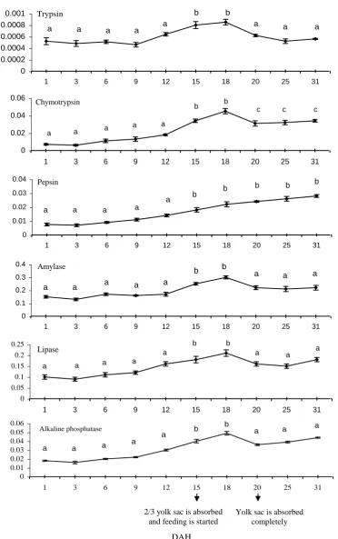
As showed at figure 2, trypsin, lipase and alkaline phosphatase activities indicated an increasing trend in the first 12 days without a significant difference (P > 0.05). From 12 to 18 days, there was an increased activity and a more significant difference
than the first 12 days (P < 0.05). After 18 days, trypsin and lipase decreased up to 25 days then increased from 25 to 31 days while alkaline phosphatase decreased up to 20 days then increased from 20 to 31 days without a significant difference (P > 0.05).
y = 67.915e0.0632x R2 = 0.9775
0 20 40 60 80 100 120 140 160
1 3 6 9 12 15 18 20 25 31
Days After Hatching
W
et
B
o
d
y
M
as
s
(m
g
)
Iranian Journal of Fisheries Sciences, 10(3), 2011 407
Figure 2: Mean value (±SD for replicate (n = 3) pooled fry) of specific activity of
digestive enzymes in O.mykiss fry during the experimental period.
Different superscripts denote significant differences (P < 0.05) within
DAH. Statistical analyses are subjected to the one way ANOVA test by spss11.5 software.
2/3 yolk sac Absorbtion and start of feeding
Sp ec if ic Activ ity ( U m g p ro tei n
-1 )
2/3 yolk sac absorbtion and start of feeding
Chymotrypsin
0 0.02 0.04 0.06
1 3 6 9 12 15 18 20 25 31
a c c c b b a a a a Pepsin 0 0.01 0.02 0.03 0.04
1 3 6 9 12 15 18 20 25 31
a b b a a a a
b b b
Amylase 0 0.1 0.2 0.3 0.4
1 3 6 9 12 15 18 20 25 31
a a a a b b a a a a a Lipase 0 0.05 0.1 0.15 0.2 0.25
1 3 6 9 12 15 18 20 25 31
a a a a b b a a a a Trypsin 0 0.0002 0.0004 0.0006 0.0008 0.001
1 3 6 9 12 15 18 20 25 31
a a a a
b b a a a a
2/3 yolk sac is absorbed and feeding is started
Yolk sac is absorbed completely Sp ec if ic Activ ity ( U m g p ro tei n -1 ) DAH Alkaline phosphatase 0 0.01 0.02 0.03 0.04 0.05 0.06
1 3 6 9 12 15 18 20 25 31
a a a b b a a a a a
408 Golchinfar et al., Assessment of digestive enzymes activity during the fry……..
Chymotrypsin activity demonstrated an increasing grade in the first 12 days but no difference statistically (P > 0.05) and showed increased slope with a more significant difference in its activity during 12 to 18 days compared to the first 12 days (P < 0.05). After 18 days, chymotrypsin activity decreased initially up to 20 days and then increased to 31 days without a significant difference (P > 0.05). The difference in chymotrypsin activity was more significant during 20 to 31 than the first 20 days (P > 0.05).
Pepsin activity revealed an increasing profile during 31 days without a significant difference in the first 12 days (P > 0.05), while after 12 to 31 days there was a more significant difference than the first 12 days (P > 0.05). Amylase activity showed a constant trend in the first 12 days without a significant difference (P > 0.05) but increased significantly from 12 to 18 days than the first 12 days (P < 0.05). After 18 days, it decreased up to 25 days and then increased up to 31 days without a significant difference (P > 0.05). Amylase activity showed a more significant difference from 18 to 31 days than 12 to 18 days (P < 0.05) but between 18 to 31 days and the first 12 days there was no significant difference (P > 0.05).
All assessments in enzyme activities showed no significant difference after 12 to 18 days (P > 0.05).
Discussion
Studies have expressed that between the age and growth in fish larvae there is a close correlation that can be related to formation and differentiation of body organs such as stomach, intestine, liver
and other organs (Govoni et al., 1986; Diaz-Lopez et al., 1997: Cara et al., 2003; Cahu et al., 2004; Faulk et al., 2007; Zamani et al., 2009). In rainbow trout a high correlation was observed between the wet body mass and the DAH where the estimated values were y = 67.915e 0.0632x, r2 = 0.9775. In addition, the assessments in different fish larvae revealed that formation and differentiation of body organs can affect some physiological changes such as enzymes secretion (Kuzmina and Gelman, 1998; Cara et al., 2003; Faulk et al., 2007). The presence of some main digestive enzymes in fish larvae in primary stages after hatching has been shown clearly in species such as,
Acipenser fulvescens (Buddington, 1985),
Acipenser transmontanus L. (Gawlicka et al., 1995), Sparus aurata (Moyano et al., 1996), Solea senegalensis kaup (Martinez et al., 1999), Oreochromis niloticus L. (Tengjaroenkul et al., 2001), Diplodus puntazzo (Suzer et al., 2007a),
Rachycentron canadum (Faulk et al., 2007) Paralabrax maculatofasciatus (Pena et al., 2009), Salmo trutta caspius (Zamani et al., 2009) and Paralabrax maculatofasciatus (Alvarez-Gonzalez et al., 2010). The present consideration showed enzyme existence in the rainbow trout fry 1 day after hatching. After 1 DAH, further development of such activities seems to be affected mainly by the progressive appearance of the digestive organs and by the response to feeding as observed in some species such as Caspian brown trout (Mojazi Amiri et al., 2005; Zamani et al, 2009). These typical results in patterns show a succession of increasing and decreasing with time as observed in
Iranian Journal of Fisheries Sciences, 10(3), 2011 409
other fish larva (Moyano et al., 1996; Martinez et al., 1999). Nevertheless, several authors have described a pattern characterized by a gradual decrease of specific activities of the some digestive enzymes, (e.g. amylase) a result that may be explained by the progressive increase in soluble proteins taking place in growing larvae as the body tissues fraction becomes steadily more important. In the present study, an increase was observed in enzyme activities at 15 DAH, which may reflect the response to feeding with a significant difference from the activity from 1 to 12 DAH (P < 0.05), since the synthesis of the main digestive enzymes in larval and juvenile fishes is greatly dependent on the food (Suzer et al., 2006). Pepsin secreted via stomach indicated to come along with increasing DAH, as seen in other fish larva (Buddington, 1985; Suzer et al., 2007a; Zamani et al., 2009). The absence of pepsin in the digestive tract of teleosts during early stages of life is thought to be compensated by micropinocytosis and intracellular digestion of proteins in the posterior intestine (Cara et al., 2003). From 12 DAH up to 31 DAH, pepsin activity showed a relatively increased profile whic could probably be explained by the development of organs such as gastric glands as observed in others (Cara et al., 2003; Alvarez-Gonzalez et al., 2008; Zamani et al., 2009). Alkaline phosphatase has been detected during larval development in different fish (Cara et al., 2003; Suzer et al., 2007b; Alvarez-Gonzalez et al., 2008; Zamani et al., 2009). According to Lojda et al., (1979), alkaline phosphatase is found primarily in cell membranes where active transport occurs and its increase in the intestine of
fish larva should indicate a more functional development of entrocytes. The presence of amylase activity has been reported in larvae of several fishes including Acipenser fulvescens
(Buddington, 1985), Sparus aurata
(Moyano et al., 1996), D.sargus (Cara et al., 2003) and Pagrus pagrus, L. (Suzer et al., 2007b) and Cyprinus carpio var. Jian (Jiang et al., 2009). Initial high levels of amylase could be better explained as a result of programmed gene expression, as suggested by Zambonino-infante and Cahu (2001). However, amylase had a constant profile in rainbow trout fry from 1 to 12 DAH (2/3 yolk sac was absorbed) but the presence of a certain amount of carbohydrate in the formulated diet given to fry at 15 DAH, could lead to enzyme secretion. Lipase has been detected during larval development in fishes including
D.sargus, A.fulvescens, S.senegalensis,
D.puntazzo, Cyprinus carpio (var. Jian) and Salmo Caspius. The major lipase in fishes appears to be a non-specific and bile salt-dependent lipase (Gjellesvik et al., 1992). Oozeki & bailey (1995) recommended the existence of two types of lipase in fish larvae, one related to yolk sac absorption (which is rich in phospholipids) and the other, whose activity is developed later, related to digestion of exogenous lipids. In the present study, lipase was also affected by major events such as the start of feeding (15 DAH). Trypsin and chymotrypsin belong to alkaline proteases with a considerable role in food digestion (Jobling, 1995). The pattern of trypsin and chymotrypsin showed a peak at the 15 DAH which can reflect the response to feeding. There is some indication that the
410 Golchinfar et al., Assessment of digestive enzymes activity during the fry……..
activities of digestive enzymes are low at first feeding and increase during the larval development. Kawai and Ikeda (1973) observed that activities of amylase, maltase and trypsin increased in bulk assays of larvae of increasing age after yolk absorption. In contrast, Hjelmeland et al. (1983) used a more sensitive and specific radio-immuno assay between hatching to yolk sac absorption and showed that trypsin and trypsinogen activities decreased to low levels for 14 days, and then increased. There may be two explanations for the increase in carbohydrolytic and proteolytic activities in fish larvae after yolk absorption and first feeding. First, the inherent enzymes available in the food may have caused an increase and second, enzymes production by the liver, pancreas and mucosal epithelium may have stimulated in response to initial food consumption and increased ration size. Results of enzymatic assessments revealed the existence of an increased profile in enzyme activity between 15 to 18 DAH and there was statistically a significant difference from the activity from 1 to 12 DAH (P < 0.05). In conclusion, enzyme assessments reveal that rainbow trout fry can receive food earlier than 15 DAH (between 12 to 15 DAH). Also, the results of our study indicate the best time for start of feeding so that useless feeding is inhibited and a suitable growth is achieved. More researches are necessary on digestive physiology (e.g. histochemical study of digestive enzymes), in order to delineate at exactly what age the fry is able to digest and absorb exogenous nutrients. One of the most important discussions in feeding is paying attention to physiologic
characterizations and understanding digestive enzymes activity from the hatching to yolk sac absorption.
Acknowledgments
The authors would like to thank the staff of Kelardasht Cold Water Fisheries Center for their help in collections of eggs and fries. We are also grateful for the most efficient technical assistance given by the personnel of the biotechnology department of Razi Vaccine and Serum Research Institute for the assay of enzymatic activity.
References
Alvarez-Gonzalez, C. A., Moyano-Lopez, F. J., Civera-Cerecedo, R.,
Carrasco-Chavez, V.,
Ortiz-Galindo, J. L. and Dumas, S., 2008.
Development of digestive enzyme activities in larvae of spotted sand bass Paralabrax maculatofasciatus: I. Biochemical analysis. Fish Physiology and Biochemistry, 34, 373–384.
Alvarez-Gonzalez, C. A., Moyano-Lopez, F. J., Civera-Cerecedo, R.,
Carrasco-Chavez, V.,
Ortiz-Galindo, J. L., Nolasco-Soria, H., Tovar-Ramırez, D. et al., 2010.
Development of digestive enzyme activity in larvae of spotted sand bass Paralabrax maculatofasciatus II: Electrophoretic analysis. Fish Physiology and Biochemistry, 36, 29– 37.
Anonymous, 2008. Iranian Fisheries Organization Annual Report. 56 P. (in Persian).
Anson, M. L., 1938. The estimation of pepsin, trypsin, papain and cathepsin
Iranian Journal of Fisheries Sciences, 10(3), 2011 411
with hemoglobin. Journal of General Physiology, 22, 79-89.
Bernfeld, P., 1951. Amylases α and β. In Methods in Enzymology, Vol.1 (Colowick, P. & Kaplan , N.O., eds), pp:149-157. New York: Academic Press.
Buddington, R. K., 1985. Digestive secretions of lake sturgeon, Acipenser fulvescens, during early development.
Journal of Fish Biology, 26, 715-723.
Cahu,C., Ronnestad,I.,Grangier,V. and
Zambonino Infante,J. L.,2004.
Expression and activities of pancreatic enzymes in developing sea bass larvae (Dicentrarchus labrax) in relation to intact and hydrolyzed dietary protein;
involvement of
cholecystokinin.Aquaculture,238(1-4), 295-308.
Cara, J. B., Moyano, F. J., Cardenas, S., Fernandez-Diaz, C. and Yuffera, M., 2003. Assessment of digestive enzyme activities during larval development of white bream. Journal of Fish Biology, 63, 48-58.
Cara, B, Moyano, F.J., Zambonino, J.L. and Fauvel, C., 2007. Trypsin and chymotrypsin as indicators of nutritional status of post-weaned sea bass larvae. Journal of Fish Biology, 70, 1798–1808.
Caruso, G., G. Denarob, M. and Genovesea, L., 2008. Temporal changes in digestive enzyme activities in the gastrointestinal tract of European eel (Anguilla anguilla) following feeding. Marine and Freshwater Behaviour and Physiology, 41(4), 215–228.
Chong, A.S.C., Hashim, R., Chow-Yang, L., and Ali, A.B. 2002. Partial
characterization and activities of proteases from the digestive tract of discus fish (Symphysodon aequifasciata). Aquaculture, 203, 321-333.
Deguara, S., Jauncey, K. and Agius, C., 2003. Enzyme activities and pH variations in the digestive tract of gilthead sea bream. Journal of Fish Biology, 62, 1033-1043.
Diaz-Lopez, M., Moyano, F.J., Garcia-Carreno, F.L.,Alarcon, F.J. and Sarasquete,M.C.,1997. Substrate SDS-PAGE determination of protease activity through larval development in sea bream. Aquaculture International, 5, 461–471.
Erlanger, B., Kokowsky, N. and Cohen, W., 1961. The preparation and properties of two new chromogenic substrates of trypsin. Archive Biochemistry and Biophysics, 95, 271-278.
Faulk, C.K., Benninghoff, A.D. And Holt, G.J., 2007. Ontogeny of the gastrointestinal tract and selected digestive enzymes in cobia Rachycentron canadum (L.). Journal of Fish Biology, 70, 567–583.
Fuente-Betancourt,
D.LG.,Garcia-Carreno, F., Del Toro, N.M.A. and
Cordova-Murueta, J.H., 2009.
Effect of pH and temperature on jumbo squid proteins. Journal of Food Biochemistry, 33, 260–272.
Gawlicka, A., Teh, S. L., Hung, S. S. O., Hinton, D. E. and De La Noue, J., 1995. Histological and histochemical changes in the digestive tract of white sturgeon larvae during ontogeny. Fish Physiology and Biochemistry, 14, 357-371.
412 Golchinfar et al., Assessment of digestive enzymes activity during the fry…….. Gawlicka, A., Parent, B., Horn, M.H.,
Ross, N., Opstad, I. and Torrissen, O.J., 2000. Activity of digestive enzymes in yolk-sac larvae of Atlantic halibut (Hippoglossus hippoglossus): indication of readiness for first feeding. Aquaculture, 184(3-4), 303-314.
Gjellesvik, D.R., Lombardo, D. and Walther, B.T., 1992. Pancreatic bile salt dependent lipase from cod (Gadus morhua): purification and properties.
Biochemica And Biophysica Acta, 1124, 123-134.
Govoni, J.J, Boehlert, G.W. and
Watanabe,Y., 1986. Physiology of digestion in fish larvae.
Environmental Biology of Fishes, 16 (1-3), 59-77
Hjelmeland, K., Huse, I., Jorgensen, T., Molvik, G. and Raa, J., 1983.
Trypsin and trypsinogen as indices of growth and survival potential of cod (Gadus morhua L.) larvae. Flodevigen Rapportserie, 3, 1-17.
Hummel, B. C. W., 1959. A modified spectrophotometric determination of chymotrypsin, trypsin and thrombin.
Canadian Journal of Biochemistry and Physiology, 37, 1393-1399.
Jiang,W.D., Feng, L., Liu, Y., Jiang, J. and Zhou, X.Q., 2009. Growth, digestive capacity and intestinal microflora of juvenile Jian carp (Cyprinus carpio var. Jian) fed graded levels of dietary inositol. Aquaculture Research, 40, 955-962.
Jobling, M., 1995. Digestion and
absorption. In:Jobling,
M.(Ed),Environmental Biology of Fishes, Chapter6.Chapman & Hall, London England. Pp, 175-210.
Kawai, S. and Ikeda, S., 1973. Studies enzymes of fishes IY. Development of the digestive enzymes of carp and black sea bream after hatching.
Bulletin of the Japanese Society of Scientific Fisheries, 39, 877-881.
Kim, B.G., Divakaran, S., Brown, C.L
and Ostrowski, A. C., 2001.
Comparative digestive enzyme ontogeny in two marine larval fishes: Pacific threadfin (Polydactylus sexfilis) and bluefin trevally (Caranx melampygus). Fish Physiology and Biochemistry, 24, 225–241.
Kolkovski, S., 2001. Digestive enzymes in fish larvae and juveniles-implications and application to formulated diets.
Aquaculture, 200, 181-201.
Kuzmina, V. V. 1996. Influance of age on digestive enzymes activity in some freshwater teleosts. Aquaculture, 148, 25-37.
Kuzmina,V. V. and Gelman, A. G., 1998. Traits in the development of the digestive function in fish. Journal of Ichthyology, 39 (1), 106-115.
Kuzmina, V. V. and Skvortsova, E. G., 2001. Activity of proteolytic enzymes of potential prey of predatory fish influence of natural and anthropogenic factors. Journal of Ichthyology, 41(3), 246-254.
Lazo, J. P., Holt, G. J. and Arnold, C. R., 2000. Ontogeny of pancreatic enzymes in larval red drum, Sciaenops ocellatus. Aquaculture Nutrition, 6, 183-192.
Lojda, Z., Gossrau, R. and Schiebler,T. H., 1979. Enzyme Histochemistry: A Laboratory
Manual.NewYork:Springer-Verlag.
Iranian Journal of Fisheries Sciences, 10(3), 2011 413 Lopez-Vasquez,K., Castro-Perez, C. A.
and Val, A. L., 2009. Digestive enzymes of eight Amazonian teleosts with different feeding habits. Journal of Fish Biology, 74, 1620–1628.
Lowry, O. H., Rosebrough, N. J., Farr, A. L and Randall, R. J., 1951.
Protein measurement with the folin phenol reagent. Journal of Biological Chemistry, 193, 265-275.
Martinez, I., Moyano, F. J., Fernandez-Diaz, C and Yufer, M., 1999.
Digestive enzyme activity during larval development of the Senegal sole (Solea senegalensis). Fish Physiology and Biochemistry, 21, 317-323.
Mojazi Amiri, B., Bahrekazemi, M., Pousti, I. and Vilaki, A. S., 2005. A histological study on the development of the digestive tract of Salmo trutta caspius (Kessleri) from hatching to parr stage. Iranian Journal of Fisheries Science, 5 (1), 63-84.
Moyano F. J., Barros A. M., Prieto, A., Cañavate J. P. and Cārdenas S., 2005. Evaluación de la ontogenia de enzimas digestivas en larvas de hurta,
Pagrus auriga (Pisces: Sparidae).
Revista Aquatic Sciences, 22, 39-47.
Moyano, F. J., Diaz, M., Alarcon, F. J. and Sarasquete, M. C., 1996.
Characterization of digestive enzyme activity during larval development of gilthead seabream (Sparus aurata).
Fish Physiology and Biochemistry, 15, 121-130.
Oozeki, Y. and Bailey, K. M., 1995.
Ontogenetic development of digestive enzymes activities in larval walleye pollock, Theragra chalcograma.
Marine Biology, 122, 177-186.
Pena, R., Dumas, S. and Rodriguez-Jaramillo, C., 2009. Development and distribution of intestinal enzymatic activity in Paralabrax maculatofasciatus larvae fed with live prey. Aquaculture Research, 40, 218-224.
Ribeiro, L., Couto, A., Olmedo, M., Alvarez-Blazquez, B., Linares, F. and Valente, L. M. P., 2008.
Digestive enzyme activity at different developmental stages of blackspot seabream, Pagellus bogaraveo.
Aquaculture Research, 39, 339-346.
Ribeiro, L., Zambonino Infante, J. L., Cahu, C. and Dinis, M. T., 1999.
Development of digestive enzymes in larvae of Solea senegalensis, Kaup 1858. Aquaculture, 179, 465–473.
Suzer, G., Aktülün, S., Çoban , D., Kamaci, H.O., Saka, Ş., Fırat, K. and Alpbaz, A., 2007a. Digestive enzyme activities in larvae of sharpsnout sea bream (Diplodus puntazzo), Comparative Biochemistry and Physiology, 148A, 470-477.
Suzer, G., Kamaci, H.O., Çoban, D., Saka, Ş., Firat, K., Özkara, B. and Özkara, A., 2007b. Digestive enzyme activity of the red porgy (Pagrus pagrus, L.) during larval development under culture conditions. Aquaculture Research, 38, 1778-1785.
Suzer, G., Firat, K. and Saka, S., 2006.
Ontogenic development of the digestive enzymes in common pandora, Pagellus erythrinus, L. larvae, Aquaculture Research, 37(15), 1565-1571.
Tengjaroenkul, B., Bonnie, J., Smith, S. A. and Chatreewongsin, U., 2001.
Ontogenic development of the
414 Golchinfar et al., Assessment of digestive enzymes activity during the fry……..
intestinal enzymes of cultured Nile tilapia, Oreochromis nilotichus.
Aquaculture, 211, 241-251.
Walter, K. and Schutt, C., 1974.
Alkaline phosphatase in serum (continous assay) in: Bergmeyer ,H.U. (Ed), Methods of enzymatic analysis,vol(2).,2nd edn. Academic Press, New York. NY. pp, 860-864.
Worthington, C. C., 1991. Worthington
enzyme manual related
Biochemical.3th Edition. Freehold, New Jersey. Pp, 212-215.
Zamani, A., Hajimoradloo, A., Madani,
R. and Farhangi, M., 2009.
Assessment of digestive enzymes activity during the fry development of the endangered Caspian brown trout
Salmo caspius. Journal of Fish Biology, 75(4), 932-937.
Zambonino-infante, J. L. and Cahu, C., 2001. Ontogeny of the gastrointestinal tract of marine fish larvae.
Comparative Biochemistry and Physiology, 130C, 477- 487.