Original Article
Study of Autonomic Function Tests in Geriatric Population
P. Vijitha *
1, M.V. Sailaja
2, N. Mallikarjuna Reddy
3.
*1 Assistant professor in Physiology, ACSR Govt. Medical College, Nellore, Andhra Pradesh, India.
2 Assistant professor in Physiology, RIMS, Kadapa, Andhra Pradesh, India.
3 Professor in Physiology, Narayana Medical College, Nellore, Andhra Pradesh, India.
Autonomic dysfunction worsens with old age. It results from an imbalance between the sympathetic and parasympathetic divisions. Autonomic function tests were carried out in 50 healthy subjects in the age group of 65 to 90 years and compared with 50 controls in the age group of 25 to 65 years. The Parasympathetic activity was assessed by heart rate response to deep breathing, valsalva maneuver and orthostatic tests. Sympathetic function was assessed by blood pressure response to cold pressor test and sustained hand grip exercise. We found a decline in sympathetic and parasympathetic function with advancing age. No gender variation in both sympathetic and parasympathetic function was observed in this study. The E: I ratio values were significantly decreased in geriatric population. (p-value 0.037). In cold pressor test, the SBP and DBP difference values were significantly reduced in geriatric population. (p-value-0.000). Heart rate response to hand grip test was reduced significantly (p-value-0.035). Autonomic status will have an important bearing on determining the therapeutic strategy and drug action in the elderly in whom there may be altered responsiveness to autonomic reflexes and receptor sensitivity.
KEY WORDS: Autonomic function, sympathetic, parasympathetic, blood pressure, cold pressor test and hand
grip exercise.
Online Access and Article Informtaion
International Journal of Integrative Medical Sciences
www.imedsciences.com
ABSTRACT
Address for correspondence: Dr. P. Vijitha, Assistant professor in Physiology, ACSR Medical College, Nellore, Andhra Pradesh, India. E-Mail: [email protected]
Int J Intg Med Sci 2015, Vol 2(3):79-86. ISSN 2394 - 4137 DOI: 10.16965/ijims.2015.105
Quick Response code
Received: 25-02-2015
Reviewed: 25-02-2015
Accepted: 16-03-2015
Published: 31-03-2015
Source of Funding: Self Conflicts of interest: None
DOI: 10.16965/ijims.2015.105
INTRODUCTION
Ageing is a natural process of merely growing order in a temporal sense and should be welcomed. The different theories can be combined into two groups, according to the first group, ageing is the cumulative result of random cell damage which goes throughout life, while the other group considers ageing is an inevitable result of our genetic programming [1]. In India 7.1% of the people are over 65 years as against 12% in U.S.A and Britain (as per 2001 census). Also in India although the percentage of aged persons to total population is low in comparison to developed countries, neverthe-less, the absolute size of aged population is
SUBJECTS AND METHODS
reflexes are based on reflex arc which consists of receptors, sensory neurons, center of integration, autonomic motor neurons and visceral effectors. The Autonomic reflexes regulate activities of smooth muscles, cardiac muscles and glands. The Hypothalamus controls and integrates the functions of both the divisions of Autonomic nervous system. During emotional states, limbic cortex controls the Autonomic nervous system [2]. The decline in various functions continues slowly and eventu-ally become considerable and functioneventu-ally sig-nificant as age advances. Thus physiologically, ageing refers to an impaired ability to maintain homeostasis in the face of external and/or internal challenges or stresses. As a result an individual becomes more vulnerable to these challenges and stresses and may finally succumb to one of these [3]. Both parasympa-thetic and sympaparasympa-thetic nervous system which constitute the autonomic nervous system (ANS) are affected by ageing. A number of tests to assess the functional status of ANS. The parasympathetic nervous system tests include beat to beat variation [4], Valsalva ratio [5] and the baro reflex sensitivity [6] and sympathetic nervous system tests include the hand grip test [7], the galvanic skin resistance [8, 9] and the cold pressor response [10]. In earlier also suggested an indication of affect of ageing on the autonomic nervous system activity so a formal study was planned to be carried out which involved a battery of autonomic tests to be done on 25-65 years age and >65years groups. The changes were noticed in some parameter of both sympathetic and parasympa-thetic function tests during comparison between 2 groups and amongst males and females of same age group.
of Hg, Diastolic blood pressure 60-90 mm of Hg and Random Blood Sugar 100-140 mg / dL from general population. <65 years, H/o Hypertension and H/o Diabetes mellitus were excluded from the study.
All subjects underwent autonomic function tests which included Deep Breathing Test assess the parasympathetic activity, by using Cardiowin system; PC based 12 channel simultaneous digital ECG, Genesis Media System Pvt Ltd. and ECG Jelly. Subject was instructed by made to lie down comfortably in supine position with head elevated to 300 and ECG electrodes were connected for recording Lead II ECG, to maintain deep breathing at a rate of six breaths per minute (allowing 5 seconds each for inspiration and expiration) maximum and minimum heart rates were recorded with each respiratory cycle. Expiration (E) to inspiration (I) ratio was determined by using the formula:
This is a cross-sectional study undertaken by me in the Department of Physiology, Narayana Medical College, and Nellore, Andhra Pradesh, India, for a period of 10 months beginning from August 2009. After Informed consent was taken, 50 were healthy elderly individuals ageds above 65 years with 50 were teaching and non-teaching staff of Narayana Medical College aged 25 to 65 years. Systolic blood pressure 100-140 mm
Maximum R-R interval during deep expiration
Minimum R-R interval during deep inspiration Normal Values of E: I ratio in different age groups [11]
Valsalva Manoeuver is a measure of parasym-pathetic and symparasym-pathetic functions [11, 12]. For the response to occur in valsalva manoeuver parasympathetic acts as afferent and efferent and sympathetic acts as a part of the efferent pathway. Therefore the valsalva ratio assesses more of parasympathetic function. By using Cardiowin system; PC based 12 channel simul-taneous digital ECG, Genesis Media System Pvt Ltd., ECG Jelly, Sphygmomanometer (Diamond) and Mouth Piece. Subject was made to lie down in a semi recumbent or sitting position, Nostrils were closed manually, Mouth piece was put into the mouth of the subject and the Mercury manometer was connected to the mouth piece. ECG machine was switched on for continuous recording. Subject was asked to exhale forcefully into the mercury manometer and asked to maintain the expiratory pressure at 40 mm of Hg for 10 – 15 seconds. ECG changes were recorded throughout the procedure, 30 seconds before and after the procedure. Valsalva ratio was calculated by using the formula:
E : I ratio =
Valsalva ratio =
Longest R-R interval after the manoeuver (Phase-IV)
Normal values of valsalva ratio in different age groups. Valsalva ratio more than 1.45 is considered to be normal. When it is 1.2 – 1.45, it is border line and if it is less than 1.2, it is regarded as abnormal. During the valsalva manoeuver failure of heart rate to increase during strain suggests a sympathetic dysfunction and failure of heart rate to slow down after the strain suggests parasympathetic dysfunction. Cold Pressor test (Cold pressure test) [11, 13, 14] the afferent limb of the reflex pathway in cold pressor test is somatic fibers whereas the efferent limb is sympathetic fibers. Beaker containing Ice cold water, Sphygmomanometer (Diamond), and Stethoscope (Littmann). Blood pressure was recorded under basal conditions. Cold water was taken in a container. Subject was asked to submerge one of his upper limbs in cold water up to the middle of the fore arm for 60 seconds. Blood pressure was recorded at the end of 60 seconds of submersion of the limb. Submersion of the limb in ice cold water increases systolic blood pressure by about 10-20 mm of Hg and diastolic blood pressure by about 10 mm of Hg.
Heart rate response to standing on changing the posture from supine to standing heart rate increases immediately by 10-20 beats per minute. This response is detected by recording ECG in supine and standing postures [11, 12, 13]. By using Cardiowin system; PC based 12 channel simultaneous digital ECG, Genesis Media System Pvt Ltd., and ECG Jelly. Subject was made to lie down in supine posture. ECG electrodes were connected from the subject to the cardiowin system. Subject was asked to relax completely for a minimum period of 10 minutes. Basal heart rate was recorded by using cardiowin system. Subject was asked to stand up immediately and change in heart rate was noted from the cardiowin, monitoring screen. Heart rate response to standing was determined by using the formula:
Heart rate in standing position – Heart rate in supine position.
Hand grip test in the hand grip test, there is a rise in heart rate and blood pressure. The blood pressure rise is due to increased sympathetic activity and heart rate rise is due to decreased
parasympathetic activity [11, 13]. By using Cardiowin system; PC based 12 channel simultaneous digital ECG, Genesis Media System Pvt Ltd., and ECG Jelly Sphygmomano-meter (Diamond), Stethoscope (Littmann), and Hand Grip Dynamometer. Subject was made to lie down in semi recumbent position. ECG electrodes were connected for lead II recording of ECG and sphygmomanometer for blood pressure measurement. Basal heart rate and blood pressure were recorded by instructed to maintain a pressure of 30% of the maximum activity in the hand grip dynamometer for about 5 minutes. Heart rate and change in SBP, DBP were recorded. The normal response is rise in DBP by > 10-15 mm of Hg and rise in heart rate by about 30% of the pretest value.
RESULTS
Table 1: Showing the E : I ratio values of means, Std.
deviation, and p-values in controls and cases.
Age in years Mean Std. Deviation P Value
Controls 25-65 years 1.1878 0.14399 0.037* Cases 65-90 Years 1.1324 0.11729 0.037* *statistically significance. <0.05 is statistically significant.
Graph 1: showing the means of E : I ratio values in
controls and cases.
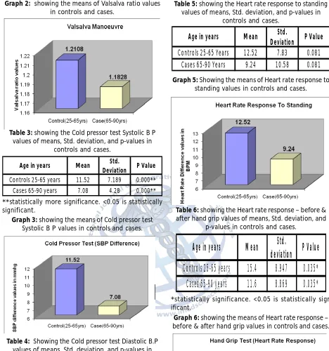
Table 2: showing the Valsalva ratio values of means,
Std. deviation, and p-values in controls and cases.
Age in years Mean Std.
Deviation P Value Controls 25-65 Years 1.2108 0.18359 0.387
Graph 2: showing the means of Valsalva ratio values
in controls and cases.
Table 3: showing the Cold pressor test Systolic B P
values of means, Std. deviation, and p-values in controls and cases.
Age in years Mean Std.
Deviation P Value
Controls 25-65 years 11.52 7.189 0.000**
Cases 65-90 years 7.08 4.28 0.000**
**statistically more significance. <0.05 is statistically significant.
Graph 3: showing the means of Cold pressor test
Systolic B P values in controls and cases.
Table 4: Showing the Cold pressor test Diastolic B.P
values of means, Std. deviation, and p-values in controls and cases.
Age in years Mean Std.
deviation P Value Control 25-65 years 11.44 8.917 0.000** Cases 65-90 Years 6.12 5.021 0.000**
**statistically more significance. <0.05 is statistically significant.
Graph 4: showing the means of Cold pressor test
Diastolic B.P values in controls and cases.
Table 5: showing the Heart rate response to standing
values of means, Std. deviation, and p-values in controls and cases.
Age in years Mean Std.
Deviation P Value
Controls 25-65 Years 12.52 7.83 0.081 Cases 65-90 Years 9.24 10.58 0.081
Graph 5: Showing the means of Heart rate response to
standing values in controls and cases.
Table 6: showing the Heart rate response – before &
after hand grip values of means, Std. deviation, and p-values in controls and cases.
Age in years
Mean
Std.
deviation
P Value
Controls 25-65 years
15.4
8.947
0.035*
Cases 65-90 years
11.6
8.869
0.035*
*statistically significance. <0.05 is statistically sign ificant.Graph 6: showing the means of Heart rate response –
before & after hand grip values in controls and cases.
Table 7: showing the Systolic blood pressure – before
& after hand grip values of means, Std. deviation, and p-values in controls and cases.
Age in years Mean Std.
Deviation P Value
Controls 25-65 years 8.84 6.3 0.71
Graph 7: showing the means of Systolic blood
pres-sure – before & after hand grip values in controls and cases.
Table 8: showing the Diastolic blood pressure – before
& after hand grip values of means, Std. deviation, and p-values in controls and cases.
Age in years Mean Std.
Deviation P Value
Controls 25-65 years 7.48 4.156 0.212 Cases 65-90 years 6.52 3.448 0.212
Graph 8: showing the means of Diastolic blood pressure
– before & after hand grip values in controls and cases.
DISCUSSION
In healthy young individuals breathing at a normal rate, the HR varies with the phases of respiration i.e., HR accelerates during inspira-tion and decelerates during expirainspira-tion, this is known as sinus arrhythmia. Sinus arrhythmia is a normal phenomenon and is due to fluctuations in parasympathetic output to the heart. Barore-ceptors are solely responsible for resting vagal tone in the normally breathing individuals. During inspiration, neuronal activity of inspira-tory neurons in the medulla besides initiating inspiration also discharge to Nucleus of tractus
contraction of the muscle that activate small fibbers in the afferent limb of the reflex arc .The response is a rise in diastolic pressure, more than 15 mm of hg and rise in the heart rate by about 30 per cent. The blood pressure rise is due to increased sympathetic activity, heart rate rise is due to decreased parasympathetic activity [11]. In our study a gradual decrease in heart rate response was observed. The results of our study are significant (p-value-=0.035.) Our findings are corroborating with S Suchiritha, A V Bharathi et al [22] and J .Gert Van Dijic at al. [23]. Heart rate response to handgrip test measures cardiac vagal tone. Decreased baroreceptor sensitivity with advancing age and parasympathetic withdrawal may explain the diminished heart rate response.
Blood pressure response to sustained hand grip was evaluated in all age groups, a rise in systolic blood pressure was observed in all subjects. These results are not statistically significant, (p value= 0.710). In our study the diastolic blood pressure response to sustained hand grip was significantly lower in all age groups. Thus, sympathetic function as assessed by sustained handgrip exercise, was reduced significantly in subjects above 60years indicating, late onset of decline in sympathetic efficiency in normal subjects, with advancing age. The results of our study are not significant, (p-value=o.212). Our findings are matching with the findings of Kaijser and Sachs [19], who observed a decreased blood pressure response to sustained handgrip test in older subjects, due to reduced effector organ sensitivity. However our results differ with earlier studies done by Vita G et al [16] S J Piha [24] and J Gert Van Dijik et al [23], who did not observe any significant decline in blood pressure response to sustained handgrip test, with advancing age.
The Valsalva maneuver has four phases; Phase 1 consists of the onset of strain. In this phase there occurs transient increase in B.P that lasts for a few seconds. This is due to increased intra thoracic pressure and mechanical compression of great vessels. However the H.R does not change much. Phase-2 is a phase of straining. In the early part of this phase venous return decreases, due to sustained rise in intrathoracic pressure. This change persists for four seconds.
In the later part of this phase, B.P returns towards normal due to increased peripheral resistance as a result of sympathetic vaso constriction. However H.R increases steadily throughout this phase due to vagal withdrawal in early phase and sympathetic activation in the latter phase. Phase-3 occurs following the release of strain in which there occurs transient decrease in BP lasting for a few seconds. This is caused by mechanical displacement of blood to pulmonary vascular bed, which was under increased intrathoracic pressure. There is little change in heart rate during the phase. Phase-4 occurs with further release of strain. The BP slowly increases due to persistent sympathetic mediated increase in peripheral resistance. This decreases HR proportionately through Baroreceptor stimulation [11]. Valsalva ratio is the ratio of the maximal tachycardia to the maximum bradycardia, induced by a standard valsalva maneuver.
The valsalva maneuver tests the integrity of both sympathetic and parasympathetic divisions of the Autonomic nervous system; However it is more of a parasympathetic test. The valsalva ratio has been widely used as a test of cardio vagal and baroreceptor function. In our study valsalva ratio showed a decline in the geriatric population. However, this was not statistically significant (p-value= 0.081), but the decreasing trend was similar to the study conducted by I A O’Brien et al (15), Dan Ziegler et al [25] and Jain A D O’Brien [15].
these results are statistically not significant, (p-value-0.081). A, I. O Brien, et all [15], Vita G. et al [16] and Gret Van Dijik et al, [23] Agelink N W et all [18] observed a linear decline in heart rate response to standing, which was not similar to our findings.
In order to see any significant variation in the autonomic response in males versus females, we compared the results of autonomic function tests in 30 males and 20 females. No significant variation was observed in autonomic function in our study. Our results are in accordance with the results of earlier studies done by Ken Umetani, M D et al [27]. Agelink M .W. et al [18], Sampo j piha et al [17] and Gautschy B et al. [28] Gender differences are most pronounced in subjects less than 30 years, heart rate variability of young male subjects being significantly greater than that of age matched female subjects. Differences disappear by the age of 50 years. The knowledge about the status of autonomic nervous system with advancing age will be useful in the management of cardiovascular, respiratory, urinary and gastro intestinal disturbances, the frequency of which increases with advancing age. The alteration of autonomic balance with advancing age will also have a bearing on drug action and in determining therapeutic strategy in the elderly.
CONCLUSION
The autonomic function tests can be used to assess the status of autonomic nervous system in the elderly, as these tests are simple, non-invasive and inexpensive. The population of elderly individuals above 60 years of age is rapidly increasing. It has been proved by many studies in our review that, regular physical ex-ercise can attenuate the age related changes in the cardiovascular autonomic functions by increasing parasympathetic outflow. Thus, the knowledge of autonomic status will have an important bearing on determining the therapeu-tic strategy and drug action in the elderly in whom there may be altered responsiveness to autonomic reflexes and receptor sensitivity.
[1]. Indukhurana, Geriatric physiology, In: Text book of medical physiology. India: Elsevier 2006; Ch 12.7: 1280-1285.
[2]. William F. Ganong, The Autonomic nervous system, In: Review of Medical Physiology, 23rd ed, India: McGraw -Hill Company 2010; Ch 17: 261-271. [3]. R.L.Bijalani, The physiology of ageing, In: Text book
of medical physiology, 2nd ed, India: Jaypee brothers
1997; Ch 1.6: 38-42.
[4] Hilsted J, Jensen SB. A simple test for
autonomic neuropathy in juvenile diabetics. Acta
Med Scand. 1979;205(5):385-7.
[5]. Levin AB. A simple test of cardiac function based upon the heart rate changes induced by the
Valsalva maneuver. Am J Cardiol. 1966 Jul;18(1):90-9.
[6]. Bennett T, Hosking DJ, Hampton JR. Baroreflex
sensitivity and responses to the Valsalva manoeuvre in subjects with diabetes mellitus. J
Neurol Neurosurg Psychiatry. 1976 Feb;39(2):178-83.
[7]. Ewing DJ, Irving JB, Kerr F, Wildsmith JA, Clarke BF.
Cardiovascular responses to sustained handgrip in normal subjects and in patients with diabetes
mellitus: a test of autonomic function. Clin Sci Mol
Med. 1974 Mar;46(3):295-306.
[8]. Heard GE. The psychologalvanic response in the study of sympathetic activity. Brit. Jour. Surg. 1964:51; 629-31.
[9]. M E Ahmed, L Delbridge, and L P Le Quesne. The role of autonomic neuropathy in diabetic foot ulceration. Jour. Neuro. Sci. 1986:47; 203-49. [10]. LeBlanc J, Dulac S, Côté J, Girard B. Autonomic
nervous system and adaptation to cold in man. J Appl Physiol. 1975 Aug;39(2):181-6.
[11]. G.K. Pal and Pravathy Pal, Autonomic function tests, In: Text book of practical physiology, India: Orient longman, 2009; Ch 40: 296-304.
[12]. Ewing D.J., Clarke B.F., Diagnosis and management of diabetic autonomic neuropathy, Br Med J Clin Res Ed Oct 1982; 285(6346): 916-8.
[13]. Ewing D.J., Clarke B.F., Autonomic neuropathy - its diagnosis and prognosis, Clin Endocrinol Metab. 1986 Nov; 15(14): 855-88.
[14]. Clarke B.F. and Ewing D.J., Cardiovascular reflex tests, In: The natural history of diabetic autonomic neuropathy, N Y State J Med. May 1982; 82(6): 903-8.
[15].I A O’Brien, P O’Hare, R J CorrallHeart rate
variability in healthy subjects: effect of age and
the derivation of normal ranges for tests ofauto-nomic function. Br Heart J. 1986 April; 55(4): 348– 354.
[16]. V ita G, Princi P, Calabro R, Toscano A, Manna
L, Messina C. Cardiovascular reflex tests.
Assessment of age-adjusted normal range. J Neurol
Sci. 1986 Oct;75(3):263-74.
How to cite this article:P. Vijitha, M.V. Sailaja, N. Mallikarjuna Reddy. Study of Autonomic Function Tests in Geriatric Population. Int J Intg Med Sci 2015;2(3):79-86. DOI:10.16965/ ijims.2015.105
[17].Piha SJ Cardiovascular responses to various autonomic tests in males and females. Clin Auton Res. 1993 Feb;3(1):15-20.
[18]. Agelink M.W., Maleessa R., and Baumann B., et al, Standardized tests of heart variability, Clin Auton. Res. Apr 2001; 11(2): 65-6.
[19].Kaijser L, Sachs C. Autonomic cardiovascular
responses in old age. Clin Physiol. 1985 Aug;5
(4):347-57.
[20].R. G. Victor, W. N. Leimbach, D. R. Seals, B. G. Wallin,and A. L. Mark, Effects of the cold pressor test on muscle sympathetic nerve activity in humans, American Heart Association 1987 Vol. 9; 5: 429-36.
[21]. Pascualy M1, Petrie EC, Brodkin K, Peskind ER, Veith
RC, Raskind MA. Effects of advanced aging on
plasma catecholamine responses to the cold pressor test. Neurobiol Aging. 1999 Nov-Dec;20(6):637-42.
[22]. S. Sucharitha, A.V. Bhrathi and Mario vaz, Effect of age and nutritional status on heart rate responses to cough and maximum handgrip, Indian J Physiol Pharmacol 2004; 48(1): 106-110.
[23]. van Dijk JG, Koenderink M, Zwinderman AH, Haan
J, Kramer CG, den Heijer JC. Autonomic nervous system tests depend on resting heart rate and blood
pressure. J Auton Nerv Syst. 1991 Jul;35(1):15-24. [24]. Piha S.J. Age related diminution of cardiovascular
autonomic responses diagnostic problems in the elderly, Cln Physiol. 1993; 13: 507-17.
[25].Ziegler D1, Laux G, Dannehl K, Spüler M, Mühlen
H, Mayer P, Gries FA. Assessment of cardiovascular autonomic function: age-related normal ranges
and reproducibility of spectral analysis, vector analysis, and standard tests of heart rate variation and blood pressure responses. Diabet Med. 1992 Mar;9(2):166-75.
[26].Umetani K1, Singer DH, McCraty R, Atkinson M.
Twenty-four hour time domain heart rate variability and heart rate: relations to age and gender over nine decades. J Am Coll Cardiol. 1998 Mar 1;31(3):593-601.
[27].Gautschy B, Weidmann P, Gnädinger MP.
Autonomic function tests as related to age and