ARTICLE OPEN ACCESS
Neurodegeneration as the presenting symptom in
2 adults with xeroderma pigmentosum
complementation group F
Niraj M. Shanbhag, MD, PhD, Michael D. Geschwind, MD, PhD, John J. DiGiovanna, MD,
Catherine Groden, MS, CRNP, Rena Godfrey, PA, Matthew J. Yousefzadeh, PhD, Erin A. Wade, BA, Laura J. Niedernhofer, MD, PhD, May Christine V. Malicdan, MD, PhD, Kenneth H. Kraemer, MD, William A. Gahl, MD, PhD, and Camilo Toro, MD
Neurol Genet2018;4:e240. doi:10.1212/NXG.0000000000000240
Correspondence Dr. Toro [email protected]
Abstract
Objective
To describe the features of 2 unrelated adults with xeroderma pigmentosum complementation group F (XP-F) ascertained in a neurology care setting.
Methods
We report the clinical, imaging, molecular, and nucleotide excision repair (NER) capacity of 2 middle-aged women with progressive neurodegeneration ultimately diagnosed with XP-F.
Results
Both patients presented with adult-onset progressive neurologic deterioration involving chorea, ataxia, hearing loss, cognitive deficits, profound brain atrophy, and a history of skin photo-sensitivity, skin freckling, and/or skin neoplasms. We identified compound heterozygous pathogenic mutations inERCC4and confirmed deficient NER capacity in skinfibroblasts from both patients.
Conclusions
These cases illustrate the role of NER dysfunction in neurodegeneration and how adult-onset neurodegeneration could be the major symptom bringing XP-F patients to clinical attention. XP-F should be considered by neurologists in the differential diagnosis of patients with adult-onset progressive neurodegeneration accompanied by global brain atrophy and a history of heightened sun sensitivity, excessive freckling, and skin malignancies.
From the Department of Neurology (N.M.S., M.D.G.), University of California San Francisco, CA; Laboratory of Cancer Biology and Genetics (J.J.D., K.H.K.), Center for Cancer Research, National Cancer Institute, National Institutes of Health; NIH Undiagnosed Diseases Program (C.G., R.G., M.C.V.M., W.A.G., C.T.), National Human Genome Research Institute, National Institutes of Health, Bethesda, MD; and Department of Molecular Medicine (M.J.Y., E.A.W., L.J.N.), Center on Aging, The Scripps Research Institute, Jupiter, FL.
Funding information and disclosures are provided at the end of the article. Full disclosure form information provided by the authors is available with the full text of this article at Neurology.org/NG.
The Article Processing Charge was funded by the NIH.
Patients with genome instability caused by defects in the DNA damage repair response illustrate the detrimental effect of cumulative DNA damage. This includes heightened cancer risk, accelerated aging, and neurodegeneration. A broad spectrum of neurologic manifestations has been associated with disorders of DNA repair, including xeroderma pigmen-tosum (XP), Cockayne syndrome (CS), ataxia telangiectasia, trichothiodystrophy, and Nijmegen breakage syndrome.1
XP is a rare autosomal recessive disorder arising from deficient DNA repair. XP is caused primarily by mutations in genes encoding components of the nucleotide excision repair (NER) DNA repair pathway. NER recognizes and repairs several types of DNA lesions, including those due to ultraviolet (UV) radiation, which results in bulky dimers at dipyrimidine sites.2 The typical result is sun damage to exposed skin and eyes. Patients with XP have a high frequency of skin cancer in re-sponse to genomic damage caused by UV radiation. Approx-imately one-fourth of patients with XP experience neurologic deterioration, and when present, shortens life expectancy.3 Cumulative toxicity by non–UV-mediated unrepaired geno-mic lesions in nonreplicating tissue explains neuronal injury.
We report 2 middle-aged Caucasian women with adult-onset chorea, ataxia, dystonia, neuropathy, and later progressive cognitive impairment, but with life-long acute skin sun-burning on minimal sun exposure, freckle-like skin lesions on sun-exposed skin, and early onset of skin cancer in 1 case. Both were found to have compound heterozygous mutations inERCC4and significantly reduced NER capacity, confirming the diagnosis of XP complementation group F (XP-F). Their presentations highlight the importance of adult neurologists considering XP-F when evaluating patients with atypical neurodegeneration.
Methods
Standard protocol approvals, registrations, and patient consents
Patients were referred by their neurologists to participate in the NIH Undiagnosed Diseases Program (UDP). They en-rolled in protocol 76-HG-0238 (ClinicalTrials.gov Identifier: NCT00369421) approved by the National Human Genome Research Institute (NHGRI) Institutional Review Board. Written informed consent was obtained according to protocol guidelines. Both patients had completed extensive clinical evaluations over the preceding decades, including inves-tigations for toxic, metabolic, infectious, autoimmune, and inflammatory processes and had undergone genetic testing for
common inherited adult-onset neurodegenerative disorders. Additional clinical details are summarized in the table.
Patient 1
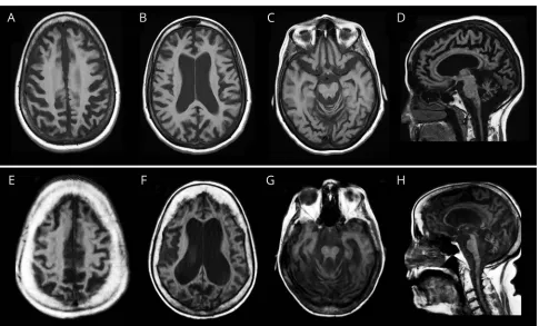
A now 60-year-old woman of Northern European descent with a history of infertility and skin photosensitivity but no skin neoplasms sought medical attention at the age 46 years with 1 year of progressive memory and balance difficulties. Her skin examination revealed moderate freckling of sun-exposed regions (figure 1A). On neurologic examination, she demon-strated mild chorea of her extremities and face, limb ataxia, absent ankle reflexes, and impaired gait. She had 1 sibling with MS but no other similarly affected relatives (figure 1B). Brain MRI showed severe global atrophy out of proportion to her recent symptom onset (figure 2, A–D). EMG and nerve con-duction studies indicated diffuse axonal sensorimotor poly-neuropathy. A sural nerve biopsy showed decreased myelinated fibers with clusters of regeneratingfibers (figure 1D). Neuro-logic progression was gradual but relentless over the next 11 years; the patient became dependent on a walker for ambulation and deteriorated in essentially all cognitive domains, with an early Mini-Mental State Examination4score of 26/30, a Mon-treal Cognitive Assessment (MoCA)5score of 18/30 at 6 years, and an MoCA score of 16/30 at 8 years after her initial visit.
Patient 2
A 52-year-old woman of Ashkenazi Jewish descent presented with a 20-year history of progressive dystonia, gait ataxia, hearing loss, and worsening cognition. Her past medical history included photosensitivity with blistering skin lesions after limited sun exposure resulting in multiple facial lentigines and basal cell carcinomas as a teenager. Because of aggressive photo-protection, she had minimal freckling but multiple scars from basal cell carcinoma resections (figure 1E). There was a mater-nal history of mild postural tremor but no similarly affected relatives (figure 1F). On neurologic examination, the patient was disoriented, partly amnestic, and struggled to follow simple commands. She had dystonic and choreiform movements of the head and neck, a coarse, irregular appendicular tremor, ataxia, and spasticity and was using a wheelchair. She had brisk reflexes and upgoing toes. Brain MRI demonstrated global cerebral at-rophy and inner table hyperostosis (figure 2, E–H). There was no electrophysiologic evidence of polyneuropathy. The patient died at age 54 years from secondary medical complications.
Results
Following singleton whole-exome sequencing, variants were prioritized based on known disease association, population
Glossary
frequency, and various in-silico pathogenicity models. Both patients had compound heterozygous mutations inERCC4 (table). Patient 1 (figure 1C) carried 2 known ERCC4 mutations associated with XP-F; NM_005236.2: c.1765C>T; p.Arg589Trp and c.2395C>T,p.Arg799Trp.6,7 Patient 2 (figure 1G) had the p.Arg799Trp allele and an ultra-rare ERCC4 truncating mutation; NM_005236.2:c.1376C>A, p.Ser459*. Segregation analysis of the families confirmed compound heterozygous allele pairs in both.
NER capacity was investigated by measuring unscheduled DNA synthesis (UDS) determining the incorporation of nucleotides into the nuclear genome of nonreplicating, UV-C irradiated patient-derivedfibroblasts (seefigure 1 legend for details). The percent repair was obtained by comparing irra-diated and unirrairra-diated patient cells to reference standards NER-proficient human fibroblasts (C5RO) and NER-deficient humanfibroblasts (XP51RO). XP51RO cells were from a patient with a homozygous p.Arg153Pro ERCC4 TableXP-F patients’molecular, clinical, and neurologic findings
Case 1 (UDP_3675) Case 2 (UDP_7356)
ERCC4(NM_005236.2) mutations c.2395C>T, p.Arg799Trp/c.1765C>T, p.Arg589Trp c.2395C>T, p.Arg799Trp/c.1376C>A, p.Ser459*
Age (y)
At NIH evaluation 53 54
Current 60 Deceased
Ethnicity Northern European Ashkenazi Jewish
History of acute skin sun-burning on minimal exposure
Yes Yes
Freckle-like skin lesions Moderate Mild
Skin cancer No ≥20 basal cell carcinomas (first by age 16 y)
Symptom prompting first neurologic evaluation; age (y)
Cognitive impairment; 46 Chorea and ataxia; 34
First subjective neurologic symptom; age “Twitchy, affected dexterity”; mid 20s “Clumsy”; late teens
Sequence of neurologic symptoms and signs Chorea/ataxia/neuropathy, cognitive impairment, and dementia
Chorea/ataxia, dystonia, spasticity, dementia
Height (cm); weight (kg); BMI 162.3; 60.2; 22.9 147; 49; 22.7
Developmental milestones Normal Normal
Education College classes College degree
Speech Ataxic dysarthria (mild) Ataxic dysarthria (mild)
Swallowing Mild dysphagia with tongue weakness and protrusion Normal swallow study
Hearing No subjective complaint of hearing loss. Audiometry not available
Neurosensory hearing loss (onset mid 30’s)
Activities of daily living Needs some assistance Fully dependent
Gait; assisting device Shuffling and ataxic; walker Ataxic and spastic; wheelchair
DTRs and plantar responses Absent ankle jerks, extensor Brisk throughout, extensor
Urinary incontinence Yes Yes
Polyneuropathy Diffuse axonal sensorimotor polyneuropathy No
Other neoplasm history No No
Keratopathy No No
Optic discs; retina Normal; normal Pale; Normal
Brain imaging Severe global atrophy Severe global atrophy and hyperostosis of the inner table
DNA repair (UDS) 26.0% ± 4.8% of normal 21.3% ± 5.1% of normal
mutation associated with a phenotype of XFE progeroid syndrome and severely reduced (<3%) UDS.8 UDS of fibroblasts from patients 1 and 2 had 26.0% ± 4.8% and 21.3% ± 5.1% of the expected NER capacity, respectively, indicating significant NER impairment (figure 1H).
Discussion
The prevalence of XP in the general population approximates 1 in 1 million, being higher in Japan because of a founder mu-tation in XPA. One-fourth of individuals with XP exhibit neurologic manifestations including acquired microcephaly, neuropathy, sensorineural hearing loss, and progressive cog-nitive impairment. In a long-term follow-up study of 89 patients with XP in the United Kingdom, three patients were diagnosed with XP-F, one of whom demonstrated neurologic features similar to our patients.9Up to 10% of all XP is XP-F.3,10XP-F is caused by mutations in the ERCC4 gene. The encoded ERCC4/XPF protein forms a tight complex with the ERCC1 protein. This heterodimer provides 59-nuclease activity for a crucial excision step of NER.2
Compared with other XP subtypes, patients with XP-F have less propensity for skin neoplasms and present later, often
past the 4th decade.10Neurodegeneration is largely reported in younger patients in complementation groups XP-A, XP-B, XP-D, and XP-G but not in XP-C or XP-E and is often her-alded by progressive neurosensory hearing loss.3Neurologic features reported in XP-F include gait disturbances, ataxia, neuropathy, chorea, sensorineural hearing loss, cognitive de-cline, and cerebral and cerebellar atrophy.6,7,10Our observa-tions support reports suggesting thatERCC4: p.Arg799Trp might be a common allele among XP-F patients with adult-onset neurodegeneration.6 Besides XP-F,ERCC4 mutations are associated with other diseases, including XFE progeria with very low UDS, Fanconi Anemia, and XP/CS.3,8
Like other reported XP-F cases with neurodegeneration, our patients manifested disparity between severity of brain atrophy by MRI at the time offirst imaging and relatively modest early cognitive deficits, consistent with compensation of a very slow neurodegenerative process over decades. This contrasts with other neurodegenerative disorders, such as Alzheimer disease, in which profound impairment usually accompanies such se-vere degree of brain atrophy.
These cases elicit consideration of several points. First, dys-function of the NER pathway can be associated with profound Figure 1Two cases of XP-F with adult-onset neurologic deterioration
neurodegeneration. Second, there is a causal role of cumula-tive unrepaired DNA damage in neurodegeneracumula-tive disorders in general. The precise type of damage leading to neuro-degeneration is unclear but may relate to stable oxidative genomic lesions. Finally, the diagnosis of XP, and particularly XP-F, should be considered in adult patients with unexplained neurodegeneration associated with global brain atrophy, es-pecially when accompanied by a history of photosensitivity, skin malignancies, and/or excessive freckling.
Author contributions
N.M. Shanbhag: wrote the initial draft of the manuscript. M.D. Geschwind, J.J. DiGiovanna, C. Groden, and R. God-frey: clinical consult and contributed to manuscript writing. M.J. Yousefzadeh: measured NER in fibroblasts, in-terpretation of data, and contributed to manuscript writing. E.A. Wade and L.J. Niedernhofer: measured NER infi bro-blasts, interpretation of data, data analysis, and contributed to manuscript writing. M.C.V. Malicdan: interpretation of data, data analysis, sample logistics, and contributed to manuscript writing. K.H. Kraemer: study design, interpretation of data, and contributed to manuscript writing. W.A. Gahl: study design, contributed to manuscript writing, and funding ac-quisition. C. Toro: study design, clinical consult, in-terpretation of data, data analysis, and wrote the initial draft of the manuscript.
Acknowledgment
The authors thank their patients, their families, and referring physicians. Dr. Dennis Landis contributed to the clinical evaluations of these patients at the NIH.
Study funding
Intramural Research Program of the National Institutes of Health (NIH) NHGRI and the NCI, Common Fund, Office of the Director; the NIH/NHLBI grant (HHSN268201400058); the NIH/NIA grant (P01AG043376): M.J.Y., E.A.W. and L.J.N.; and the Michael J. Homer Family Fund: M.D.G.
Disclosure
Dr. Shanbhag has received research support from the NIH NINDS and Alzheimer’s Association. Dr. Geschwind has re-ceived funding for travel and/or speaker honoraria from Oakstone Publishing, Inc; has served on the editorial board of Dementia & Neuropsychologia; serves or has served as a con-sultant to Advanced Medical Inc, Best Doctors Inc, Grand Rounds, Gerson Lehrman Group Inc, Guidepoint Global, MEDACorp, LCN Consulting, Optio Biopharma Solutions, various medical-legal consulting, Biohaven Pharmaceuticals Inc, Teva Pharmaceuticals, and Quest Diagnostics; and receives or has received research support from the NIH/NIA (R01 AG031189) PI 2013-2018, Alliance Biosecure, Michael J. Homer Family Fund, CurePSP, and Tau Consortium. Dr. Figure 2Representative axial and sagittal sections of T1-weighted brain MRIs
DiGiovanna serves or has served on scientific sdvisory boards of the Medical and Scientific Advisory Board of the Foun-dation for Ichthyosis and Related Skin Types; holds patents with regard to the use of synthetic peptides to disrupt the cytoskeleton and therapeutic uses of hmgn1 and hmgn2; consults or has consulted for the US Food and Drug Ad-ministration; and is employed by the National Cancer Institute and NIH. Dr. Groden, Dr. Godfrey, and Dr. You-sefzadeh report no disclosures. Dr. Wade is or has been employed by The Scripps Research Institute and The Na-tional Institute on Aging. Dr. Niedernhofer holds patents with regard to a rapid test to measure DNA repair capacity of an individual; has received research support from the NIH/ NHLBI, NIH/NIA, and Glenn Award for Aging Research. Dr. Malicdan serves or has served on the editorial board of BMC Musculoskeletal Disorders; holds patents with regard to therapeutic pharmaceutical agent for diseases associated with decrease in function of gne protein, food composition, and food additive, Gne-/-hGNED176VTg as a mouse model for DMRV/HIBM and CANNABINOID RECEPTOR MEDI-ATING COMPOUNDS; and is employed by the NIH Intramural Research Program. Dr. Kraemer serves or has served on the editorial board ofPhotodermatology and Pho-toimmunology. Dr. Gahl has received funding for travel and/or speaker honoraria from Cystinosis Research Network; serves or has served on the editorial board ofMolecular Genetics and Metabolism; receives or has received licensing royalties from
ManNAc; and receives research support from the NIH. Dr. Toro is a full-time employee of the NIH and receives funding from the NIH intramural program. Full disclosure form in-formation provided by the authors is available with the full text of this article at Neurology.org/NG.
Received January 22, 2018. Accepted infinal form April 23, 2018.
References
1. McKinnon PJ. DNA repair deficiency and neurological disease. Nat Rev Neurosci 2009;10:100–112.
2. Sijbers AM, de Laat WL, Ariza RR, et al. Xeroderma pigmentosum group F caused by a defect in a structure-specific DNA repair endonuclease. Cell 1996; 86:811–822.
3. Kraemer KH, DiGiovanna JJ. Xeroderma pigmentosum. In: Adam MP, Ardinger HH, Pagon RA, et al, editors. GeneReviews((R)). Seattle: University of Washington; 1993–2016.
4. Folstein MF, Folstein SE, McHugh PR.“Mini-mental state.”A practical method for grading the cognitive state of patients for the clinician. J Psychiatr Res 1975;12: 189–198.
5. Nasreddine ZS, Phillips NA, Bedirian V, et al. The Montreal Cognitive Assessment, MoCA: a brief screening tool for mild cognitive impairment. J Am Geriatr Soc 2005; 53:695–699.
6. Gregg SQ, Robinson AR, Niedernhofer LJ. Physiological consequences of defects in ERCC1-XPF DNA repair endonuclease. DNA Repair (Amst) 2011;10:781–791. 7. Moriwaki S, Nishigori C, Imamura S, et al. A case of xeroderma pigmentosum
complementation group F with neurological abnormalities. Br J Dermatol 1993;128: 91–94.
8. Niedernhofer LJ, Garinis GA, Raams A, et al. A new progeroid syndrome reveals that genotoxic stress suppresses the somatotroph axis. Nature 2006;444:1038–1043. 9. Fassihi H, Sethi M, Fawcett H, et al. Deep phenotyping of 89 xeroderma
pigmen-tosum patients reveals unexpected heterogeneity dependent on the precise molecular defect. Proc Natl Acad Sci USA 2016;113:E1236–E1245.
DOI 10.1212/NXG.0000000000000240
2018;4;
Neurol Genet
Niraj M. Shanbhag, Michael D. Geschwind, John J. DiGiovanna, et al.
pigmentosum complementation group F
Neurodegeneration as the presenting symptom in 2 adults with xeroderma
This information is current as of June 8, 2018
Services
Updated Information &
http://ng.neurology.org/content/4/3/e240.full.html
including high resolution figures, can be found at:
References
http://ng.neurology.org/content/4/3/e240.full.html##ref-list-1
This article cites 9 articles, 1 of which you can access for free at:
Subspecialty Collections
http://ng.neurology.org//cgi/collection/all_genetics
All Genetics
following collection(s):
This article, along with others on similar topics, appears in the
Permissions & Licensing
http://ng.neurology.org/misc/about.xhtml#permissions
its entirety can be found online at:
Information about reproducing this article in parts (figures,tables) or in
Reprints
http://ng.neurology.org/misc/addir.xhtml#reprintsus
Information about ordering reprints can be found online:
reserved. Online ISSN: 2376-7839.
Published by Wolters Kluwer Health, Inc. on behalf of the American Academy of Neurology.. All rights an open-access, online-only, continuous publication journal. Copyright Copyright © 2018 The Author(s).