Open Access
Software
Meta-DiSc: a software for meta-analysis of test accuracy data
Javier Zamora*
1, Victor Abraira
1, Alfonso Muriel
1, Khalid Khan
2and
Arri Coomarasamy
2Address: 1Clinical Biostatistics Unit, Ramón y Cajal Hospital, Madrid, Ctra. Colmenar km 9.100 Madrid 28034, Spain and 2University of
Birmingham and Birmingham Women's Hospital, Edgbaston, Birmingham, UK
Email: Javier Zamora* - [email protected]; Victor Abraira - [email protected]; Alfonso Muriel - [email protected]; Khalid Khan - [email protected]; Arri Coomarasamy - [email protected]
* Corresponding author
Abstract
Background: Systematic reviews and meta-analyses of test accuracy studies are increasingly being recognised as central in guiding clinical practice. However, there is currently no dedicated and comprehensive software for meta-analysis of diagnostic data. In this article, we present Meta-DiSc, a Windows-based, user-friendly, freely available (for academic use) software that we have developed, piloted, and validated to perform diagnostic meta-analysis.
Results: Meta-DiSc a) allows exploration of heterogeneity, with a variety of statistics including chi-square, I-squared and Spearman correlation tests, b) implements meta-regression techniques to explore the relationships between study characteristics and accuracy estimates, c) performs statistical pooling of sensitivities, specificities, likelihood ratios and diagnostic odds ratios using fixed and random effects models, both overall and in subgroups and d) produces high quality figures, including forest plots and summary receiver operating characteristic curves that can be exported for use in manuscripts for publication. All computational algorithms have been validated through comparison with different statistical tools and published meta-analyses. Meta-DiSc has a Graphical User Interface with roll-down menus, dialog boxes, and online help facilities.
Conclusion: Meta-DiSc is a comprehensive and dedicated test accuracy meta-analysis software. It has already been used and cited in several meta-analyses published in high-ranking journals. The software is publicly available at http://www.hrc.es/investigacion/metadisc_en.htm.
Background
Accurate diagnosis forms the basis of good clinical care, as without it one can neither prognosticate correctly nor choose the right treatment. Indeed, a wrong diagnosis can harm patients by exposing them to inappropriate or sub-optimal therapy [1]. Thus studies of diagnostic accuracy, and particularly their systematic reviews and meta-analy-ses, are being recognised as instrumental in underpinning evidence-based clinical practice. Initiatives such as STARD
[2] and developments within the Cochrane Collaboration [3] to accept protocols and reviews of test accuracy studies highlight the emphasis being given to evidence-based diagnosis.
Currently, there is only one test accuracy meta-analysis package, Meta-Test [4], which addresses some of the unique statistical issues related to test accuracy, such as pooling of sensitivities and specificities and summary Published: 12 July 2006
BMC Medical Research Methodology 2006, 6:31 doi:10.1186/1471-2288-6-31
Received: 31 March 2006 Accepted: 12 July 2006
This article is available from: http://www.biomedcentral.com/1471-2288/6/31
© 2006 Zamora et al; licensee BioMed Central Ltd.
receiver operating characteristics (sROC) analysis. How-ever, it is a DOS-based application with an interface that many find difficult to use, and integrate into Windows-based applications. Moreover, it lacks crucial analytical tools such as pooling of likelihood ratios (LRs), tests for heterogeneity and meta-regression facilities.
We, therefore, developed, piloted and validated a compre-hensive, Windows-based test accuracy meta-analysis soft-ware, Meta-DiSc, which is presented in this article, with a worked example.
Implementation
Meta-DiSc software was created in Microsoft Visual Basic 6, and some mathematical routines have been linked from the NAG C mathematical library [5]. The software is distributed as a single file, downloadable freely from URL: http://www.hrc.es/investigacion/metadisc_en.htm. Its installation is simple, guided by onscreen instructions. The programme has a user-friendly interface with roll-down menus, dialog boxes and online HTML compiled help files. These help files include a user manual and a description of the implemented statistical methods.
Meta-DiSc allows data entry into its datasheet in three dif-ferent ways: a) directly by typing data into the datasheet using the keyboard, b) copying from another spreadsheet (e.g. Microsoft Excel) and pasting into Meta-DiSc datash-eet, or c) importing text files from other sources (for exam-ple, in the comma delimited format). Several variables can be defined in the datasheet, including study identifi-ers, accuracy data from each study (true positives, false positives, true negatives and false negatives) and study level co-variates, such as those defining population spec-trum or methodological quality of the studies.
Once the data have been entered into the datasheet of Meta-DiSc, various statistical analyses can be imple-mented (Figure 1). The implementation of these statistical procedures needs to be carefully thought through and judicious, as it may be inappropriate (or indeed mislead-ing) to use all the procedures (particularly statistical pool-ing) in all reviews. Meta-DiSc provides analysts with adequate tools to assess the appropriateness of pooling. Readers interested in details of these methods are referred to statistical methods section of the help files (also avail-able as a PDF standalone document [6] and to existing texts and guidelines on diagnostic meta-analysis [7-10].
Describing the results of individual studies
When describing accuracy results from several studies, it is important to get an indication of the magnitude and pre-cision of the accuracy estimates derived from each study, as well as to assess the presence or absence of inconsisten-cies in accuracy estimates across studies (heterogeneity).
As accuracy estimates are paired and often inter-related (sensitivity and specificity, or LR positive and LR nega-tive), it is necessary to report these simultaneously [11]. One accuracy measure that combines these paired meas-ures is diagnostic odd ratio (dOR) [12], which has limited clinical use, although useful in procedures like meta-regression (see below).
Meta-DiSc computes accuracy estimates and confidence intervals from individual studies and shows results either as numerical tabulations or graphical plots in two for-mats: a) forest plots, for sensitivities, specificities, LRs or dOR, with respective confidence intervals; and b) plots of individual study results in ROC space, with or without an sROC curve.
Exploring heterogeneity (threshold effect)
Exploring heterogeneity is a critical issue to a) understand the possible factors that influence accuracy estimates, and b) to evaluate the appropriateness of statistical pooling of accuracy estimates from various studies. One of the pri-mary causes of heterogeneity in test accuracy studies is threshold effect, which arises when differences in sensitiv-ities and specificsensitiv-ities or LRs occur due to different cut-offs or thresholds used in different studies to define a positive (or negative) test result. When threshold effect exists, there is a negative correlation between sensitivities and specificities (or a positive correlation between sensitivities and 1-specificities), which results in a typical pattern of "shoulder arm" plot in a sROC space [8]. It is worth not-ing that correlation between sensitivity and specificity could arise due to a number of reasons other than thresh-old (e.g. partial verification bias, different spectrum of patients or different settings).
Available tools in Meta-DiSc
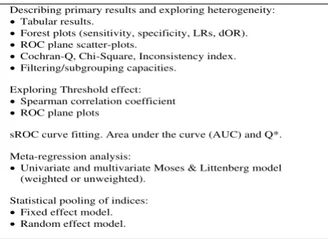
Figure 1
Available tools in Meta-DiSc. Tools implemented in the software Meta-DiSc to perform different steps of meta-analy-sis of diagnostic tests accuracy.
Describing primary results and exploring heterogeneity:
• Tabular results.
• Forest plots (sensitivity, specificity, LRs, dOR).
• ROC plane scatter-plots.
• Cochran-Q, Chi-Square, Inconsistency index.
• Filtering/subgrouping capacities.
Exploring Threshold effect:
• Spearman correlation coefficient
• ROC plane plots
sROC curve fitting. Area under the curve (AUC) and Q*.
Meta-regression analysis:
• Univariate and multivariate Moses & Littenberg model (weighted or unweighted).
Statistical pooling of indices:
• Fixed effect model.
Meta-DiSc allows assessment for threshold effect in three different ways: a) visual inspection of relationship between pairs of accuracy estimates in forest plots. If threshold effect is present, the forest plots will show increasing sensitivities with decreasing specificities, or vice versa. The same inverse relationship will be apparent with LR positive and LR negative; b) representation of accuracy estimates from each study in a sROC space – a typical "shoulder arm" pattern would suggest presence of threshold effect; and c) computation of Spearman correla-tion coefficient between the logit of sensitivity and logit of 1-specificity. A strong positive correlation would suggest threshold effect.
Exploring for heterogeneity (other than threshold effect)
Apart from variations due to threshold effect, there are several other factors that can result in variations in accu-racy estimates amongst different test accuaccu-racy studies in a review. These reasons include chance as well as variations in study population (e.g. severity of disease and co-mor-bidities), index test (differences in technology, assays, operator etc.), reference standard, and the way a study was designed and conducted [13]. Since such heterogeneity is almost always present in accuracy systematic reviews, test-ing for the presence and the extent of heterogeneity of results between primary studies, prior to undertaking any meta-analysis, is a critical part of any diagnostic review, as is exploration of the possible causes of heterogeneity [14].
Meta-DiSc allows users to test for heterogeneity amongst various studies in two different ways: a) Visual inspection of forest plots of accuracy estimates. If the studies are rea-sonably homogeneous, the accuracy estimates from indi-vidual studies will lie along a line corresponding to the pooled accuracy estimate. Large deviations from this line will indicate possible heterogeneity; b) statistical tests, including Chi-square and Cochran-Q, which are automat-ically implemented during analysis to evaluate if the dif-ferences across the studies are greater than expected by chance alone. A low p-value will suggest presence of het-erogeneity beyond what could be expected by chance alone. In addition to these heterogeneity statistics, Meta-DiSc computes the inconsistency index (I-squared) which has been proposed as a measure to quantify the amount of heterogeneity [15].
Meta-regression
If substantial heterogeneity is found to be present from the analyses detailed above, then reasons for such hetero-geneity can be explored by relating study level co-variates (e.g., population, test, reference standard or methodolog-ical features) to an accuracy measure, using meta-regres-sion techniques. The accuracy measure that is normally used is dOR, as it is a unitary measure of diagnostic per-formance that encompasses both sensitivity and
specifi-city or both LR positive and LR negative. Using dOR as a global measure of accuracy is a suitable method to com-pare the overall diagnostic accuracy of different tests [13]. However, its use is limited because it cannot be used directly in clinical practice and, furthermore, possible opposing effects of a study characteristic on sensitivity or specificity may be masked by using dOR.
Meta-DiSc implements meta-regression using a generali-zation of Littenberg and Moses Linear model [8,13] weighted by inverse of the variance or study size or unweighted. Random effects between studies can be esti-mated by different methods and added to the weighting scheme [16]. Estimations of coefficients of the model are performed by least squares method as implemented in NAG mathematical routines. The outcome variable is ln(dOR) which is related via a linear model to any number of study level covariates, and optionally includ-ing the variable representinclud-ing threshold effect [13]. The outputs from meta-regression modelling in Meta-DiSc are the co-efficients of the model, as well as ratio of dOR (rdOR) with respective confidence intervals. If a particular study level co-variate is significantly associated with diag-nostic accuracy, then its co-efficient will have a low p-value, and the rdOR will give a measure of magnitude of the association.
More advanced meta-regression techniques such as Hier-archical sROC model [17] and bivariate analysis of sensi-tivity and specificity [18] has been developed. These methods overcome some of the statistical shortcomings inherent to Littenberg and Moses model [8,19].
Statistical pooling
Statistical pooling is not always appropriate or necessary in every systematic review of test accuracy studies. How-ever, when used appropriately, pooling can provide useful summary information. The necessary precondition for simple pooling (weighted averaging) of each of sensitivi-ties, specificisensitivi-ties, LR positives and LR negatives, is that the studies and results are reasonably homogeneous (i.e. no substantial heterogeneity, including threshold effect, is present). If heterogeneity due to threshold effect were present, the accuracy data can be pooled by fitting a sROC curve and summarising that curve by means of the Area Under the Curve (AUC) or using other statistics such as the Q* index [19] (i.e. the point of the curve in which sen-sitivity equals specificity). If there is heterogeneity due to sources other than threshold effect, then pooling should only be attempted within homogeneous subsets, which would normally have been defined a priori.
fixed or random effect [10,20] models. The output from these analyses are presented numerically in tables, and graphically as forest plots. Pooled estimates are provided with their respective confidence intervals; b) It imple-ments several ways to fit a sROC curve when threshold effect is present. Default option is to compute a symmet-rical sROC curve after fitting the linear model proposed by Littenberg and Moses. However, users can choose differ-ent options to fit this curve, for example, combining indi-vidual dORs by the Mantel-Haenszel or the DerSimonian Laird methods [10,20] to estimate an overall dOR, and then fitting an sROC curve. When the dOR changes with diagnostic threshold, the sROC curve is asymmetrical. Meta-DiSc allows the user to check for asymmetry of the sROC curve, and fit an asymmetrical sROC curve if appro-priate. Finally, Meta-DiSc allows estimation of AUC and the Q* index, along with their standard errors, as a sum-mary measure of global accuracy which also aids inter-test comparisons; c) Meta-DiSc allows pooling of various summary measures within subgroups defined by study level co-variates with the help of a filter utility.
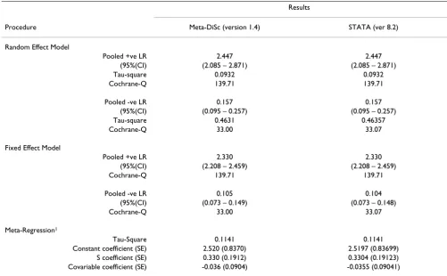
Wherever possible, the results of the above statistical pro-cedures were validated using different general purpose sta-tistical software such as STATA (ver 8.2) and SAS (8.2) using actually published and simulated data sets (Table 1).
Results
We illustrate the various procedures that Meta-DiSc implements in a case-study of ultrasound test in the diag-nosis of uterine pathology [21,22]. Ultrasound measure-ment of the lining of the uterus (endometrium) can predict pathology such as endometrial hyperplasia (a pre-cancerous condition) or cancer. The greater the thickness of endometrium, the more likely that the target condition is present. Various thresholds (such as 3, 4 or 5 mm etc) have been used to define a positive ultrasound result.
A systematic review of test accuracy studies identified 57 studies. Figure 2 shows a datasheet in Meta-DiSc which has been loaded with information from these 57 studies. The information includes study identifiers, accuracy data,
Table 1: Validation of statistical procedures. Validation of different statistical procedures using a simulated data-set. Results of Meta-DiSc (version 1.4) are compared with those obtained with metan (version 1.86) and metareg (version 1.06) STATA commands. Prior to the analyses, all four cells of all studies were added with 1/2 to avoid division by zero when computing some indices or standard errors. Meta-DiSc and STATA data-set are provided as additional files [see Additional file 1] and [see Additional file 2].
Results
Procedure Meta-DiSc (version 1.4) STATA (ver 8.2)
Random Effect Model
Pooled +ve LR 2.447 2.447
(95%(CI) (2.085 – 2.871) (2.085 – 2.871)
Tau-square 0.0932 0.0932
Cochrane-Q 139.71 139.71
Pooled -ve LR 0.157 0.157
(95%(CI) (0.095 – 0.257) (0.095 – 0.257)
Tau-square 0.4631 0.46357
Cochrane-Q 33.00 33.07
Fixed Effect Model
Pooled +ve LR 2.330 2.330
(95%(CI) (2.208 – 2.459) (2.208 – 2.459)
Cochrane-Q 139.71 139.71
Pooled -ve LR 0.105 0.104
(95%(CI) (0.073 – 0.149) (0.073 – 0.148)
Cochrane-Q 33.00 33.07
Meta-Regression1
Tau-Square 0.1141 0.1141
Constant coefficient (SE) 2.520 (0.8370) 2.5197 (0.83699) S coefficient (SE) 0.330 (0.1912) 0.3304 (0.19123) Covariable coefficient (SE) -0.036 (0.0904) -0.0355 (0.09041)
thresholds, and some study level co-variates (such as hor-mone replacement therapy use).
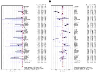
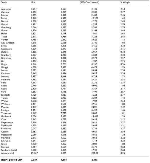
As the first step in the analysis, we have used Meta-DiSc to present accuracy measures from each individual study in forest plots for sensitivities (figure 3a), specificities (figure 3b), LRs (figures 4a and 4b) and dOR (figure 5). All these indices can also be represented in tabular form as shown in table 2. Although the forest plots and the tables contain a pooled summary at the bottom, at this early stage in the analysis, it is recommended that the plots are used to obtain a general overview of the accuracy estimates from each study, and the interpretation of the pooled summary is left to later stages of analysis.
The next step is the representation of sensitivity against 1-specificity from each study in a ROC space (figure 6), which can be used for exploration for threshold effect. The
pattern of the points in this plot suggest a "shoulder-arm" shape, indicating the possibility of threshold effect. We, therefore, performed a Spearman rank correlation as a ther test for threshold effect, and found that there was fur-ther indication of threshold effect (Table 3, Spearman correlation coefficient = 0.394; p = 0.006). Having found some clues about the presence of threshold effect, we now focus on a subgroup of 21 studies that used a singular threshold of >5 mm to define test positivity. Although an explicit threshold of 5 mm was used in these studies, there can still be an implicit threshold effect due to, for exam-ple, variation in the interpretation of the test results. Therefore, within this subgroup with an explicit threshold of 5 mm, it is still recommended that the above explora-tions for threshold effect are undertaken. We performed such analyses for this subgroup in Meta-DiSc, and found no evidence of further threshold effect (data not shown). There are a number of other more advanced methods not
Meta-Disc datasheet
Figure 2
implemented in Meta-DiSc that allow to incorporate explicitly information about tests thresholds defined between or within studies [17].
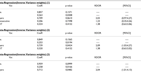
As the next step, heterogeneity arising from factors other than threshold effect is explored. We performed a visual exploration of the forest plots of accuracy measures for these 21 studies as well as statistical tests for heterogeneity (Meta-DiSc output not shown). In addition, possible sources of heterogeneity across the studies were explored using meta-regression analysis with the following co-vari-ates as predictor variables: use or non-use of hormone replacement therapy (HRT); technique of ultrasound measurement (single or double layer); and population enrolment (consecutive or other). Results are shown in Table 4, which suggest that the number of layers is strongly associated with accuracy. The double layer tech-nique is associated with two times higher accuracy
com-pared to single layer measurement (rdOR = 2.04; 95% CI: 1.01–4.13; p = 0.048)
The final step in the analysis is pooling if this is consid-ered appropriate. We illustrate pooling of the LRs for neg-ative test results in one homogenous subgroup of studies of non-HRT users, with a test threshold of ≤ 5 mm, and using a single layer technique (Figure 7). Finally, we dem-onstrate sROC curve fitting in the presence of threshold effect for the whole data-set in Figure 8.
Discussion and conclusion
Meta-DiSc allows description of individual study results; exploration of heterogeneity with a variety of statistics including chi-square, I-squared and Spearman correlation tests; implements meta-regression techniques to explore the relationships between study characteristics and accu-racy estimates; performs statistical pooling of sensitivities,
Forest plot
Figure 3
Forest plot. Forrest plot of sensitivities (3a) and specificities (3b) from test accuracy studies of ultrasound in the prediction of endometrial cancer.
A B
Specificity
0 0,2 0,4 0,6 0,8 1
Auslender 0,51 (0,42 - 0,61) Zannoni 0,53 (0,49 - 0,57) Bakour 0,49 (0,38 - 0,60) Botsis 0,88 (0,80 - 0,93) Fistonic 0,19 (0,12 - 0,29) Garuti 0,33 (0,28 - 0,38) Granberg 0,52 (0,49 - 0,55) Guner 0,47 (0,39 - 0,55) Haller 0,26 (0,16 - 0,39) Tsuda 0,63 (0,55 - 0,71) Varner 0,54 (0,25 - 0,81) Abu Ghazzeh 0,38 (0,28 - 0,49) Briley 0,51 (0,43 - 0,58) Cacciatore 0,27 (0,14 - 0,43) DeSilva 0,74 (0,60 - 0,86) Granberg 0,76 (0,70 - 0,82) Grigoriou 0,67 (0,60 - 0,73) Gu 0,27 (0,11 - 0,50) Gupta 0,64 (0,52 - 0,75) Hänggi 0,79 (0,67 - 0,87) Ivanov 0,58 (0,46 - 0,69) Karlsson 0,65 (0,54 - 0,75) Loverro 0,84 (0,74 - 0,91) Malinova 0,49 (0,38 - 0,60) Merz 0,43 (0,28 - 0,59) Nasri 0,66 (0,52 - 0,78) Nasri 0,61 (0,50 - 0,72) Pertl 0,27 (0,19 - 0,35) Suchocki 0,12 (0,06 - 0,20) Taviani 0,54 (0,37 - 0,70) Weber 0,39 (0,29 - 0,50) Wolman 0,64 (0,49 - 0,77) Moreles 0,61 (0,53 - 0,68) Rudigoz 0,74 (0,59 - 0,86) Todorova 0,50 (0,16 - 0,84) Gruboeck 0,88 (0,80 - 0,94) Chan 0,62 (0,47 - 0,75) Degenhardt 0,66 (0,55 - 0,75) Dijkhuizen 0,49 (0,36 - 0,62) Brolmann 0,53 (0,39 - 0,66) Ceccini 0,71 (0,66 - 0,76) Masearetti 0,58 (0,33 - 0,80) Mortakis 0,58 (0,45 - 0,69) Schramm 0,50 (0,42 - 0,58) Smith 0,54 (0,37 - 0,69) Osmers 0,50 (0,43 - 0,57) Seelbach-Göbel 0,44 (0,36 - 0,51) Altuncu et al. 0,97 (0,85 - 1,00)
Specificity (95% CI)
Pooled Specificity = 0,54 (0,53 to 0,55) Chi-square = 646,82; df = 47 (p = 0,0000) Inconsistency (I-square) = 92,7 %
B
Sensitivity
0 0,2 0,4 0,6 0,8 1
Auslender 1,00 (0,79 - 1,00) Zannoni 0,98 (0,90 - 1,00) Bakour 1,00 (0,72 - 1,00) Botsis 1,00 (0,63 - 1,00) Fistonic 1,00 (0,77 - 1,00) Garuti 0,98 (0,91 - 1,00) Granberg 1,00 (0,97 - 1,00) Guner 1,00 (0,82 - 1,00) Haller 1,00 (0,79 - 1,00) Tsuda 0,93 (0,68 - 1,00) Varner 1,00 (0,16 - 1,00) Abu Ghazzeh 1,00 (0,03 - 1,00) Briley 1,00 (0,48 - 1,00) Cacciatore 1,00 (0,40 - 1,00) DeSilva 0,33 (0,01 - 0,91) Granberg 1,00 (0,63 - 1,00) Grigoriou 1,00 (0,86 - 1,00) Gu 1,00 (0,59 - 1,00) Gupta 0,67 (0,09 - 0,99) Hänggi 0,86 (0,64 - 0,97) Ivanov 1,00 (0,69 - 1,00) Karlsson 0,93 (0,68 - 1,00) Loverro 1,00 (0,86 - 1,00) Malinova 1,00 (0,95 - 1,00) Merz 1,00 (0,77 - 1,00) Nasri 1,00 (0,59 - 1,00) Nasri 1,00 (0,54 - 1,00) Pertl 0,95 (0,74 - 1,00) Suchocki 1,00 (0,88 - 1,00) Taviani 1,00 (0,16 - 1,00) Weber 0,98 (0,91 - 1,00) Wolman 1,00 (0,40 - 1,00) Moreles 0,91 (0,71 - 0,99) Rudigoz 0,78 (0,40 - 0,97) Todorova 1,00 (0,16 - 1,00) Gruboeck 0,82 (0,48 - 0,98) Chan 1,00 (0,80 - 1,00) Degenhardt 0,86 (0,71 - 0,95) Dijkhuizen 1,00 (0,63 - 1,00) Brolmann 1,00 (0,69 - 1,00) Ceccini 0,94 (0,70 - 1,00) Masearetti 1,00 (0,29 - 1,00) Mortakis 1,00 (0,59 - 1,00) Schramm 0,62 (0,42 - 0,79) Smith 1,00 (0,40 - 1,00) Osmers 1,00 (0,87 - 1,00) Seelbach-Göbel 0,95 (0,83 - 0,99) Altuncu et al. 0,83 (0,36 - 1,00)
Sensitivity (95% CI)
specificities, likelihood ratios and diagnostic odds ratios, using fixed and random effects models, both overall and in subgroups; and produces high quality figures, includ-ing forest plots and summary receiver operatinclud-ing character-istic curves that can be exported for use in manuscripts for publication.
Meta-DiSc is an evolving software. As new diagnostic meta-analytic methods become established over time, they will be implemented into the program in the future. For example, bivariate method of pooling sensitivity and specificity [18] is currently being developed. We will care-fully follow the progress in this field. Once accepted as an established meta-analytic method, it will be implemented in Meta-DiSc. On similar lines, methods of data extraction from individual studies that only provide accuracy meas-ures are currently being developed within our depart-ment. Once these methods have been verified, we will
implement this option to assist systematic reviewers in extracting 2-by-2 tables from such studies.
Meta-DiSc is a comprehensive and dedicated test accuracy meta-analysis software. All computational algorithms in it have been validated through comparison with different statistical tools and published meta-analyses. Its use and citation in several meta-analyses published in high-rank-ing journals is evidence of external validation of its high quality [23-28].
Availability and requirements
The software is publicly available at http://www.hrc.es/ investigacion/metadisc_en.htm.
Operating system: The software runs on Windows based personal computers (Windows 95 or higher) with Pen-tium-class processor or equivalent, with minimum of 32
Forest plot
Figure 4
Forest plot. Forrest plot of likelihood ratios for positive (4a) and negative (4b) test results from studies of ultrasound in the prediction of endometrial cancer.
A B
Positive LR
0,01 1 100,0
Auslender 1,99 (1,62 - 2,45) Zannoni 2,09 (1,92 - 2,28) Bakour 1,89 (1,49 - 2,41) Botsis 7,36 (4,44 - 12,21) Fistonic 1,20 (1,04 - 1,38) Garuti 1,47 (1,36 - 1,59) Granberg 2,07 (1,93 - 2,21) Guner 1,83 (1,57 - 2,14) Haller 1,32 (1,12 - 1,56) Tsuda 2,52 (1,96 - 3,22) Varner 1,79 (0,84 - 3,83) Abu Ghazzeh 1,21 (0,54 - 2,75) Briley 1,85 (1,40 - 2,47) Cacciatore 1,24 (0,88 - 1,75) DeSilva 1,31 (0,24 - 6,96) Granberg 3,94 (2,93 - 5,28) Grigoriou 2,95 (2,43 - 3,57) Gu 1,31 (0,96 - 1,79) Gupta 1,85 (0,78 - 4,35) Hänggi 4,00 (2,47 - 6,47) Ivanov 2,27 (1,69 - 3,05) Karlsson 2,65 (1,94 - 3,63) Loverro 5,96 (3,65 - 9,73) Malinova 1,96 (1,59 - 2,42) Merz 1,70 (1,29 - 2,24) Nasri 2,74 (1,83 - 4,10) Nasri 2,40 (1,71 - 3,37) Pertl 1,29 (1,11 - 1,50) Suchocki 1,12 (1,03 - 1,22) Taviani 1,80 (0,98 - 3,30) Weber 1,62 (1,37 - 1,90) Wolman 2,48 (1,56 - 3,96) Moreles 2,31 (1,85 - 2,90) Rudigoz 2,98 (1,64 - 5,43) Todorova 1,67 (0,73 - 3,81) Gruboeck 7,04 (3,69 - 13,42) Chan 2,54 (1,78 - 3,64) Degenhardt 2,52 (1,86 - 3,41) Dijkhuizen 1,86 (1,39 - 2,49) Brolmann 2,02 (1,49 - 2,74) Ceccini 3,27 (2,65 - 4,02) Masearetti 2,06 (1,10 - 3,87) Mortakis 2,21 (1,60 - 3,06) Schramm 1,24 (0,90 - 1,71) Smith 1,94 (1,25 - 3,00) Osmers 1,96 (1,70 - 2,27) Seelbach-Göbel 1,68 (1,45 - 1,94) Altuncu et al. 29,17 (4,09 - 208,02)
Positive LR (95% CI)
Random Effects Model Pooled Positive LR = 2,09 (1,88 to 2,31) Cochran-Q = 506,06; df = 47 (p = 0,0000) Inconsistency (I-square) = 90,7 %
Tau-square = 0,0992 Negative LR
0,01 1 100,0
Auslender 0,06 (0,00 - 0,88) Zannoni 0,03 (0,00 - 0,24) Bakour 0,08 (0,01 - 1,28) Botsis 0,06 (0,00 - 0,94) Fistonic 0,17 (0,01 - 2,70) Garuti 0,05 (0,01 - 0,35) Granberg 0,01 (0,00 - 0,13) Guner 0,05 (0,00 - 0,83) Haller 0,11 (0,01 - 1,75) Tsuda 0,11 (0,02 - 0,71) Varner 0,31 (0,02 - 4,09) Abu Ghazzeh 0,65 (0,06 - 7,30) Briley 0,16 (0,01 - 2,35) Cacciatore 0,37 (0,03 - 5,30) DeSilva 0,90 (0,40 - 2,03) Granberg 0,07 (0,00 - 1,08) Grigoriou 0,03 (0,00 - 0,47) Gu 0,22 (0,01 - 3,50) Gupta 0,52 (0,10 - 2,61) Hänggi 0,18 (0,06 - 0,52) Ivanov 0,08 (0,01 - 1,18) Karlsson 0,10 (0,02 - 0,69) Loverro 0,02 (0,00 - 0,36) Malinova 0,01 (0,00 - 0,23) Merz 0,08 (0,00 - 1,21) Nasri 0,10 (0,01 - 1,40) Nasri 0,12 (0,01 - 1,69) Pertl 0,20 (0,03 - 1,36) Suchocki 0,14 (0,01 - 2,31) Taviani 0,31 (0,02 - 3,96) Weber 0,04 (0,01 - 0,29) Wolman 0,16 (0,01 - 2,19) Moreles 0,15 (0,04 - 0,56) Rudigoz 0,30 (0,09 - 1,03) Todorova 0,33 (0,02 - 4,55) Gruboeck 0,21 (0,06 - 0,72) Chan 0,04 (0,00 - 0,70) Degenhardt 0,21 (0,09 - 0,47) Dijkhuizen 0,11 (0,01 - 1,69) Brolmann 0,09 (0,01 - 1,31) Ceccini 0,09 (0,01 - 0,59) Masearetti 0,22 (0,02 - 2,99) Mortakis 0,11 (0,01 - 1,60) Schramm 0,76 (0,46 - 1,24) Smith 0,19 (0,01 - 2,63) Osmers 0,04 (0,00 - 0,56) Seelbach-Göbel 0,12 (0,03 - 0,46) Altuncu et al. 0,17 (0,03 - 1,03)
Negative LR (95% CI)
Forrest plot
Figure 5
Forrest plot. Forest plot of diagnostic odds ratios (dOR) from test accuracy studies of ultrasound in the prediction of endometrial cancer.
Diagnostic Odds Ratio
0,01 1 100,0
Auslender 34,78 (2,04 - 593,79) Zannoni 62,15 (8,55 - 451,56) Bakour 22,47 (1,28 - 393,49) Botsis 115,48 (6,32 - 2.109,25) Fistonic 7,00 (0,40 - 123,10) Garuti 29,25 (4,00 - 213,73) Granberg 246,16 (15,26 - 3.970,34) Guner 34,36 (2,04 - 578,15) Haller 11,91 (0,68 - 209,19) Tsuda 23,75 (3,04 - 185,50) Varner 5,77 (0,23 - 143,37) Abu Ghazzeh 1,86 (0,07 - 46,84) Briley 11,26 (0,61 - 206,72) Cacciatore 3,39 (0,17 - 68,12) DeSilva 1,46 (0,12 - 17,56) Granberg 53,86 (3,05 - 950,71) Grigoriou 98,32 (5,90 - 1.638,95) Gu 5,91 (0,29 - 119,06) Gupta 3,54 (0,31 - 40,93) Hänggi 22,00 (5,71 - 84,78) Ivanov 29,00 (1,64 - 513,45) Karlsson 25,74 (3,23 - 205,10) Loverro 258,78 (14,83 - 4.514,43) Malinova 135,80 (8,15 - 2.264,08) Merz 21,90 (1,23 - 391,31) Nasri 28,85 (1,56 - 531,94) Nasri 20,60 (1,12 - 378,06) Pertl 6,56 (0,84 - 51,00) Suchocki 7,96 (0,46 - 138,73) Taviani 5,81 (0,26 - 128,90) Weber 39,29 (5,22 - 295,45) Wolman 15,81 (0,81 - 310,35) Moreles 15,43 (3,50 - 68,07) Rudigoz 9,92 (1,80 - 54,49) Todorova 5,00 (0,18 - 136,32) Gruboeck 34,20 (6,45 - 181,32) Chan 56,54 (3,21 - 994,48) Degenhardt 12,22 (4,35 - 34,30) Dijkhuizen 16,46 (0,91 - 297,75) Brolmann 23,38 (1,31 - 418,59) Ceccini 37,28 (4,86 - 285,93) Masearetti 9,47 (0,43 - 208,76) Mortakis 20,41 (1,12 - 371,17) Schramm 1,64 (0,73 - 3,68) Smith 10,38 (0,53 - 205,27) Osmers 55,00 (3,31 - 913,67) Seelbach-Göbel 14,26 (3,34 - 60,84) Altuncu et al. 170,00 (9,11 - 3.172,49)
Diagnostic OR (95% CI)
Random Effects Model
Pooled Diagnostic Odds Ratio = 17,48 (11,59 to 26,37) Cochran-Q = 70,56; df = 47 (p = 0,0147)
Table 2: Tabulation of Likelihood ratio for positive test result (LR+) with respective 95% confidence intervals from all test accuracy studies included in systematic review of ultrasound for prediction of endometrial cancer.
Study LR+ [95% Conf. Iterval.] % Weight
Auslender 1,994 1,623 -2,449 2,54
Zannoni 2,092 1,919 -2,280 2,77
Bakour 1,895 1,490 -2,408 2,45
Botsis 7,360 4,437 -12,208 1,69
Fistonic 1,200 1,045 -1,378 2,69
Garuti 1,471 1,358 -1,593 2,78
Granberg 2,066 1,935 -2,206 2,79
Guner 1,834 1,569 -2,144 2,65
Haller 1,321 1,118 -1,561 2,63
Tsuda 2,517 1,964 -3,225 2,43
Varner 1,795 0,842 -3,826 1,13
Abu Ghazzeh 1,215 0,538 -2,745 1,03
Briley 1,855 1,396 -2,465 2,33
Cacciatore 1,239 0,877 -1,752 2,15
DeSilva 1,306 0,245 -6,957 0,34
Granberg 3,937 2,933 -5,284 2,30
Grigoriou 2,946 2,430 -3,572 2,57
Gu 1,307 0,956 -1,787 2,25
Gupta 1,846 0,783 -4,350 0,96
Hänggi 4,000 2,472 -6,473 1,76
Ivanov 2,273 1,691 -3,054 2,30
Karlsson 2,649 1,936 -3,627 2,24
Loverro 5,957 3,648 -9,729 1,73
Malinova 1,963 1,591 -2,421 2,53
Merz 1,697 1,287 -2,236 2,35
Nasri 2,740 1,833 -4,096 1,98
Nasri 2,400 1,711 -3,367 2,17
Pertl 1,293 1,115 -1,499 2,67
Suchocki 1,120 1,027 -1,222 2,77
Taviani 1,802 0,983 -3,304 1,44
Weber 1,618 1,374 -1,904 2,64
Wolman 2,481 1,556 -3,956 1,80
Moreles 2,312 1,845 -2,896 2,49
Rudigoz 2,981 1,638 -5,426 1,46
Todorova 1,667 0,729 -3,808 1,01
Gruboeck 7,036 3,689 -13,422 1,35
Chan 2,543 1,779 -3,635 2,12
Degenhardt 2,516 1,856 -3,411 2,27
Dijkhuizen 1,859 1,389 -2,489 2,31
Brolmann 2,017 1,487 -2,736 2,27
Ceccini 3,267 2,655 -4,021 2,54
Masearetti 2,059 1,096 -3,866 1,38
Mortakis 2,213 1,602 -3,058 2,22
Schramm 1,241 0,899 -1,714 2,22
Smith 1,938 1,252 -3,001 1,88
Osmers 1,964 1,699 -2,271 2,68
Seelbach-Göbel 1,680 1,455 -1,940 2,68
Altuncu et al. 29,167 4,089 -208,02 0,25
(REM) pooled LR+ 2,087 1,881 -2,315
Heterogeneity chi-squared = 506,06 (d.f.= 47) p = 0,000 Inconsistency (I-square) = 90,7%
No. studies = 48. Filter OFF
Table 4: Results of meta-regression analysis for predicting the presence or absence of endometrial carcinoma with variables: use or non-use of hormone replacement therapy (HRT); technique of ultrasound measurement (single or double layer); and population enrolment (consecutive or other).
Meta-Regression(Inverse Variance weights) (1)
Var. Coeff. p-value RDOR [95%CI]
Cte. 0,857 0,1571 ----
----S 0,263 0,0208 ----
----Layers 0,709 0,0610 2,03 (0,97;4,27)
Consecutive 0,206 0,7398 1,23 (0,35;4,26)
HRT 0,324 0,4152 1,38 (0,63;3,06)
Meta-Regression(Inverse Variance weights) (2)
Var. Coeff. p-value RDOR [95%CI]
Cte. 0,849 0,1565 ----
----S 0,253 0,0194 ----
----Layers 0,739 0,0424 2,09 (1,03;4,27)
HRT 0,320 0,4152 1,38 (0,63;3,02)
Meta-Regression(Inverse Variance weights) (3)
Var. Coeff. p-value RDOR [95%CI]
Cte. 0,959 0,0999 ----
----S 0,258 0,0166 ----
----Layers 0,712 0,0482 2,04 (1,01;4,13)
Table 3: Results of Spearman rank correlation of sensitivity against (1 – specificity) to assess the threshold effect in all test accuracy studies included in systematic review of ultrasound for prediction of endometrial cancer.
Var. Coeff. Std. Error T p-value
A 2.412 0.292 8.266 0.0000
b(1) 0.187 0.101 1.857 0.0697
Spearman correlation coefficient: 0,394 p-value = 0,006 (Logit(TPR) vs Logit(FPR) Moses' model (D = a + bS)
Unweighted regression Tau-squared estimate = 0,3540
(Convergence is achieved after 2 iterations) Restricted Maximum Likelihood estimation (REML) No. studies = 48
Forrest plot
Figure 7
Forrest plot. Forrest plots of Likelihood ratios for positive (7a) and negative (7b) test results in one homogenous sub-group of studies of non-HRT users, with a test threshold of ≤ 5 mm, and using a single layer technique.
A
B
Positive LR
0,01 1 100,0
Abu Ghazzeh 1,21 (0,54 - 2,75) Briley 1,85 (1,40 - 2,47) Cacciatore 1,24 (0,88 - 1,75) Grigoriou 2,95 (2,43 - 3,57) Gu 1,31 (0,96 - 1,79) Gupta 1,85 (0,78 - 4,35) Hänggi 4,00 (2,47 - 6,47) Ivanov 2,27 (1,69 - 3,05) Karlsson 2,65 (1,94 - 3,63) Loverro 5,96 (3,65 - 9,73) Malinova 1,96 (1,59 - 2,42) Nasri 2,74 (1,83 - 4,10) Suchocki 1,12 (1,03 - 1,22) Taviani 1,80 (0,98 - 3,30) Weber 1,62 (1,37 - 1,90) Wolman 2,48 (1,56 - 3,96) Positive LR (95% CI)
Random Effects Model
Pooled Positive LR = 2,08 (1,59 to 2,72) Cochran-Q = 240,88; df = 15 (p = 0,0000) Inconsistency (I-square) = 93,8 % Tau-square = 0,2593
Negative LR
0,01 1 100,0
Abu Ghazzeh 0,65 (0,06 - 7,30) Briley 0,16 (0,01 - 2,35) Cacciatore 0,37 (0,03 - 5,30) Grigoriou 0,03 (0,00 - 0,47) Gu 0,22 (0,01 - 3,50) Gupta 0,52 (0,10 - 2,61) Hänggi 0,18 (0,06 - 0,52) Ivanov 0,08 (0,01 - 1,18) Karlsson 0,10 (0,02 - 0,69) Loverro 0,02 (0,00 - 0,36) Malinova 0,01 (0,00 - 0,23) Nasri 0,10 (0,01 - 1,40) Suchocki 0,14 (0,01 - 2,31) Taviani 0,31 (0,02 - 3,96) Weber 0,04 (0,01 - 0,29) Wolman 0,16 (0,01 - 2,19) Negative LR (95% CI)
Random Effects Model
Pooled Negative LR = 0,14 (0,08 to 0,25) Cochran-Q = 15,95; df = 15 (p = 0,3853) Inconsistency (I-square) = 6,0 % Tau-square = 0,0794
ROC Space
Figure 6
ROC Space. Representation of sensitivity against (1-specifi-city) in Receiver Operating Characteristics space for each study of ultrasound in the prediction of endometrial cancer.
Sensitivity ROC space
1-specificity
0 0,2 0,4 0,6 0,8 1
0 0,1 0,2 0,3 0,4 0,5 0,6 0,7 0,8 0,9 1
sROC curve
Figure 8
sROC curve. Receiver operating characteristics curve for all studies included in systematic review of ultrasound for prediction of endometrial cancer.
Sensitivity SROC Curve
1-specificity
0 0,2 0,4 0,6 0,8 1
0 0,1 0,2 0,3 0,4 0,5 0,6 0,7 0,8 0,9 1
Symmetric SROC
MB of RAM and minimum of 20 MB of hard disk space. SVGA color monitor; minimum 800 × 600 screen resolu-tion and 256 colors.
Licence: Freeware for academic use.
Competing interests
The author(s) declare that they have no competing inter-ests.
Authors' contributions
JZ conceived the idea. AM, VA and JZ developed the soft-ware. AC and KSK tested the software on a number of reviews and gave suggestions for improvements. All authors participated in preparing this manuscript.
Additional material
Acknowledgements
This work has been partly funded by Spanish Health Ministry Grants no PI02/0954, G03/090 and PI04/1055.
References
1. Thomson R, McElroy H, Sudlow M: Guidelines on anticoagulant treatment in atrial fibrillation in Great Britain: variation in content and implications for treatment. BMJ 1998, 316:509-513.
2. Bossuyt PM, Reitsma JB, Bruns DE, Gatsonis CA, Glasziou PP, Irwig LM, Lijmer JG, Moher D, Rennie D, de Vet HC: Towards complete and accurate reporting of studies of diagnostic accuracy: The STARD Initiative. Radiology 2003, 226:24-28.
3. Collaboration C: Methods Groups Newsletter. http://www
cochrane org/newslett/MGNews-2004 pdf 2006 [http://
www.cochrane.org/newslett/MGNews-2004.pdf].
4. Lau J: Meta-Test. Boston: New England Medical Center; 1997. 5. The NAG C Library, Mark 6. Oxford: Numerical Algorithms
Group; 2004.
6. Zamora J, Muriel A, Abraira V: Meta-DiSc Statistical Methods. 2006 [ftp://ftp.hrc.es/pub/programas/metadisc/ MetaDisc_StatisticalMethods.pdf].
7. Irwig L, Tosteson ANA, Gatsonis C, Lau J, Colditz G, Chalmers TC, Mosteller F: Guidelines for Metaanalyses Evaluating Diagnos-tic-Tests. Annals of Internal Medicine 1994, 120:667-676.
8. Moses LE, Shapiro D, Littenberg B: Combining independent stud-ies of a diagnostic test into a summary ROC curve: data-ana-lytic approaches and some additional considerations. Stat Med 1993, 12:1293-1316.
9. Deville WL, Buntinx F, Bouter LM, Montori VM, de Vet HC, van der Windt DA, Bezemer PD: Conducting systematic reviews of diagnostic studies: didactic guidelines. BMC Med Res Methodol
2002, 2:9.
10. Deeks JJ: Systematic reviews of evaluations of diagnostic and screening tests studies. In Systematic reviews in health care:
meta-analysis in context 2nd Edition edition. Edited by: Egger M, Davey SG
and Altman DG. BMJ Books; 2001.
11. Honest H, Khan KS: Reporting of measures of accuracy in sys-tematic reviews of diagnostic literature. Bmc Health Services Research 2002, 2:.
12. Glas AS, Lijmer JG, Prins MH, Bonsel GJ, Bossuyt PM: The diagnos-tic odds ratio: a single indicator of test performance. J Clin Epi-demiol 2003, 56:1129-1135.
13. Lijmer JG, Bossuyt PM, Heisterkamp SH: Exploring sources of het-erogeneity in systematic reviews of diagnostic tests. Stat Med
2002, 21:1525-1537.
14. Dinnes J, Deeks J, Kirby J, Roderick P: A methodological review of how heterogeneity has been examined in systematic reviews of diagnostic test accuracy. Health Technol Assess 2005, 9:1-113.
15. Higgins JP, Thompson SG: Quantifying heterogeneity in a meta-analysis. Stat Med 2002, 21:1539-1558.
16. Thompson SG, Sharp SJ: Explaining heterogeneity in meta-anal-ysis: a comparison of methods. Stat Med 1999, 18:2693-2708. 17. Rutter CM, Gatsonis CA: A hierarchical regression approach to
meta-analysis of diagnostic test accuracy evaluations. Stat Med 2001, 20:2865-2884.
18. Reitsma JB, Glas AS, Rutjes AWS, Scholten RJPM, Bossuyt PM, Zwind-erman AH: Bivariate analysis of sensitivity and specificity pro-duces informative summary measures in diagnostic reviews.
Journal of Clinical Epidemiology 2005, 58:982-990.
19. Walter SD: Properties of the summary receiver operating characteristic (SROC) curve for diagnostic test data. Stat Med 2002, 21:1237-1256.
20. DerSimonian R, Laird N: Meta-analysis in clinical trials. Control Clin Trials 1986, 7:177-188.
21. Gupta JK, Chien PF, Voit D, Clark TJ, Khan KS: Ultrasonographic endometrial thickness for diagnosing endometrial pathology in women with postmenopausal bleeding: a meta-analysis.
Acta Obstet Gynecol Scand 2002, 81:799-816.
22. Khan KS, Kunz R, Kleijnen J, Antes G: Case study 4: Reviewing evidence on test accuracy. In Systematic Review to Support
Evi-dence-based Medicine 2003 edition. London, The Royal Society of
Medicine; 2003:109-119.
23. Morgan M, Kalantri S, Flores L, Pai M: A commercial line probe assay for the rapid detection of rifampicin resistance in Mycobacterium tuberculosis: a systematic review and meta-analysis. BMC Infectious Diseases 2005, 5:62.
24. Flores L, Pai M, Colford JM, Riley LW: In-house nucleic acid amplification tests for the detection of Mycobacterium tuberculosis in sputum specimens: analysis and meta-regression. BMC Microbiol 2005, 5:55.
25. Gisbert J, Abraira V: Accuracy of Helicobacter pylori Diagnos-tic Tests in Patients with Bleeding PepDiagnos-tic Ulcer: A System-atic Review and Meta-analysis. The American Journal of Gastroenterology 2006, 101:848-863.
26. Shiga T, Wajima Z, Inoue T, Sakamoto A: Predicting difficult intu-bation in apparently normal patients: a meta-analysis of bed-side screening test performance. Anesthesiology 2005, 103:429-437.
27. Zijlstra JM, van der Werf G, Hoekstra OS, Hooft L, Huijgens PC: F-fluoro-deoxyglucose positron emission tomography for post-treatment evaluation of malignant lymphoma: a systematic review. Haematologica 2006, 91:522-9.
28. Goodacre S, Sutton AJ, Sampson FC: Meta-Analysis: The Value of Clinical Assessment in the Diagnosis of Deep Venous Thrombosis. Annals of Internal Medicine 2005, 143:129-139.
Pre-publication history
The pre-publication history for this paper can be accessed here:
http://www.biomedcentral.com/1471-2288/6/31/prepub
Additional File 1
Meta-Disc data set. This file contains simulated data. It is provided to help users to validate statistical procedures shown in table 1.
Click here for file
[http://www.biomedcentral.com/content/supplementary/1471-2288-6-31-S1.dsc]
Additional File 2
STATA data set. This file contains simulated data. It is provided to help users to validate statistical procedures shown in table 1.
Click here for file