R E S E A R C H
Open Access
Towards the creation of decellularized organ
constructs using irreversible electroporation
and active mechanical perfusion
Michael B Sano
1, Robert E Neal II
1, Paulo A Garcia
1, David Gerber
2, John Robertson
1,3, Rafael V Davalos
1,4** Correspondence: [email protected] 1
School of Biomedical Engineering and Sciences, Virginia Tech - Wake Forest University, Blacksburg, VA, USA
Abstract
Background:Despite advances in transplant surgery and general medicine, the number of patients awaiting transplant organs continues to grow, while the supply of organs does not. This work outlines a method of organ decellularization using non-thermal irreversible electroporation (N-TIRE) which, in combination with reseeding, may help supplement the supply of organs for transplant.
Methods:In our study, brief but intense electric pulses were applied to porcine livers while under active low temperature cardio-emulation perfusion. Histological analysis and lesion measurements were used to determine the effects of the pulses in decellularizing the livers as a first step towards the development of extracellular scaffolds that may be used with stem cell reseeding. A dynamic conductivity numerical model was developed to simulate the treatment parameters used and determine an irreversible electroporation threshold.
Results:Ninety-nine individual 1000 V/cm 100-μs square pulses with repetition rates between 0.25 and 4 Hz were found to produce a lesion within 24 hours post-treatment. The livers maintained intact bile ducts and vascular structures while demonstrating hepatocytic cord disruption and cell delamination from cord basal laminae after 24 hours of perfusion. A numerical model found an electric field threshold of 423 V/cm under specific experimental conditions, which may be used in the future to plan treatments for the decellularization of entire organs. Analysis of the pulse repetition rate shows that the largest treated area and the lowest
interstitial density score was achieved for a pulse frequency of 1 Hz. After 24 hours of perfusion, a maximum density score reduction of 58.5 percent had been achieved. Conclusions:This method is the first effort towards creating decellularized tissue scaffolds that could be used for organ transplantation using N-TIRE. In addition, it provides a versatile platform to study the effects of pulse parameters such as pulse length, repetition rate, and field strength on whole organ structures.
Background
Over the past fifty years, organ transplantation has become a standard care for patients diagnosed with end stage organ failure including cirrhosis and renal failure. Liver transplantation is very successful, with 90 and 75% survival rates after 1 and 5 years, respectively. Unfortunately, the number of patients with cirrhosis, chronic viral hepati-tis and hepatocellular carcinoma has steadily increased, leading to unmet demands for
organ transplantation [1]. According to the United Network of Organ Sharing (UNOS), there are over 108,000 candidates in the US alone currently waiting for organ trans-plants including kidney, liver, heart, and lung. In 2009, there were fewer than 7,000 liver transplants from both living and deceased donors [2].
Despite advances in transplant surgery and general medicine, the number of patients awaiting transplant organs continues to grow, while organ supply does not. Organ supply is constrained by obstacles that impede acquisition, such as the requirement for organ removal coincident with brainstem death necessitating the use of hospital resources to maintain artificial life support. As a result, organ donation may be proble-matic when intensive care resources are strained[3]. In addition, life support for poten-tial organ donations has been ethically debated[4,5] and donation refusal is common in regions where social, cultural, and religious pressures constrain organ procurement.
The increasing gap between organ donation and supply to severely-ill patients has fostered an increased interest in alternative organ sources[6]. For the development and differentiation of full organs suitable for human transplant, structures that provide microvasculature for the delivery of nutrients to all cells must be developed[7-9]. Traditional top-down manufacturing techniques are currently unable to produce a hierarchical vascular structure scale which can span the more than 4 orders of magni-tude of human organs[10]. Microfabrication techniques can replicate some features of the complex architecture of mammalian microvasculature, but current processes fail to extend into the macro-scale[11]. Thus, structures which have features spanning multi-ple length scales are currently only fabricated through biological mechanisms and the relatively new field of biofabrication has developed, with the goal of utilizing and manipulating these processes [12].
Decellularization of existing tissues extends the concept of biofabrication by taking advantage of the body’s natural programming to create a complete tissue, including a functional vascular network. Rat liver extracellular matrix constructs have been created using chemical decellularization and reseeding [13-15]. Decellularized rat hearts, reseeded with multiple cell types, can contract and have the ability to generate pump-ing pressures [16]. Challenges to chemical decellularization techniques include the potential for detergents to damage extracellular matrix components [17,18] the poten-tial to create and deposit toxins [13,17], and the inherent difficulty of scaling these techniques up from small rat organs to larger organs [14]. These challenges must be overcome before decellularized organs can successfully be translated to the clinical setting.
Hypothermic oxygenated perfusion (HOPE) is a method of whole organ preservation which mechanically delivers an oxygenated, nutrient-rich blood substitute to an entire organ at sub-physiological temperatures [26,27]. This method has been successfully demonstrated to improve the preservation quality and transplant success rates of kidneys which have undergone warm ischemia [28,29]; with research striving to reach 72 hour preservation times [30]. In addition, Schon et al.[31] and Brockmannet al.
[32] have demonstrated the ability to prolong organ quality using normothermic perfusion, a process in which the perfused fluid is held at or near physiological temperatures. These methods of organ preservation can be used to isolate N-TIRE tis-sue ablation effects from the immune response observedin vivo and the natural degra-dation of tissue post mortem.
Electroporation is a non-linear biophysical process in which the application of pulsed electric fields leads to an increase in permeability of cells, presumably through the creation of nanoscale pores in the lipid bilayer [33]. At low pulsing energy, this perme-ability is reversible and cellular health and function is maintained. Once a critical elec-tric field intensity threshold is surpassed (approximately 500 [34] to 700 V/cm [35] for ninety 50 μs pulses at 4 Hz in brain and eight 100μs pulses at 1 Hz in liver, respec-tively), the cell membrane is unable to recover and cell death is induced in a precise and controllable manner with sub-millimeter resolution [36,37]. This process is referred to as non-thermal irreversible electroporation (N-TIRE) [38]. N-TIRE does not rely on thermal mechanisms [38] and preserves the structure of the underlying extracellular matrix as well as nerve conduits and bile ducts [39]. Since N-TIRE cell death does not require any drugs, there should not be any creation or deposition of toxins when killing the cells from this technique.
Recently, we and others have determined, through the use of translational laboratory models, that capitalizing on the ability of N-TIRE to destroy cells without destroying the extracellular matrix might make N-TIRE a viable means for scaffold creation via organ decellularization [40,41]. We hypothesize that viable decellularized tissue scaf-folds can be obtained using non-thermal irreversible electroporation (N-TIRE) on organs under continuous perfusion.
Machine-perfused porcine livers were treated with N-TIRE using external plate or needle electrodes within one hour of organ harvest and establishment of active perfu-sion. At varying time points after electroporation, livers were removed from perfusion, immediately after which samples were collected, preserved in 10% neutral buffered for-malin, prepared for histology, and their microscopic structure was examined. Examina-tion of the N-TIRE treated and control (untreated) regions of tissue demonstrated that N-TIRE was capable of decellularizing large volumes of tissue when performed in con-junction with active organ perfusion, suggesting that N-TIRE may be a viable method of decellularization for tissue engineering applications.
Methods
Tissue
flushed with lactated Ringer’s solution (LRS) to remove blood/clots before placement on the perfusion system.
Perfusion
The VasoWave™Perfusion System (Smart Perfusion, Denver, NC) was used to perfuse the livers for 4 and 24 hours. This system produces a cardioemulating pulse wave to generate physiological systolic and diastolic pressures and flow rates within the organ. The system is capable of controlling the oxygen content of the perfusate above and below physiological norms. A perfusate, consisting of modified LRS, was delivered to the portal vein and hepatic artery and recycled back into the system via the hepatic vein. All livers were under active machine perfusion within one hour post-mortem and the perfusate was held at 4°C
Electroporation
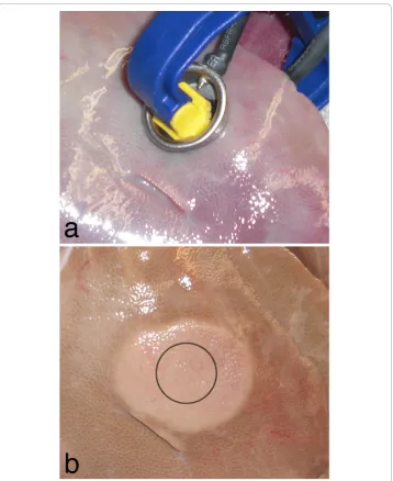
The ECM 830 Square Wave BTX Electroporation System (Harvard Apparatus, Cam-bridge, MA) was used to deliver low-energy pulses to the liver tissue while it was on ice undergoing active perfusion with the solution maintained at 21°C. Two metal plate electrodes, 2 cm in diameter, were attached to a pair of ratcheting vice grips (38 mm, Irwin Quick-Grips) using Velcro. High voltage wire was used to connect the electrodes to the BTX unit. The electrodes were clamped gently to the liver and the center-to-center distance between the electrodes was measured. The voltage output on the BTX unit was adjusted such that the approximate applied electric field was 1000 V/cm. Then, ninety-nine individual 100-μs square pulses were administered at repetition rates of 0.25, 0.5, 1.0 and 4.0 Hz. Repetition rates trials were performed at random and repeated a minimum of three times. Sham controls were performed by placing the electrodes over the tissue without delivering any pulses. Since needle electrodes are typically employed in clinical applications of IRE, two additional trials were performed using needle electrodes separated by 0.5 cm, inserted into the tissue approximately 1 cm, using a voltage-to-distance ratio of 1500 V/cm at rates of 1 and 4 Hz. The experi-mental setup for plate electrodes is illustrated in Figure 1a. All N-TIRE treatments were completed within two hours post mortem. The surface lesion created at each treatment site was measured at the end of the 24 hour perfusion period. Statistical analysis of the lesion diameters was conducted using JMP 8.0 (SAS Institute Inc., South Cary, NC) via Student’s t-test with a 0.1alevel. Histological images were imported into ImageJ (Ver-sion 1.43u, NIH, USA). For each sample, a binary image was created using the threshold tool based on a sample selected within an acellular region. An average pixel value for each image was calculated using the measure RGB plugin and a density score was cre-ated by normalizing these values to 1; where 1 corresponds to regions filled with cells and extracellular material and 0 to a region completely devoid of material. Samples were analyzed for statistical significance in JMP via Student’s t-test with a maximum 0.05
alevel and a minimum of 6 samples for each treatment group.
Tissue preparation
tissues were trimmed and processed for routine paraffin embedding, then sectioned at 4 micrometers, and stained with hematoxylin-eosin (H&E) or Masson’s trichrome stain. Tissue sections were evaluated by a veterinary pathologist who had no knowl-edge of the N-TIRE treatment parameters.
Numerical model
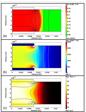
Numerical modeling can be used to predict the electric field distribution, and thus pro-vide insight into the N-TIRE treatment regions in tissue [42,43]. This has been chosen as the method to correlate lesion volume with electric field in the liver. The methods for predicting N-TIRE areas are similar to the ones described by Sel et al.[35]. In order to understand the effective electric field threshold to induce N-TIRE in the liver,
finite element simulations were conducted using Comsol Multiphysics 3.5a (Comsol, Stockholm, Sweden). The numerical model was constructed using 2 cm diameter plates, each 1 mm thick, placed above and below the tissue. The model was generated in an axis symmetric platform and the conductivity changes incorporated the effects of electroporation and temperature as described by Garcia et al. [34], with identical parameters from [35] and its physical setup may be seen in Figure 2. The electric field distribution is given by solving the Laplace equation:
∇ ⋅ ∇
(
)
=0wheresis the electric conductivity of the tissue andis the potential. The electrical boundary condition along the tissue that is in contact with the energized electrode is
= Vo. The electrical boundary condition at the interface of the other electrode is= 0.
The boundaries where the analyzed domain is not in contact with an electrode are treated as electrical insulation.
Conductivity changes due to electroporation and temperature have been modeled to calculate the dynamic conductivity according to the following equation:
dynamic
dc delta range o
normE dc
flc hs normE E E T T _
,
(
)
= 0⎡⎣1+ 2
(
−)
+((
−)
⎤⎦wheres0 is the baseline conductivity.flc2hsis a smoothed heavyside function with a
continuous second derivative that ensures convergence of the numerical solution. This function is defined in Comsol, and it changes from zero to one whennormE_dc-Edelta
= 0 over the range Erange. However, any continuous step function may be used to
model the conductivity change, depending on the application, such as the sigmoidal ones proposed in [35,44]. In theflc2hs function that mimics the sigmoidal ones, nor-mE_dcis the magnitude of the electric field, andEdelta is the magnitude of the electric
field at which the transition occurs over the range,Erange. In the simulations, we used Edelta= 580 V/cm andErange = ±120 V/cm. These values were selected from the
litera-ture in which models incorporated conductivity changes due to electroporation and were validated with real-time measurements in rat and rabbit liver [35,45]. The baseline tissue conductivity was set to 0.067 S/m [35], and N-TIRE affected tissue was considered to increase by a factor of 3.6 as determined by Selet al.[35,46], reaching a final conduc-tivity of 0.241 S/m. The electric field within the tissue domain was first determined using a conductivity of 0.067 S/m, adjusted to incorporate the dynamic conductivity, and reevaluated to determine the final electric field distribution. This numerical model was solved for the pulse parameters that produced the maximum thermal effects (i.e. 1500 V/cm at 4 Hz) used on the livers in order to obtain a simulation of the electric field to which the tissue was exposed using the 1.5% °C-1(Δs/s/ΔT) temperature coeffi-cient in electrical conductivity. The temperature was calculated with the Penne’s bioheat equation with the additional joule heating term [47] and with the values of the liver tissue heat capacity (cp= 3.6 kJ·kg-1K-1), thermal conductivity (k = 0.512 W·m-1K-1),
density (r= 1050 kg·m-3), blood perfusion per unit volume (wb= 1 kg·m-3s-1), and the
heat capacity of blood (cp= (3.64 kJ·kg-1K-1) taken from the literature [48,49]. The outer
surface of the analyzed liver domain and top electrode surface was mathematically con-sidered to have proportional loss to air due to convective heat transfer, h = 10W/(m2· K), as in [50] withT∞= 21°C. The electrode-tissue boundaries were treated as continuity. The N-TIRE electric field thresholds were then found by measuring lesion dimensions and determining the electric field value at this region in the model after the completion of the 99 pulses and thus incorporating all the thermal effects as well.
Results
produced from an applied voltage of 1500 V may be seen, taken 4 hours after treat-ment. Numerically modeled, this lesion size was produced within the region of tissue experiencing an electric field of 423 +/- 147 V/cm (average ± standard deviation). The results of the numerical model for this trial may be seen in Figure 2.
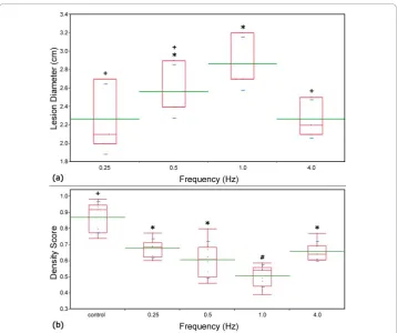
The average applied voltage to distance ratio between the plates for the frequency trials was 962 V/cm, corresponding to an average applied voltage and tissue thickness of 696.9 +/- 141.7 V and 7.3 +/- 1.5 mm, respectively. Lesions from these trials developed over 22 hours post-treatment, and were 2.5 cm in diameter on average (125% electrode diameter); with a minimum lesion of 2 cm occurring at 0.25 Hz and 936 V/cm, and maximum lesion of 3.2 cm occurring at 1.0 Hz and 950 V/cm. Though not dramatically significant, the results suggest that lesion sizes were on average greatest at 1 Hz and decreased as the frequency increased or decreased. The lesions which developed after treatments applied at 0.25 and 4 Hz were statistically smaller (a= 0.1) than those which developed for treatments applied at 1 Hz (Figure 3). Future studies will investigate the role of pulse parameters such as repetition rate, duration, magnitude and number on lesion volume.
Analysis of the treated tissue reveals a uniform treatment region that extended cylindrically through the tissue with no visible damage distal to the treatment regions.
This resulted in calculated treated volumes between 1.97 cm3 and 6.37 cm3 for corresponding tissue thicknesses of 0.628 and 0.792 cm.
Histological examination 24 hours post-treatment indicates that treated regions exhibit cell death (Figure 4b) compared to controls (Figure 4a). Hepatic acini in pigs are bordered by connective tissue, which contains blood vessels and biliary structures, and have a prominent cord architecture terminating in a hepatic venule. In areas adja-cent to energy delivery, hepatic cell cords were well preserved, with mildly vacuolated hepatocytes (an expected finding at 24-hour ex vivomachine perfusion cycle). Sinusoi-dal structure in untreated areas is open, reflecting the flow of perfusate between hepa-tic artery/portal vein and hepahepa-tic vein. N-TIRE treatment disrupts hepahepa-tic cords and induces cell degeneration (Figure 4b). Preservation of major acinar features, including connective tissue borders and blood vessels, is evident. In zones of N-TIRE treatment, cell cords were indistinct and membranes lining sinusoids are fragmented to varying degrees.
Pigs, like humans, have substantial septation of liver acini by thin bands of fibrous connective tissue that run between portal triads. This macrostructure had an effect on the distribution of lesions induced by electroporation. Lesions are confined within structural acini in a manner that at the edges of the electroporation field acini with lesions could border normal or nearly normal acini. Thus, the bands of connective tissue act as insulation for the electrical pulsing, an important observation when con-sidering procedures for treating focal liver lesions with electroporation or for evolving an intact connective tissue/duct/vascular matrix for subsequent tissue engineering. Figure 5a shows a portion of untreated porcine liver with normal sinusoidal cell cords arrayed from portal tracts to central vein. Cell morphology is well preserved. Some vascular congestion with red blood cells is noted and there is also mild centrilobular biliary stasis. Mildly damaged porcine acini are observed in regions subjected to elec-troporation from needle electrodes (Figure 5b). The center of the acinus shows disrup-tion of cord architecture and some cell degeneradisrup-tion and clumping. A higher magnification view of this area is shown in Figure 5c, where cellular changes are more readily appreciated. These treated regions display mild lesions consisting of hepatocytic
cord disruption and cells delaminating from cord basal laminae. Mild biliary stasis is noted (dark pigment).
Administration of N-TIRE treatment, either with needle electrodes or with plate electrodes produced lesions in some hepatic acini that are distinctive. The severity of lesions within individual acini ranges from mild to moderately severe. Mild lesions consisted of small clumps of hepatocytes that detach from basal membranes. These cells show a loss of organization of fine intracellular structure and clumping of cyto-plasm/organelles (Figure 5b-c).
Moderately severe lesions are readily discerned (Figure 6). Cells affected by the N-TIRE procedure show varying degrees of cell swelling and karyolysis (Figure 6b). Within individual acini, most cells are affected. In some acini, frank nuclear pyknosis and cellular degeneration is seen, with small clumps of hyperchromic cells unattached from basal membranes. In some acini, centrilobular biliary stasis is noted, with aggre-gation of bile pigments in distal sinusoidal spaces. In all cases, as noted, bridging bands of connective tissue, with intact bile ducts and vascular structures are seen, even immediately bordering acini with significant N-TIRE-induced tissue damage.
The density score for control samples was 0.87 +/- 0.0097 corresponding to approxi-mately 87% of the histological tissue containing cells and extracellular material. Each treatment group had a statistically significant different density score versus the control (a = 0.01). The lowest density score of 0.509 +/- 0.069, was obtained for N-TIRE
Figure 5IRE treatments result in hepatocytic cord disruption and cell delamination. (a) A section of untreated liver after 24 hours of perfusion. Sections of the same liver treated with 90, 1500 V/cm, 100μs pulses at 4 Hz using needle electrodes after 24 hours of perfusion at (b) 10× and (c) 20× magnification.
treatments where pulses were applied at a rate of 1.0 Hz. Additionally, treatments applied at 1.0 Hz resulted in a statistically significant lower density score as compared to all other treatment groups (a= 0.05).
Discussion
To the best of our knowledge, this is the first work reporting the effect of non-thermal irreversible electroporation in an actively perfused organ. This effort is the first step towards creating decellularized tissue scaffolds that could be used for organ transplan-tation. This paper is aimed as a proof of concept to show that the cells may be removed, and therefore we targeted our study towards treating centimeter-scale regions of tissue. However, because N-TIRE procedures are dependent on the electric field to which a region of tissue is exposed, and thermal effects are mitigated by brief pulses with intervals between pulses, it is possible to scale up N-TIRE procedures to treat larger regions of tissue and organs.
The clearance of cellular debris was analyzed in this study using an image analysis algorithm as a preliminary method to determine the effectiveness of this technique. A more comprehensive study will include staining for primary and secondary antibo-dies, apoptotic markers, and DNA [16] and analysis of these samples via electron microscopy. Assays to determine the quantity of sulfated glycosaminoglycans, elastin, and collagen will be used as a measure of success of this method to preserve the important proteins in the extracellular matrix [13]. Additionally, biodegradation evalua-tion [13] and analysis of the vascular structure [14] must be completed before cell seeding and animal studies can be conducted.
The results reported here were localized to volumes of tissue up to 6.37 cm3 for a single N-TIRE treatment. This can readily be expanded into much larger volumes by performing multiple treatments with the goal of creating decellularized structures for partial and full liver transplants. Analysis of the pulse repetition rate shows that the largest treated area and the lowest density score was achieved for a pulse frequency of 1 Hz. After 24 hours of perfusion, a maximum density score reduction of 58.5 percent had been achieved and cellular debris remained within the tissue construct. Since cell viability in the treatment regions was minimal, this is likely due to the combination of three factors; adhesion of cellular debris to the extracellular matrix, low physiological flow rates and pressures at the lobule level, and possible damage to the microvascula-ture by the N-TIRE treatments.
Although electroporation has been shown to preserve major blood vessels, vascular occlusion after electroporation has been reported in the literature under multiple treat-ment regitreat-ments including work done by Edd et al. [51] (a single 20 ms, 1000 V/cm pulse), Sersaet al. [52] (eight, 100μs, 1300 V/cm pulses at 1 Hz) and Nuccitelliet al.
but essentially transient and reversible decline in blood flow [56]. This phenomenon may have occurred during the course of perfusion ex-vivo, though it was not directly observed, and it may have an effect on the clearance of cellular debris from the vascu-lar network.
Additionally, the branching network of vessels within the liver produces a system with low pressures and fluid velocities at the capillary level and within individual lobules. The combination of physiological geometry and the loss of fine capillary struc-ture caused by the N-TIRE treatments may have resulted in local sheer stresses which were not significant enough to fully clear cellular debris from the tissue. Removal of this debris is essential in minimizing immune response of recipients. Future work will focus on optimizing treatment and perfusion protocols to minimize disruption of the microvasculature network while enhancing the clearance of debris. This may include the continuous application of sub 1000 V/cm pulses at 1 Hz and perfusion at higher than physiological pressures which we believe will enhance the decellularization pro-cess. Recently developed chemical decellularization processes require perfusion cycles of up to 72 hours [14] for the complete removal of cellular material from a rat liver matrix and extension of N-TIRE treatment and perfusion cycles to these durations may be necessary to achieve complete decellularization.
Both external plate electrodes and needles placed within the tissue produced clearly delineated regions of cell death. Plate electrodes produced circular surface lesions, which when appeared cylindrical in shape in sectioned samples and extended between the top and bottom electrodes. Sections of tissue treated with needle electrodes pro-duced oval shaped surface lesions which extended through the tissue.
Needle punctures damaged the tissue structure and provided an alternative path for fluid to flow. Rather than returning through the vasculature, some perfusate escaped the organ through the punctures hindering the perfusion process. Due to this, treatment of an entire organ using needle electrodes is likely not possible and external electrodes appear to be the best method of inducing N-TIRE in large tissue volumes.
In N-TIRE areas, cell death was directly related to energy delivered. Close to elec-trode placements, over 90% of the cells were degenerate and in varying stages of lysis. In the more reversibly energized zone, cell disruption was 20-30% of cells. Other cells may have been degenerate or leaky, but not morphologically abnormal. We have observed that the machine perfusion system can mobilize large amounts of cellular debris, a significant benefit for tissue engineering.
In addition to producing decellularized tissue scaffolds, this method provides an ideal platform to study the effects of pulse parameters such as pulse length, repetition rate, and field strength on whole organ structures. Additionally, since we have direct control over the electrical properties of the perfusate, this could serve as a model for examin-ing the effects of N-TIRE on diseased or cancerous organs with unique electrical or physical properties.
on decellularized collagen matrices and implanted into athymic mice, developed nephron-like structures after 8 weeks [9]. In addition, five millimeter thick porous polyvinyl-alcohol (PVA) constructs, implanted in mice and then injected with hepato-cytes, developed liver-like morphology over the course of one year [7]. However, cell survival and proliferation in each of these structures was limited to a few millimeters from a nutrient source [7].
The resulting scaffolds from N-TIRE plus perfusion maintain the vasculature neces-sary for perfusion into structures far beyond the nutrient diffusion limit that exists for non-vascularized structures. Since the temperature of the perfusate used can be as low as 4°C, thermal aspects associated with Joule heating are negligible. This provides an ideal platform in which to explore the effects on the cells and tissue of electric fields in isolation from the effects of thermal damage. Additionally, the low temperature of the organ compared to in vivoapplications may allow for the application of much higher voltages to attain appropriate electric fields for decellularizing thicker structures without inducing thermal damage. This is important since the thickness of a human liver can exceed 10 cm in some regions.
When planning to decellularize tissues and organs undergoing active perfusion, the treatment region of decellularized tissue may be predicted through numerical model-ing. From the lesion sizes and numerical model used here, when decellularizing an entire organ for a transplantable scaffold, the protocol should expose all of the tissue to an electric field of 423 +/- 147 V/cm. This will ensure complete cell death, allowing comprehensive reseeding of the scaffold with the desired cells, thus minimizing the effects of recipient rejection. The threshold found here is slightly lower than the approximate 500-700 V/cm values reported in previous investigations [34,35]. This may be a result of the unique pulse parameters used (e.g., pulse number) or an inher-ent increased sensitivity of the cells to the pulses when under perfusion. The variability in the electric field threshold may result from the multiple inhomogeneous characteris-tics of the tissue anatomy and structure, such as the vascular system and tissue thick-ness, leading to lesions that were not perfectly circular.
The continuous active machine perfusion methods utilized here in the decellulariza-tion process may also be advantageous for recellularizadecellulariza-tion. Once the decellularizadecellulariza-tion process is complete, it should be possible to reseed the scaffold without risking damage attendant with removing the newly-created scaffold from the perfusion system. Since the arterial and venous supplies are individually addressable, multiple cell types can be delivered simultaneously to different regions of the organ. Similarly, retrograde perfu-sion through the biliary system may be the ideal pathway in which to deliver hepato-cytes for the reseeding process.
the period of perfusion and conditions of perfusion with the cardioemulation system has clear advantages for this gradual, evolutionary approach to decellularization and eventual recellularization of liver.
Conclusions
This study investigated the ability to develop decellularized tissue scaffolds using N-TIRE on organs undergoing active perfusion. Porcine livers were harvested and placed under active mechanical perfusion while N-TIRE electrical pulses were applied using plate and needle electrodes. Livers were removed from the perfusion system and the resultant lesions and control regions were examined histologically and a density score improvement of 58.5 percent was observed. Through numerical modeling of the electric field distribution from the pulse applications, it was found that an N-TIRE threshold of 423 +/- 147 V/cm may be used to predict the affected area. The continu-ous active machine perfusion method utilized during the decellularization process in this study provides the necessary platform for scaffold recellurization, a vital aspect required for practical organ transplantation techniques.
Acknowledgements
This work was supported by the Coulter Foundation. The authors would like to thank Charles Aardema for his technical assistance with the VasoWave™Perfusion System.
Author details 1
School of Biomedical Engineering and Sciences, Virginia Tech - Wake Forest University, Blacksburg, VA, USA. 2Department of Surgery, School of Medicine, University of North Carolina, Chapel Hill, NC, USA.3Department of Biomedical Sciences & Pathobiology, Virginia-Maryland Regional College of Veterinary Medicine, Blacksburg, VA, USA. 4Wake Forest Institute of Regenerative Medicine, Wake Forest University, Winston-Salem, NC, USA.
Authors’contributions
RVD and JR conceived and designed the experiments. MBS, REN, and PAG performed and analyzed the experiments and numerical modeling. DG supplied critically important intellectual content in revising the manuscript. All authors read and approved the final manuscript.
Competing interests
Davalos has a patent pending on this technology.
Received: 21 August 2010 Accepted: 10 December 2010 Published: 10 December 2010
References
1. Chan SC, Fan ST, Lo CM, Liu CL, Wei WI, Chik BHY, Wong J:A Decade of Right Liver Adult-to-Adult Living Donor Liver Transplantation - the Recipient Mid-Term Outcomes.Annals of Surgery2008,248:411-418.
2. United Network of Organ Sharing.[http://www.unos.org].
3. Fabre:Report of the British Transplantation Society Working Party on Organ Donation.1995.
4. Feest TG, Riad HN, Collins CH, Golby MGS, Nicholls AJ, Hamad SN:Protocol for Increasing Organ Donation after Cerebrovascular Deaths in District General-Hospital.Lancet1990,335:1133-1135.
5. Riad H, Nicholls A:An Ethical Debate - Elective Ventilation of Potential Organ Donors.British Medical Journal1995,
310:714-715.
6. Perera M, Mirza DF, Elias E:Liver Transplantation: Issues for the Next 20 Years.Journal of Gastroenterology and Hepatology2009,24:S124-S131.
7. Kaufmann PM, Kneser U, Fiegel HC, Kluth D, Herbst M, Rogiers X:Long-Term Hepatocyte Transplantation Using Three-Dimensional Matrices.Transplantation Proceedings1999,31:1928-1929.
8. Atala A:Experimental and Clinical Experience with Tissue Engineering Techniques for Urethral Reconstruction.
Urologic Clinics of North America2002,29:485.
9. Atala A:Engineering Organs.Current Opinion in Biotechnology2009,20:575-592.
10. Khademhosseini A, Langer R, Borenstein J, Vacanti JP:Microscale Technologies for Tissue Engineering and Biology.
Proceedings of the National Academy of Sciences of the United States of America2006,103:2480-2487.
11. Whitesides GM, Ostuni E, Takayama S, Jiang XY, Ingber DE:Soft Lithography in Biology and Biochemistry.Annual Review of Biomedical Engineering2001,3:335-373.
12. Albrecht DR, Sah RL, Bhatia SN:Geometric and Material Determinants of Patterning Efficiency by Dielectrophoresis.
Biophysical Journal2004,87:2131-2147.
14. Uygun BE, Soto-Gutierrez A, Yagi H, Izamis ML, Guzzardi MA, Shulman C, Milwid J, Kobayashi N, Tilles A, Berthiaume F, Hertl M, Nahmias Y, Yarmush ML, Uygun K:Organ Reengineering through Development of a Transplantable Recellularized Liver Graft Using Decellularized Liver Matrix.Nat Med2010,16:814-U120. 15. Baptista P, Orlando G, Mirmalek-Sani S, Siddiqui M, Atala A, Soker S:Whole Organ Decellularization - a Tool for
Bioscaffold Fabrication and Organ Bioengineering.31st Annual International IEEE EMBS Conference; Minneapolis, Minnesota, USA2009, 6526-6529.
16. Ott HC, Matthiesen TS, Goh SK, Black LD, Kren SM, Netoff TI, Taylor DA:Perfusion-Decellularized Matrix: Using Nature’s Platform to Engineer a Bioartificial Heart.Nat Med2008,14:213-221.
17. Samouillan V, Dandurand-Lods J, Lamure A, Maurel E, Lacabanne C, Gerosa G, Venturini A, Casarotto D, Gherardini L, Spina M:Thermal Analysis Characterization of Aortic Tissues for Cardiac Valve Bioprostheses.Journal of Biomedical Materials Research1999,46:531-538.
18. Kawazoye S, Tian SF, Toda S, Takashima T, Sunaga T, Fujitani N, Higashino H, Matsumura S:The Mechanism of Interaction of Sodium Dodecyl-Sulfate with Elastic Fibers.Journal of Biochemistry1995,117:1254-1260.
19. Keeffe EB:Liver Transplantation: Current Status and Novel Approaches to Liver Replacement.Gastroenterology2001,
120:749-762.
20. Cardona K, Korbutt GS, Milas Z, Lyon J, Cano J, Jiang W, Bello-Laborn H, Hacquoil B, Strobert E, Gangappa S, Weber CJ, Pearson TC, Rajotte RV, Larsen CP:Long-Term Survival of Neonatal Porcine Islets in Nonhuman Primates by Targeting Costimulation Pathways.Nat Med2006,12:304-306.
21. Hering BJ, Wijkstrom M, Graham ML, Harstedt M, Aasheim TC, Jie T, Ansite JD, Nakano M, Cheng J, Li W, Moran K, Christians U, Finnegan C, Mills CD, Sutherland DE, Bansal-Pakala P, Murtaugh MP, Kirchhof N, Schuurman HJ:Prolonged Diabetes Reversal after Intraportal Xenotransplantation of Wild-Type Porcine Islets in Immunosuppressed Nonhuman Primates.Nat Med2006,12:301-303.
22. Yamada K, Yazawa K, Shimizu A, Iwanaga T, Hisashi Y, Nuhn M, O’Malley P, Nobori S, Vagefi PA, Patience C, Fishman J, Cooper DKC, Hawley RJ, Greenstein J, Schuurman HJ, Awwad M, Sykes M, Sachs DH:Marked Prolongation of Porcine Renal Xenograft Survival in Baboons through the Use of Alpha 1,3-Galactosyltransferase Gene-Knockout Donors and the Cotransplantation of Vascularized Thymic Tissue.Nat Med2005,11:32-34.
23. Chari RS, Collins BH, Magee JC, Dimaio JM, Kirk AD, Harland RC, McCann RL, Platt JL, Meyers WC:Brief Report -Treatment of Hepatic-Failure with Ex-Vivo Pig-Liver Perfusion Followed by Liver-Transplantation.New England Journal of Medicine1994,331:234-237.
24. Makowka L, Cramer DV, Hoffman A, Breda M, Sher L, Eirashreha G, Tuso PJ, Yasunaga C, Cosenza CA, Duwu G:The Use of a Pig-Liver Xenograft for Temporary Support of a Patient with Fulminant Hepatic-Failure.Transplantation1995,
59:1654-1659.
25. Yang YG, Sykes M:Xenotransplantation: Current Status and a Perspective on the Future.Nature Reviews Immunology 2007,7:519-531.
26. Dutkowski P, de Rougemont O, Clavien PA:Machine Perfusion for‘Marginal’Liver Grafts.American Journal of Transplantation2008,8:917-924.
27. Monbaliu D, Brassil J:Machine Perfusion of the Liver: Past, Present and Future.Current Opinion in Organ Transplantation2010,15:160-166.
28. Olschewski P, Gass P, Ariyakhagorn V, Jasse K, Hunold G, Menzel M, Schoning W, Schmitz V, Neuhaus P, Puhl G:The Influence of Storage Temperature During Machine Perfusion on Preservation Quality of Marginal Donor Livers.
Cryobiology2010,60:337-343.
29. Stegemann J, Hirner A, Rauen U, Minor T:Use of a New Modified Htk Solution for Machine Preservation of Marginal Liver Grafts.Journal of Surgical Research2010,160:155-162.
30. Yamamoto N, Konishi Y, Wakashiro S, Takayasu T, Tatsumi Y, Shimahara Y, Tanaka K, Yamaoka Y, Ozawa K:72-Hour Preservation of Porcine Liver by Continuous Hypothermic Perfusion with Uw Solution in Comparison with Simple Cold-Storage.Journal of Surgical Research1991,51:288-292.
31. Schon MR, Kollmar O, Wolf S, Schrem H, Matthes M, Akkoc N, Schnoy NC, Neuhaus P:Liver Transplantation after Organ Preservation with Normothermic Extracorporeal Perfusion.Annals of Surgery2001,233:114-123. 32. Brockmann J, Reddy S, Coussios C, Pigott D, Guirriero D, Hughes D, Morovat A, Roy D, Winter L, Friend PJ:
Normothermic Perfusion a New Paradigm for Organ Preservation.Annals of Surgery2009,250:1-6. 33. Weaver JC:Electroporation of Biological Membranes from Multicellular to Nano Scales.Ieee Transactions on
Dielectrics and Electrical Insulation2003,10:754-768.
34. Garcia PA, Rossmeisl JH Jr, Neal R II, Ellis TL, Olson JD, Henao-Guerrero N, Robertson J, Davalos RV:Intracranial Nonthermal Irreversible Electroporation: In Vivo Analysis.J Membr Biol2010,236:127-136.
35. Sel D, Cukjati D, Batiuskaite D, Slivnik T, Mir LM, Miklavcic D:Sequential Finite Element Model of Tissue Electropermeabilization.IEEE Trans Biomed Eng2005,52:816-827.
36. Lee EW, Chen C, Prieto VE, Dry SM, Loh CT, Kee ST:Advanced Hepatic Ablation Technique for Creating Complete Cell Death: Irreversible Electroporation.Radiology2010,255:426-433.
37. Edd JF, Horowitz L, Davalos RV, Mir LM, Rubinsky B:In Vivo Results of a New Focal Tissue Ablation Technique: Irreversible Electroporation.IEEE Trans Biomed Eng2006,53:1409-1415.
38. Davalos RV, Mir LM, Rubinsky B:Tissue Ablation with Irreversible Electroporation.Annals of Biomedical Engineering 2005,33:223-231.
39. Maor E, Ivorra A, Leor J, Rubinsky B:The Effect of Irreversible Electroporation on Blood Vessels.Technology in Cancer Research & Treatment2007,6:307-312.
40. Davalos RV:Irreversible Electroporation to Create Tissue Scaffolds.United States patent2009.
41. Phillips M, Maor E, Rubinsky B:Nonthermal Irreversible Electroporation for Tissue Decellularization.Journal of Biomechanical Engineering-Transactions of the Asme2010,132.
42. Miklavcic D, Semrov D, Mekid H, Mir LM:A Validated Model of in Vivo Electric Field Distribution in Tissues for Electrochemotherapy and for DNA Electrotransfer for Gene Therapy.Biochimica et Biophysica Acta2000,1523:73-83. 43. Miklavcic D, Beravs K, Semrov D, Cemazar M, Demsar F, Sersa G:The Importance of Electric Field Distribution for
44. Pavselj N, Bregar Z, Cukjati D, Batiuskaite D, Mir LM, Miklavcic D:The Course of Tissue Permeabilization Studied on a Mathematical Model of a Subcutaneous Tumor in Small Animals.IEEE Trans Biomed Eng2005,52:1373-1381. 45. Cukjati D, Batiuskaite D, Andre F, Miklavcic D, Mir LM:Real Time Electroporation Control for Accurate and Safe in
Vivo Non-Viral Gene Therapy.Bioelectrochemistry2007,70:501-507.
46. Miklavcic D, Snoj M, Zupanic A, Kos B, Cemazar M, Kropivnik M, Bracko M, Pecnik T, Gadzijev E, Sersa G:Towards Treatment Planning and Treatment of Deep-Seated Solid Tumors by Electrochemotherapy.BioMedical Engineering OnLine2010,9:10.
47. Davalos RV, Mir LM, Rubinsky B:Tissue Ablation with Irreversible Electroporation.Ann Biomed Eng2005,33:223-231. 48. Duck FA:Physical Properties of Tissues: A Comprehensive Reference BookSan Diego: Academic Press; 1990.
49. Deng ZS, Liu J:Blood Perfusion-Based Model for Characterizing the Temperature Fluctuations in Living Tissue.Phys A STAT Mech Appl2001,300:521-530.
50. Lackovic I, Magjarevic R, Miklavcic D:Three-Dimensional Finite-Element Analysis of Joule Heating in Electrochemotherapy andin VivoGene Electrotransfer.IEEE Trns Dielectr Electr Insul2009,16:1338-1347. 51. Edd JF, Horowitz L, Davalos RV, Mir LM, Rubinsky B:In Vivo Results of a New Focal Tissue Ablation Technique:
Irreversible Electroporation.Ieee Transactions on Biomedical Engineering2006,53:1409-1415.
52. Sersa G, Jarm T, Kotnik T, Coer A, Podkrajsek M, Sentjurc M, Miklavcic D, Kadivec M, Kranjc S, Secerov A, Cemazar M:
Vascular Disrupting Action of Electroporation and Electrochemotherapy with Bleomycin in Murine Sarcoma.British Journal of Cancer2008,98:388-398.
53. Nuccitelli R, Chen X, Pakhomov AG, Baldwin WH, Sheikh S, Pomicter JL, Ren W, Osgood C, Swanson RJ, Kolb JF, Beebe SJ, Schoenbach KH:A New Pulsed Electric Field Therapy for Melanoma Disrupts the Tumor’s Blood Supply and Causes Complete Remission without Recurrence.International Journal of Cancer2009,125:438-445.
54. Gehl J, Skovsgaard T, Mir LM:Vascular Reactions to in Vivo Electroporation: Characterization and Consequences for Drug and Gene Delivery.Biochim Biophys Acta-Gen Subj2002,1569:51-58.
55. Kanthou C, Kranjc S, Sersa G, Tozer G, Zupanic A, Cemazar M:The Endothelial Cytoskeleton as a Target of Electroporation-Based Therapies.Molecular Cancer Therapeutics2006,5:3145-3152.
56. Jarm T, Cemazar M, Miklavcic D, Sersa G:Antivascular Effects of Electrochemotherapy: Implications in Treatment of Bleeding Metastases.Expert Review of Anticancer Therapy2010,10:729-746.
57. Sersa G, Krzic M, Sentjurc M, Ivanusa T, Beravs K, Kotnik V, Coer A, Swartz HM, Cemazar M:Reduced Blood Flow and Oxygenation in Sa-1 Tumours after Electrochemotherapy with Cisplatin.British Journal of Cancer2002,87:1047-1054. 58. Sersa G, Cemazar M, Miklavcic D, Chaplin DJ:Tumor Blood Flow Modifying Effect of Electrochemotherapy with
Bleomycin.Anticancer Research1999,19:4017-4022.
59. Folkman J:Self-Regulation of Growth in 3 Dimensions.Journal of Experimental Medicine1973,338.
60. Kaufman DS, Hanson ET, Lewis RL, Auerbach R, Thomson JA:Hematopoietic Colony-Forming Cells Derived from Human Embryonic Stem Cells.Proceedings of the National Academy of Sciences of the United States of America2001,
98:10716-10721.
doi:10.1186/1475-925X-9-83
Cite this article as:Sanoet al.:Towards the creation of decellularized organ constructs using irreversible electroporation and active mechanical perfusion.BioMedical Engineering OnLine20109:83.
Submit your next manuscript to BioMed Central and take full advantage of:
• Convenient online submission
• Thorough peer review
• No space constraints or color figure charges
• Immediate publication on acceptance
• Inclusion in PubMed, CAS, Scopus and Google Scholar
• Research which is freely available for redistribution