R E S E A R C H A R T I C L E
Open Access
Two-year topographic and densitometric
outcomes of accelerated (45 mW/cm
2
)
transepithelial corneal cross-linking for
keratoconus: a case-control study
Jinrong Huang
3,4†, Yang Shen
1,2†, Weijun Jian
1,2, Haipeng Xu
1,2, Meiyan Li
1,2, Jing Zhao
1,2, Xingtao Zhou
1,2*and
Hongfei Liao
4*Abstract
Background:Conventional corneal cross-linking is effective for retarding the progression of keratoconus. However the long-term efficacy and safety of accelerated (45 mW/cm2) transepithelial corneal cross-linking (ATE-CXL) on progressive keratoconus (KC) treatment is not fully understood. The purpose of this study is to evaluate the 2-year changes in corneal topographic parameters and densitometry values after ATE-CXL for KC. Methods: Twenty-five progressive eyes of 25 KC patients (KC group) and 25 eyes of 25 myopes without KC (control group) were enrolled. Corneal topography and densitometry values were evaluated pre-operatively and at 6, 12 and 24 months post-operatively in the KC group.
Results: The mean values of flat keratometry (K1), steep keratometry (K2), mean keratometry (Km), corneal astigmatism (CA), maximum keratometry (Kmax), central corneal thickness (CCT), thinnest corneal thickness (TCT), anterior corneal elevation (ACE) and posterior corneal elevation (PCE) all remained unchanged over time (all P values > 0.05). The densitometry values of the anterior, central, posterior and total layers over the annular diameters 0 mm to 2 mm (Φ0-2 mm) and Φ2–6 mm all decreased significantly (all P values < 0.05). At post-operative month 24, except for the densitometry value of the posterior layer (Φ0-2 mm), which was significantly lower than that of the control group (post hoc P= 0.010), all densitometry values obtained from the remaining locations of the KC eyes were equal to those of the control group (All post hoc P values > 0.05). Subgroups with Km≥50.30D or ACE ≥35.3μm progressed significantly when compared with those with Km < 50.30D (F = 8.167, P= 0.004) or ACE< 35.3μm (F = 5.207, P= 0.022).
Conclusions: K1, K2, Km, CA, Kmax, CCT, TCT, ACE, and PCE values may remain stable but severer KC patients tend to have poorer long-term outcomes. The densitometry values of the full corneal thickness (total layer over Φ0-2 mm and Φ2–6 mm) may decrease to normal levels at 2 years after ATE-CXL for KC.
Keywords: Keratoconus, Accelerated transepithelial corneal cross-linking, Corneal topographic parameters, Densitometry
* Correspondence:[email protected];[email protected]
†Jinrong Huang and Yang Shen contributed equally to this work. 1
The Eye and ENT Hospital of Fudan University, 19 Baoqing Road, Xuhui District, Shanghai 200031, China
4Department of Ophthalmology, The Affiliated Eye Hospital of Nanchang
University, 463 Bayi Avenue, Nanchang 330006, Jiangxi Province, China Full list of author information is available at the end of the article
© The Author(s). 2018Open AccessThis article is distributed under the terms of the Creative Commons Attribution 4.0
International License (http://creativecommons.org/licenses/by/4.0/), which permits unrestricted use, distribution, and
Introduction
Keratoconus (KC) is a chronic ectatic corneal disease characterized by progressive corneal thinning and steeping which lead to irregular astigmatism and visual impairment [1]. Treatment options, including spectacles, rigid gas per-meable contact lenses (RGP) [2], intrastromal corneal rings (ICR) [3], keratoplasty [4, 5] and riboflavin ultraviolet-A (UV-A) corneal cross-linking (CXL) [6], are available for improving visual acuity or (and) retarding the progression of KC. Spectacles and RGP correct refractive error but do not change the course of the disease. CXL is effective for slowing or stopping the progression of KC [7]. Literatures have reported that CXL results in in-creased collagen fiber diameter [8] and enhances biomechanical stiffness [9] through the induction of in-creased formation of covalent bonds within or between collagen fibers in the corneal stroma [10]. Greenstein SA [11] reported a significant increase in corneal densitom-etry after conventional (3 mW/cm2) corneal crosslinking (C-CXL), and transient sub-epithelial haze is frequently detected after C-CXL [12, 13]. Accelerated (45 mW/cm2) trans-epithelial corneal cross-linking with pulsed UV (ATE-CXL) is a novel and minimally invasive corneal cross-linking procedure. Higher irradiance, pulsed UV light is used to dramatically shorten treatment duration as compared with the C-CXL (3 mW/cm2) procedure. As the epithelium layer remains intact during ATE-CXL, risk of infection is minimized, and milder ocular surface inflam-matory reactions may be triggered, and hence, post-opera-tive complications may be reduced [14]. One of our previous studies [15] investigated one-year outcomes of corneal topographic parameters and corneal densitometry after ATE-CXL for KC. Our results demonstrated de-creased corneal densitometry and stable keratometry values at post-operative year 1 [15]. Corneal densitometry, which is also known as corneal backscattering, is consid-ered as a proxy for corneal transparency [16]. It is import-ant to determine whether CXL would lead to loss of transparency when assess the safety of CXL protocols. However the long-term effects of ATE-CXL on corneal densitometry and keratometry remain unknown. In the current study, we investigated 2-year changes in corneal topographic parameters and densitometry values after ATE-CXL for KC.
Methods Ethics statement
This prospective, non-randomized and cohort study was approved by the Ethics Committee of the Eye and ENT Hospital of Fudan University (ethical approval number: ky2012–017. Date of approved by ethic committee: 2012-08-16) and was carried out following the tenets of the Declaration of Helsinki. After a detailed explanation of the study, written informed consent for the treatment
was obtained from the patients or their legal representa-tives before the procedure.
Patients
Twenty-five progressive KC patients (age 25.4 ± 6.0 years, 18 male and 7 female, Amsler-Krumeich classifica-tion stages 1–4, without stromal scarring) and 25 my-opes matched for gender and age (within ±2 years) were enrolled in this study. The worse eye (the eye with more advanced keratoconus) of each KC patient underwent ATE-CXL and was followed up for 2 years.
Ophthalmologic examinations
For KC patients, manifest refraction spherical equivalent (MRSE), best spectacles corrected distance visual acuity (BSCDVA), slit-lamp anterior segment and fundus exami-nations, and endothelial cell density (SP-2000P; Topcon Corporation, Japan) were assessed pre-operatively. A 3-dimensional anterior segment analyzer (Pentacam HR, Type 70,900; Oculus, Germany) was applied to evaluate the corneal topographic parameters, including flat kerato-metry value (K1), steep keratokerato-metry value (K2), mean keratometry value (Km), corneal astigmatism (CA), maximum keratometry value (Kmax), central corneal thickness (CCT), thinnest corneal thickness (TCT), anter-ior corneal elevation (ACE) and posteranter-ior corneal eleva-tion (PCE), as well as corneal densitometry values, pre-operatively and at 6, 12 and 24 months post-operatively. All corneal densitometry values and cor-neal topographic parameters were only measured once in the control group. Exclusion criteria of ATE-CXL in-cluded TCT of less than 340μm, endothelial cell density of less than 2000 cells per mm2, severe dry eye, corneal scarring, autoimmune diseases or pregnancy [12,17,18].
Surgical techniques
1 s off) for 5 min and 20 s, for a total energy dose of 7.2 J/ cm2. Balanced salt solution was administered every 40 s for ocular surface protection during the irradiation. A bandage contact lens (Acuvue Oasys, Inc., Jacksonville, FL) was placed on the cornea after the procedure and was left in place for 3 days.
Post-operative topical medication regimens
Each patient was instructed to instill topical antibiotics (ofloxacin ophthalmic solution 0.5%; Santen Pharma-ceutical Co, Ltd.; 4 times per day for 7 days), artificial tears (TearsNaturale; Alcon Laboratories, Inc., Fort Worth, TX; 4 times per day for 20 days) and topical ste-roids (fluorometholone 0.1%; Santen Pharmaceutical Co, Ltd.; 8 times per day initially, with gradually reduced fre-quencies for a period of 20 days), post-operatively.
Statistical evaluation
Statistical analysis was performed using SPSS 20 software (SPSS Inc., IBM). Kolmogorov-Smirnov test was conducted to assess the normality of these vari-ables. Mixed linear model with Bonferroni-adjusted post hoc comparisons were performed to evaluate the changes in corneal topographic parameters and corneal densitometry over time, as well as the differ-ence in corneal densitometry between groups. The potential correlations between the corneal topo-graphic parameters and the densitometry values were evaluate by using Pearson correlation test. Cut-off P values were 0.05.
Results
In KC group, all procedures were uneventful (n= 25). Two patients failed to attend the follow-up of post-operative month 6 (n= 23). At post-operative month 12, 1 patient withdrew from the study, and 1
additional patient did not come for the visit (n = 23). At post-operative month 24, another 2 patients failed to at-tend the last visit (n= 22). The reasons for loss of follow-up included inconvenient traffic and personal af-fairs (work- or school-related problems). No statistically significant difference was detected in the mean values of age between the two groups (t=−0.440,P= 0.664).
Changes in main corneal topographic parameters after ATE-CXL
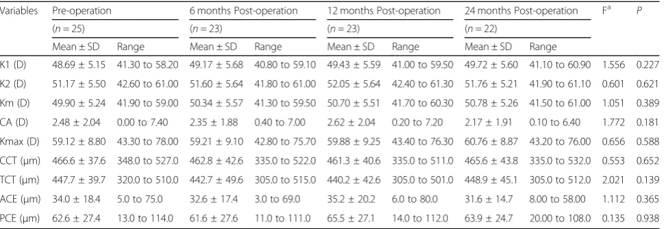
The pre-operative and post-operative corneal topo-graphic parameters, including K1, K2, Km, Kmax, CA, ACE, PCE, CCT and TCT are listed in the Table 1. All these variables remained stable over time (AllPvalues > 0.05). At post-operative month 24 (n= 22), 12 out of 25 (48%) treated eyes had deceased Kmax values, 3 out of 25 (12%) eyes had < 1 diopter (D) increase in the Kmax values, and the remaining 7 (28%) eyes showed progres-sion (≥1 diopter (D) increase in the Kmax value within 2 years). During the follow-ups, the mean values of K1, Km and Kmax increased non-statistically from 48.69D to 49.72D (F = 1.556, P= 0.227), from 49.90D to 50.78D (F = 1.051, P= 0.389) and from 59.12D to 60.76D (F = 0.656, P= 0.588), respectively. K2 and CA rose mod-estly at post-operative month 12 and then fell slightly at post-operative month 24 (F = 0.601, P= 0.621 and F = 1.772, P= 0.181, respectively). The mean values of CCT and TCT both decreased slightly at post-operative month 12 when compared with the pre-operative values, and then increased modestly at post-operative month 24 (F = 1.167,P= 0.344 and F = 0.711, P= 0.556, respectively). The mean values of ACE and PCE fluctu-ated from 31.6μm to 35.2μm and from 61.6μm to 65.5μm (F = 1.091, P= 0.372 and F = 0.025, P= 0.994), respectively.
Table 1Changes in Main Corneal Topographic Parameters
Variables Pre-operation 6 months Post-operation 12 months Post-operation 24 months Post-operation Fa P
(n= 25) (n= 23) (n= 23) (n= 22)
Mean ± SD Range Mean ± SD Range Mean ± SD Range Mean ± SD Range
K1 (D) 48.69 ± 5.15 41.30 to 58.20 49.17 ± 5.68 40.80 to 59.10 49.43 ± 5.59 41.00 to 59.50 49.72 ± 5.60 41.10 to 60.90 1.556 0.227 K2 (D) 51.17 ± 5.50 42.60 to 61.00 51.60 ± 5.64 41.80 to 61.00 52.05 ± 5.64 42.40 to 61.30 51.76 ± 5.21 41.90 to 61.10 0.601 0.621 Km (D) 49.90 ± 5.24 41.90 to 59.00 50.34 ± 5.57 41.30 to 59.50 50.70 ± 5.51 41.70 to 60.30 50.78 ± 5.26 41.50 to 61.00 1.051 0.389 CA (D) 2.48 ± 2.04 0.00 to 7.40 2.35 ± 1.88 0.40 to 7.00 2.62 ± 2.04 0.20 to 7.20 2.17 ± 1.91 0.10 to 6.40 1.772 0.181 Kmax (D) 59.12 ± 8.80 43.30 to 78.00 59.21 ± 9.10 42.80 to 75.70 59.88 ± 9.25 43.40 to 76.30 60.76 ± 8.87 43.20 to 76.00 0.656 0.588 CCT (μm) 466.6 ± 37.6 348.0 to 527.0 462.8 ± 42.6 335.0 to 522.0 461.3 ± 40.6 335.0 to 511.0 465.6 ± 43.8 335.0 to 532.0 0.553 0.652 TCT (μm) 447.7 ± 39.7 320.0 to 510.0 442.7 ± 49.6 305.0 to 515.0 440.2 ± 42.6 305.0 to 501.0 448.9 ± 45.1 305.0 to 512.0 2.021 0.139 ACE (μm) 34.0 ± 18.4 5.0 to 75.0 32.6 ± 17.4 3.0 to 69.0 35.2 ± 20.2 6.0 to 80.0 31.6 ± 14.7 8.00 to 58.00 1.112 0.365 PCE (μm) 62.6 ± 27.4 13.0 to 114.0 61.6 ± 27.6 11.0 to 111.0 65.5 ± 27.1 14.0 to 112.0 63.9 ± 24.7 20.00 to 108.0 0.135 0.938
Changes in corneal densitometry values after ATE-CXL
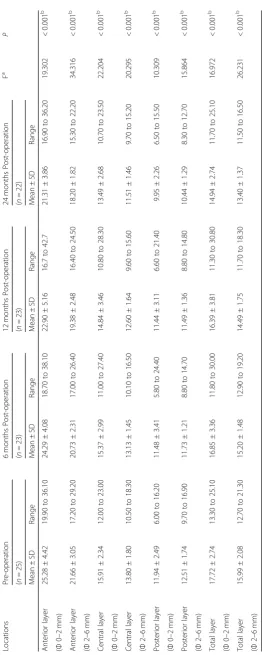
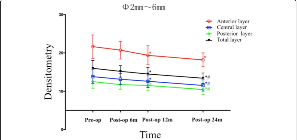
As listed in Table 2, the densitometry values of the an-terior layer, the central layer, the posan-terior layer, and the total layer over Φ0 to 2 mm, as well as over Φ2 to 6 mm, all decreased significantly over time (AllPvalues < 0.001). Bonferroni post hoc comparisons showed densi-tometry values dramatically decreased in the anterior layer (post hoc P= 0.002), the central layer (post hoc P = 0.002), the posterior layer (post hocP= 0.003) and the total layer (post hoc P= 0.001) over Φ 0 to 2 mm at post-operative month 24, when compared with the pre-operative values (Fig. 1). As illustrated in Fig.2, the mean densitometry values of the anterior layer, central layer, posterior layer and total layer overΦ2 to 6 mm all fell remarkably at post-operative month 12 (post hoc P = 0.005, post hocP= 0.003, post hoc P = 0.003 and post hoc P = 0.003, respectively), when compared with the pre-operative values. Except for the densitometry value obtained from the anterior layer over Φ2–6 mm, which changed insignificantly (post hoc P= 0.052), the densi-tometry values obtained from the central layer, the pos-terior layer and the total layer over Φ2–6 mm all significantly decreased at post-operative month 24 when compared with the values obtained at post-operative month 12 (post hoc P= 0.001, post hoc P= 0.002 and post hocP= 0.003, respectively).
As demonstrated in Table3, mixed linear model analysis revealed significant difference in densitometry values be-tween groups (AllP values≤ 0.001). Bonferroni-adjusted post hoc comparisons detected that the densitometry values obtained in the anterior layer, the central layer, the posterior layer and the total layer, over bothΦ0-2 mm and
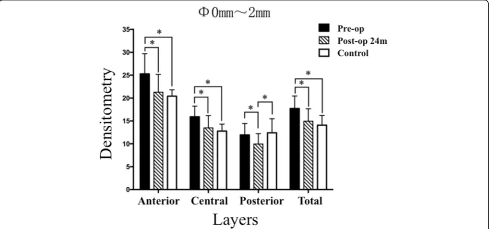
Φ2–6 mm at post-operative month 24, were significantly decreased when compared with the values obtained pre-operatively (All post hocPvalues < 0.001). Except for the densitometry value of the posterior layer over Φ0-2 mm, obtained at post-operative month 24, was signifi-cantly lower than that of the control group (post hocP= 0.010), the densitometry values of the remaining locations (Figs.3and 4) were similar to the values obtained in the control group (All post hoc P values > 0.05).
Correlations between topographic parameters and densitometry values
At post-operative month 24, corneal densitometry values of the anterior layer, central layer, posterior layer and total layer overΦ0-2 mm decreased by 4.09 ± 4.66, 2.47 ± 2.93, 2.00 ± 2.44 and 2.85 ± 3.15, respectively. Over
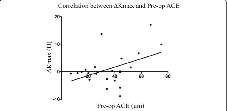
Φ2–6 mm, those values decreased by 3.44 ± 2.60, 2.27 ± 1.61, 2.12 ± 1.56 and 2.12 ± 1.56, respectively. The incre-ment of Kmax value (ΔKmax) was 1.09 ± 6.20D, however the decrements of ACE (ΔACE) and PCE (ΔPCE) were 4.4 ± 11.6μm and 1.5 ± 14.1μm, respectively. Pearson correlation test only revealed a significant correlation
between ΔKmax and pre-operative ACE value (R= 0.436,P= 0.043), (Fig. 5). Further analysis showed that when all the patients were divided into two subgroups with the cut-off median preoperative Km of 50.30D (Subgroup 1 (n= 13): preoperative Km≥50.30D; Sub-group 2 (n= 12): preoperative Km < 50.30D). Log-rank test showed that the Subgroup 1 progressed more sig-nificantly (7 of 13 cases progressed) when compared with the Subgroup 2 (none of 12 progressed), (F = 8.167,
P= 0.004).
When these cases were divided into two subgroups with the cut-off median preoperative ACE of 35.3μm (Sub-group 1 (n = 12): ACE ≥35.3μm, Subgroup 2 (n= 13): ACE < 35.3μm), Log-rank tests detected that the Sub-group 1 progressed (5 of 12 cases progressed) more re-markably when compared with the Subgroup 2 (2 of 13 progressed), (F = 5.207,P= 0.022).
Discussion
A normal cornea is transparent and maintains a smooth and stable curvature [19]. Keratoconus compromises corneal biomechanical stability, leading to progressively increasing corneal keratometry and decreasing corneal thickness.
C-CXL is effective for retarding the progression of KC [20]. However, disadvantages to the standard therapy in-clude the long procedure duration, the removal of the corneal epithelium, and the risks of infection and haze [12,21,22], which may lead to increased corneal densi-tometry and compromised transparency [11]. In the present study, we found the mean values of K1, K2, Km, Kmax, CA, CCT, TCT, ACE and PCE were unchanged over time, indicating that corneal structure remained stable during the 24-month follow-up. Moreover, as we expected [15], the pre-operative densitometry values of the full thickness of the cornea over annular regions
Φ0-2 mm and Φ2–6 mm were higher than those of the control group (Figs. 3 and 4), but at post-operative month 24, these values remarkably decreased to normal levels. Strong evidence has shown that corneal collagen diameter increases and the gap between corneal collagen fasciculi narrows significantly after corneal crosslinking using riboflavin and UVA [8]. We hypothesize that the decreased corneal backscattering (densitometry) values are mainly due to the more tightly arranged corneal col-lagen fasciculi after ATE-CXL. In addition, we did not reveal any correlations between corneal densitometry values and ΔACE, or between the corneal densitometry values andΔPCE. These results all imply that the densi-tometric variation might have little relationship with the changes in corneal shape.
irradiation for 30 min), demonstrated that both Kmax and Km readings kept stable, and ACE and PCE values continued to decrease up to 5 years after C-CXL [23]. Trans-epithelial corneal cross-linking (TE-CXL) pro-cedure avoids epithelial removal. Magli A et al. [24] compared C-CXL and TE-CXL in progressive KC
pediatric patients. They found TE-CXL provided simi-lar efficacy and fewer complications than C-CXL at 1-year follow-up. Accelerated accelerated corneal cross-linking (A-CXL) protocols increase the power in-tensity of UV-A (9mw/cm2, 18mw/cm2, 30mw/cm2and 45mw/cm2) and shorten the exposure time to achieve
Fig. 2The densitometry values overΦ2–6 mm of the anterior layer, central layer, posterior layer, and total layer of the corneas measured before and at 6, 12 and 24 months after ATE-CXL. (The asterisk refers to a significant difference that was detected when compared with the preoperative value. The octothorpe refers to a significant difference that was detected when compared with the value obtained at post-operative month 12)
Fig. 1The densitometry values overΦ0-2 mm of the anterior layer, central layer, posterior layer, and total layer of the corneas measured before and at 6, 12 and 24 months after ATE-CXL (The asterisk refers to a significant difference that was detected when compared with the
equal dosage. Meanwhile, A-CXL procedures could be performed with or without epithelium [15, 25]. A meta-analysis [26] concluded that, in general, ATE-CXL is effective and safe for KC stabilization, however, C-CXL re-sults in greater flattening of corneal curvature. In the present study, we noticed that at post-operative month 24, 12 of 25 eyes (48%) had decreased Kmax values, 3 eyes (12%) had less than 1.00D shift in Kmax values, but the remaining 7 eyes had progressive KC (Kmax value in-creased more than 1.00D), implying that ATE-CXL may have insufficient efficacy in some specific KC patients. Interestingly, further analysis revealed thatΔKmax values were positively correlated with pre-operative ACE values (R= 0.436,P= 0.043), moreover, the Log-rank tests also
showed that the subgroups with Km≥50.30D or ACE ≥35.3μm progressed significantly when compared with those with Km < 50.30D or ACE< 35.3μm, indicating that severer KC patients (Amsler-Krumeich classification above Stage 2) may tend to have poorer outcomes (greater progression). Olivo-Payne A et al. [27] also reported that trans-epithelial accelerated corneal cross-linking demon-strated poorer long-term outcomes in severer cases, which was consistent to our findings.
CXL achieves its effect through increased formation of crosslinks in the corneal stroma, thus enhancing corneal biomechanical stiffness [28]. In severe KC, the cornea is generally thinner and less biomechanically stable [29] compared with mild KC. We hypothesize that, in
Fig. 3The mean densitometry values overΦ0-2 mm of preoperation, 24-month postoperation, and the control group (The asterisk refers to that a significant difference was detected between the two groups)
Table 3Differences in Corneal Densitometry Between Groups
Locations Pre-operation
(n= 25)
24 months Post-operation Control Fa P
(n= 22) (n= 25)
Mean ± SD Mean ± SD Mean ± SD
Anterior layer (Φ0–2 mm) 25.28 ± 4.42 21.31 ± 3.86 20.44 ± 1.35 14.608 < 0.001b Anterior layer (Φ2–6 mm) 21.66 ± 3.05 18.20 ± 1.82 17.07 ± 2.71 22.590 < 0.001b Central layer (Φ0–2 mm) 15.91 ± 2.34 13.49 ± 2.68 12.78 ± 1.53 17.215 < 0.001b Central layer (Φ2–6 mm) 13.80 ± 1.80 11.51 ± 1.46 12.22 ± 1.66 20.404 < 0.001b Posterior layer (Φ0–2 mm) 11.94 ± 2.49 9.95 ± 2.26 12.39 ± 3.09 9.130 =0.001b
Posterior layer (Φ2–6 mm) 12.51 ± 1.74 10.44 ± 1.29 10.33 ± 1.47 21.574 < 0.001b
Total layer (Φ0–2 mm)
17.72 ± 2.74 14.94 ± 2.74 14.07 ± 2.14 15.995 < 0.001b
Total layer (Φ2–6 mm)
15.99 ± 2.08 13.40 ± 1.37 13.33 ± 1.19 22.801 < 0.001b
ΦAnnular diameters,a
Mixed linear model analysis,b
advanced KC, there is greater stromal degeneration and fewer intact corneal collagen fibrils are available to be cross-linked [30] with ATE-CXL; hence, the ef-fect of ATE-CXL on KC control in severe KC patients may be limited. Alternatively, more crosslinking for-mation (stronger treatment) may be required in se-vere cases to stabilize the biomechanically unstable cornea.
The current study had some limitations. First, we only assessed the 2-year changes in topographic parameters and densitometry values following ATE-CXL. The pho-topic, mesopic and scotopic visual acuity and higher order aberrations were not evaluated. It would be pru-dent to investigate the effects of ATE-CXL on visual quality in further studies. Second, the sample size is rela-tively small, however, we mainly investigate the changes
Fig. 5The correlation betweenΔKmax and pre-operative ACE (R= 0.436,P= 0.043)
in topographic and densitometric parameters before and after ATE-CXL, moreover, the KC patients and the my-opes were matched for gender and age and thus, the se-lection bias should be limited.
Conclusions
Topographic parameters, including K1, K2, Km, CA, Kmax, CCT, TCT, ACE, and PCE values, may all remain stable while the densitometry values of the total layer (overΦ0mm–2 mm andΦ2mm–6 mm) may decrease to normal levels at 2 years after ATE-CXL for KC. Patients with more severe KC may tend to have poorer out-comes. Further studies are essential to shed light on the optimal patient selection criteria for ATE-CXL.
Abbreviations
ACE:Anterior corneal elevation; ATE-CXL: Accelerated transepithelial corneal cross-linking; CA: Corneal astigmatism; CCT: Central corneal thickness; C-CXL: Conventional corneal crosslinking; K1: The anterior simulated keratometer readings (Sim Ks) on a ring in 15° around the corneal apex along the flat meridian; K2: The anterior simulated keratometer readings (Sim Ks) on a ring in 15° around the corneal apex along the steep meridian; KC: Keratoconus; Km: The mean keratometry; Kmax: The steepest radius of the anterior corneal surface; PCE: Posterior corneal elevation; TCT: The thinnest corneal thickness;Φ: Annular diameter
Acknowledgments
Not applicable.
Funding
National Natural Science Foundation of China (Grant No.81570879) National Natural Science Foundation of China (Grant No.81500753) National Natural Science Foundation of China (Grant No.81600762) The funders had no role in study design, data collection and analysis, decision to publish, or preparation of the manuscript.
Availability of data and materials
All data used and analyzed in this study are available upon request from the co-first authors Jingrong Huang and Yang Shen.
Authors’contributions
JRH and YS were the major contributors (contribute equally) in writing the manuscript; JRH, YS, and WJJ collected the data. HPX, MYL and JZ analyzed and interpreted the data; XTZ and HFL designed the study (contribute equally); XTZ operated the surgeries. All authors read and approved the final manuscript.
Ethics approval and consent to participate
This study was approved by the Ethics Committee of the Eye and ENT Hospital of Fudan University (ethical approval number: ky2012–017. Date of approved by ethic committee: 2012-08-16), and was carried out following the tenets of the Declaration of Helsinki. After a detailed explanation of the study, written informed consent for the treatment was obtained from the patients or their legal representatives before the procedure.
Consent for publication
Not applicable.
Competing interests
The authors declare that they have no competing interests.
Publisher’s Note
Springer Nature remains neutral with regard to jurisdictional claims in published maps and institutional affiliations.
Author details
1The Eye and ENT Hospital of Fudan University, 19 Baoqing Road, Xuhui
District, Shanghai 200031, China.2The Key Lab of Myopia, National Health
and Family Planning Commission of the People’s Republic of China, Shanghai, China.3Department of Ophthalmology, The First Affiliated Hospital
of Jiangxi Medical College, Shang Rao, Jiangxi Province, China.4Department
of Ophthalmology, The Affiliated Eye Hospital of Nanchang University, 463 Bayi Avenue, Nanchang 330006, Jiangxi Province, China.
Received: 8 April 2018 Accepted: 5 December 2018
References
1. Galvis V, Sherwin T, Tello A, Merayo J, Barrera R, Acera A. Keratoconus: an inflammatory disorder? Eye (Lond). 2015;29(7):843–59.
2. Mrazovac D, BarisićKutija M, Vidas S, et al. Contact lenses as the best conservative treatment of newly diagnosed keratoconus--epidemiological retrospective study. Coll Antropol. 2014;38(4):1115–8.
3. Hellstedt T, Mäkelä J, Uusitalo R, Emre S, Uusitalo R. Treating keratoconus with intacs corneal ring segments. J Refract Surg. 2005;21(3):236–46. 4. Richard JM, Paton D. Gasset AR. A comparison of penetrating keratoplasty
and lamellar keratoplasty in the surgical management of keratoconus. Am J Ophthalmol. 1978;86(6):807–11.
5. Wood TO. Lamellar transplants in keratoconus. Am J Ophthalmol. 1977; 83(4):543–5.
6. Cannon DJ, Foster CS. Collagen crosslinking in keratoconus. Invest Ophthalmol Vis Sci. 1978;17(1):63–5.
7. Wollensak G, Spoerl E, Seiler T. Riboflavin/ultraviolet-a-induced collagen crosslinking for the treatment of keratoconus. Am J Ophthalmol. 2003; 135(5):620–7.
8. Wollensak G, Wilsch M, Spoerl E, Seiler T. Collagen fiber diameter in the rabbit cornea after collagen crosslinking by riboflavin/ UVA. Cornea. 2004; 23(5):503–7.
9. Beshtawi IM, O'Donnell C, Radhakrishnan H. Biomechanical properties of corneal tissue after ultraviolet-A-riboflavin crosslinking. J Cataract Refract Surg. 2013;39(3):451–62.
10. Mencucci R, Marini M, Paladini I, et al. Effects of riboflavin/UVA corneal cross-linking on keratocytes and collagen fibres in human cornea. Clin Exp Ophthalmol. 2010;38(1):49–56.
11. Greenstein SA, Fry KL, Bhatt J, et al. Natural history of corneal haze after collagen crosslinking for keratoconus and corneal ectasia: scheimpflug and biomicroscopic analysis. J Cataract Refract Surg. 2010;36(12):2105–14. 12. Caporossi A, Mazzotta C, Baiocchi S, et al. Long-term results of riboflavin
ultraviolet a corneal collagen cross-linking for keratoconus in Italy: the Siena eye cross study. Am J Ophthalmol. 2010;149(4):585–93.
13. Koller T, Mrochen M, Seiler T. Complication and failure rates after corneal crosslinking. J Cataract Refract Surg. 2009;35(8):1358–62.
14. Leccisotti A, Islam T. Transepithelial corneal collagen cross-linking in keratoconus. J Refract Surg. 2010;26(12):942–8.
15. Shen Y, Jian W, Sun L, et al. One-year follow-up of changes in corneal densitometry after accelerated (45 mW/cm2) Transepithelial corneal collagen cross-linking for keratoconus: a retrospective study. Cornea. 2016; 35(11):1434–40.
16. Otri AM, Fares U, Al-Aqaba MA, Dua HS. Corneal densitometry as an indicator of corneal health. Ophthalmology. 2012;119(3):501–8.
17. Vinciguerra P, Randleman JB, Romano V, et al. Transepithelial iontophoresis corneal collagen cross-linking for progressive keratoconus: initial clinical outcomes. J Refract Surg. 2014;30(11):746–53.
18. Chan TC, Chow VW, Jhanji V, et al. Different topographic response between mild to moderate and advanced keratoconus after accelerated collagen cross-linking. Cornea. 2015;34(8):922–7.
19. Freegard TJ. The physical basis of transparency of the normal cornea. Eye (Lond). 1997;11(Pt 4):465–71.
20. Ng AL, Chan TC, Cheng AC. Conventional versus accelerated corneal collagen cross-linking in the treatment of keratoconus. Clin Exp Ophthalmol. 2016;44(1):8–14.
21. Spoerl E, Mrochen M, Sliney D, Trokel S, Seiler T. Safety of UVA-riboflavin crosslinking of the cornea. Cornea. 2007;26(4):385–9.
23. Hashemi H, Seyedian MA, Miraftab M, Fotouhi A, Asgari S. Corneal collagen cross-linking with riboflavin and ultraviolet a irradiation for keratoconus: long-term results. Ophthalmology. 2013;120(8):1515–2.
24. Magli A, Forte R, Tortori A, Capasso L, Marsico G, Piozzi E. Epithelium-off corneal collagen cross-linking versus transepithelial cross-linking for pediatric keratoconus. Cornea. 2013;32(5):597–601.
25. Shetty R, Pahuja NK, Nuijts RM, et al. Current Protocols of Corneal Collagen Cross-Linking: Visual, Refractive, and Tomographic Outcomes. Am J Ophthalmol. 2015;160(2):243–9.
26. Medeiros CS, Giacomin NT, Bueno RL, Ghanem RC, Moraes HV Jr, Santhiago MR. Accelerated corneal collagen crosslinking: technique, efficacy, safety, and applications. J Cataract Refract Surg. 2016;42(12):1826–35. 27. Olivo-Payne A, Serna-Ojeda JC, Hernandez-Bogantes E, et al. Trans-epithelial
accelerated corneal cross-linking for keratoconus in children. Int J Ophthalmol. 2017;10(12):1919–21.
28. Peyman A, Kamali A, Khushabi M, et al. Collagen cross-linking effect on progressive keratoconus in patients younger than 18 years of age: a clinical trial. Adv Biomed Res. 2015;23(4):245.
29. Bao F, Geraghty B, Wang Q, Elsheikh A. Consideration of corneal biomechanics in the diagnosis and management of keratoconus: is it important? Eye Vis (Lond). 2016;3:18.
30. Krachmer JH, Feder RS, Belin MW. Keratoconus and related