Journal of Applied Biotechnology Reports, Volume 1, Issue 4, Autumn 2014; 155-159
Improvement of Thermal Stability of DFPase by In silico Methods
Morteza Mirzaei1, Ali Mohammad Latifi1*, Rahim Jafari2
Abstract
Introduction
Enzymes are biological catalyst with a growing interest in a wide range of human interest. The ability to catalyze a specific reaction with enantiomeric compounds at desired temperature or pH are of their importance in medicine and many industrial applications [1, 2]. One of the most important aspects of an enzyme which should be noticed is its thermal or global stability at high temperatures in which its activity should be retained at a reasonable condition. Many examples of directed evolution for thermal stabilization are available with different methods leading to stable enzyme, including the introduction of electrostatic interactions, metal bonding residues and disulfide bridges [1-5]. Diisopropyl-fluorophosphatase (DFPase) from Loligo vulgaris (EC 3.1.8.2) is a hydrolyzing enzyme that acting on ester bonds of phosphoric-triester. It has a β-propeller structure with 6 propeller blades consisting of 314 amino acids and two calcium binding sites within a central tunnel. There is a growing interest in using of DFPase in environmental bioremediation of toxic organophosphate which make its stability an important issue on field application [6-9]. There are several designing software available for modifying an enzyme to improve a desired enzyme feature. The point which should be noticed is that they have their own limited capability to bring a trustable design in which offering tens of choices to be selected for experimental step. Most of designing programs are not able to calculate the effect of mutation on folding or local structural changes affected by replacing different amino acid at a specific location. Structural conformers resulted from a single or multiple mutations might be drastically different with that of assumed in a fixed backbone design.
A combinatorial method has been applied with outstanding result which could be beneficial in in silico rational design. Several disulfide bonds have been introduced to DFPase by disulfide design program. The effect of point mutations of Cys to the structural free energy has been calculated and then structural modeling of point mutations performed on the selected structures. Conformers resulted from structural modeling of mutations have been re-evaluated by disulfide design program. Finally, selected structures which passed all the filtering steps were subjected to a 10 ns MD simulation at different temperatures to study their flexibility and several other structural features at elevated temperatures.
In the present study, thermal stability of DFPase, as a model, has been investigated after several informational processing to narrow down the numerous candidates for mutation experimentally.
Materials and Methods
Input structure
Structure of DFPase from Loligo vulgaris has been obtained from protein data bank (PDB code 1PJX, 0.85Å resolution) [10]. Structural investigations were performed by Swiss-PDBviewer 4.0.1 and Pymol 1.7 [11,12].
Disulfide bond design
Calculation of disulfide bonds have been done by Disulfide By Design (DBD) version 1.20 [13]. The structures with the highest score have been selected after calculation of structural geometry, torsion angles and energy generated. Those mutations with lowest energy which have no interference with active site or calcium binding sites have been selected for free energy calculation step.
Efficiency of enzymes which are used in industrial or environmental applications is highly dependent on their thermal stability. In this study, the stability of DFPase has been evaluated after introducing disulfide bonds to the structure. The results obtained from a series of protein design software were subjected to molecular dynamics simulation at different temperature to test the performance of such combinatorial procedure. Amount several designs, mutation M5 showed desirable thermostability via molecular dynamics simulation and normal mode analysis. As it clearly depicted, such in silico structural investigations would be resulted in reducing the numerous choices of experimental options as it was undergone a series of computational evaluation previously.
Keywords: Rational Design, DFPase, Enzyme Stability, Molecular Dynamics Simulation, Normal Mode Analysis
1. Applied Biotechnology Research Center, Baqiyatallah University of Medical Sciences, Tehran, Iran
2. Department of Nanobiotechnology, Faculty of Biological Sciences, Tarbiat Modares University, Tehran, Iran
* Corresponding Author Ali Mohammad Latifi
Applied Biotechnology Research Center, Baqiyatallah University of Medical Sciences, Tehran, Iran
Calculation of free energy of mutated structures
The free energies of mutated structures (ΔΔG) with both
fixed flexible backbone have been calculated as single point mutation to evaluate the effect of Cys replacement to the structure by ERIS web server. Those structures with the lowest energy (negative energy value) have been selected for structural modeling [14, 15].
Flexible backbone structure modeling
Modeling of point mutations has been done by flexible backbone protein structure modeling with Backrub method [16] at Rosseta Backrub server [17]. In this method a single amino acid residue will be substituted and the neighboring residues within a radius of 6 Å of the mutated residue will be allowed to change their side-chain conformations. Finally, 10 structural conformers generated for every mutation have been selected for re-evaluation of disulfide bond establishment.
Molecular Dynamics Simulations
Those mutated structures in which passed all the selection steps were subjected to a 10 ns MD simulation in explicit water solvent at different temperatures. MD simulations have been done by Gromacs molecular dynamic simulation package 3.3 with GROMOS96 force field [18,
19]. System was designed with periodic boundary
condition with SPC water model and a hydration layer of 10 Å around DFPase. Charge neutralization has been done at their sites by adding Cl- ions and the Ca2+ ions are placed to the structure. Appropriate disulfide bonds have been introduced according to the designs and then minimization of structures was performed in 2000 steps of steepest descent and 1000 steps of conjugate gradient. Production MD was performed in the NPT ensemble at 300 and 335°K (27 and 60°C), using an external bath. The LINCS algorithm was used to constrain bond lengths with time steps of 2 fs. Electrostatic interactions were calculated using the Particle Mesh Ewald summation (PME) method. Non-bonded interactions were calculated at cutoff distance 10 Å. Constant pressure and temperature were applied by Brendsen thermostat with coupling time and constant of 1.0 and 0.1 ps respectively.
Root mean square deviation (RMSD), B-factor and radius of gyration as three important structural features of thermal
stabilization have been investigated at mentioned
temperatures. Finally, the mutated selected structures were subjected to further MDs at higher temperatures at 355 and 365°K (80 and 90°C).
Normal Mode Analysis
Normal mode analysis of DFPase was performed by
Elastic Network Model (ElNemo) server [20]. Five
lowest-frequency modes with a high degree of collectivity (modes 7–11) were calculated with server’s default values.
Results of NMA were reported as Ca distance fluctuation maps and mean square displacement. Ten snapshots were extracted from each mode for further structural analysis.
Results
Eight disulfide bonds out of 100 choices have been selected according the structural energy resulted from Disulfide by Design which is listed in Table 1. Free energy of structures for both fixed and flexible backbone have
been calculated by ERIS for those selected designs from the previous step. The results of free energy calculation are listed in Table 2.
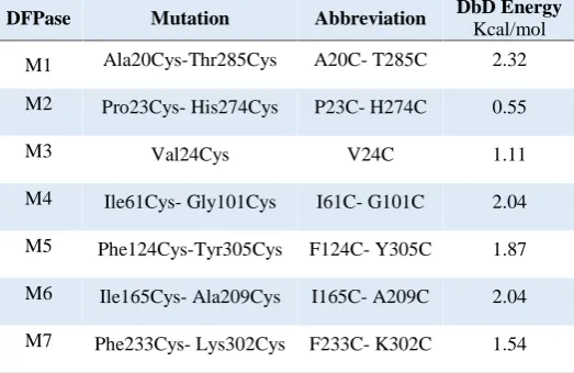
Table 1. List of structural mutation abbreviations and the energy of the mutated structure based on the DBD calculations.
DFPase Mutation Abbreviation DbD Energy
Kcal/mol
M1 Ala20Cys-Thr285Cys A20C- T285C 2.32
M2 Pro23Cys- His274Cys P23C- H274C 0.55
M3 Val24Cys V24C 1.11
M4 Ile61Cys- Gly101Cys I61C- G101C 2.04
M5 Phe124Cys-Tyr305Cys F124C- Y305C 1.87
M6 Ile165Cys- Ala209Cys I165C- A209C 2.04 M7 Phe233Cys- Lys302Cys F233C- K302C 1.54
Table 2. list of structural energies of selected mutations calculated by ERIS.
Mutations Abbreviation DBD Energy
ERIS Fixed backbone
Eris Flexible Backbone
M1 A20C- T285C 2.32 -0.87 -3.6
M 2 P23C- H274C 0.55 1.74 4.42
M 3 V24C 1.11 1.89 4.31
M 4 I61C- G101C 2.04 -11.75 -14.29
M 5 F124C- Y305C 1.87 -4.29 -7.22
M 6 I165C- A209C 2.04 2.35 5.47
M 7 F233C- K302C 1.54 1.18 -0.65
Ten rotamers resulted from Backrub method for each mutation were re-evaluated for probing the disulfide bond establishment. The disulfide bond has not been established for mutations M2, M4, M6 and M8. Finally, MD simulations for 10 ns have been performed for mutations M1, M3, M5 and M7.
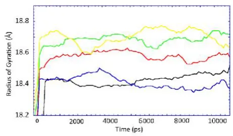
RMSD and B-factor of DFPase for native (at 300 and 335°K) and four selected mutations have been depicted at figure 1 and 2, showing the amount of backbone RMSD and B-factor at 335°K.
showing the stability of mutation M5 at 80 and 90°C. Normal mode analysis of DFPase showed that there is also a significant match between B-factor from molecular Dynamics and from normal mode calculation. B-factors are computed based on the first 100 normal modes and are scaled to match the overall B-factors in the submitted model (Fig. 6).
Figure 1. RMSD of DFPase of native (black), mutation M1 (red), M3 (green), M5 (blue) and M7 (yellow).
Figure 2. B-factor of DFPase of native (black), mutation M1 (red), M3 (green), M5 (blue) and M7 (yellow).
Figure 3. radius of gyration of DFPase of native (black), mutation M1 (red), M3 (green), M5 (blue) and M7 (yellow).
Figure 4. RMSD of DFPase for native at 300 °K (black) 335 °K (red) mutation M5 at 335 °K (green), at 355 °K (blue) and 365 °K (yellow).
Figure 5. Radius of gyration of DFPase for native at 300°K
(black) 335 K (red) mutation M5 at 300 °K (blue), at 335°K (green), at 355 °K (yellow) and 365 °K (dark green).
Figure 6. Overall B-factors calculated for the native DFPase (red line) and M5 (green line).
Discussion
aproximately unpreturbed during 10ns of simulation rather than other designs. The position of disulfide bond at
mutation M5 is accros the central tunnel joining α23 to
c-terminal part of DFPase. This bridge is joing Cys 124 to 305 which is enforce the two blades from different domains (Fig. 7).
Figure 7. Structure of DFPase at disulfide bond position, front view (left), slab view (right).
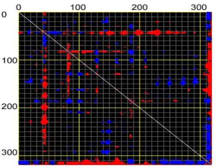
NMA graphs revealed that the disulfide bond of mutation M5 did not alter the natural normal modes of native protein. Such motions are also called harmonic motions which is necessary for the enzymatic reaction of the DFPase. However the amount of B-factor reduced for the mutated DFPase but the pattern was not altered regarding to the native protein. Analysis of catalytic Glu 37 and His 287 showed that the flexibility and B-factor resulted from molecular dynamics simulation did not showed any alteration in this position during the elevation of the temperature from 300 to 365°K. In accordance with the MD simulations, normal mode analysis also confirmed the results for the mutation M5 for the catalytic amino acids. The distance fluctuation graph (Fig. 8) also confirmed the region of fluctuations which is the active site residue is remained intact in mutation M5.
Figure 8.Distance fluctuation map of DFPase for Cα at mode 8. Flexible blocks are colored for increase (blue) and decrease (red) of distance fluctuations.
The flexibility of the C-terminal region is higher than the other parts of the structure. This region is consists of a
small α-helix connected to a freely moving β-like structure. There are some other local flexibility is observer in regions 180-185 and 220-225 which are consist of
β-turn and a loop respectively. The mentioned flexible regions are also observed through NMA results of the structure.
Conclusions
Finally, as it observed, a thermostable designed DFPase has been resulted from series of bioinformatics and computational procedure as a combinatorial in silico method with a desired output which it is stable at 80°C. Such a rational combinatorial procedure, however in one hand is time consuming method but on the other hand reducing numerous possible experimental designs at laboratory. In traditional methods of protein engineering most of designs are based on fixed backbone while the chance of obtaining desirable results would be decreased as the effect of mutations on folding or local conformations neglected. Considering the mentioned effect of such alterations on local structural conformation, turning the designing methods to flexible backbone conjugated with MD simulations may improve rational designs more accurate.
Prediction of the engineered structure would be possible while output of such methods subjected to MD simulations. Despite of limitations in MD simulation of large macromolecules in natural conditions [21], it seems that it is a powerful tool providing a convergent scheme in decreasing the time and cost of laboratory experiments.
References
1. Eijsink, V.G., Bjørk, A., Gåseidnes, S., Sirevåg, R., Synstad, B., Burg, B.V.D., Vriend, G., Rational engineering of enzyme stability. J Biotech, 2004, Vol. 113, pp. 105-120.
2. Eijsink, V.G., Gåseidnes, S., Borchert, T.V., van den Burg, B., Directed evolution of enzyme stability. Biomol Eng, 2005, Vol. 22, pp. 21-30.
3. Kumar, S., Nussinov, R., Salt bridge stability in monomeric proteins. J Mol Biol, 1999, Vol. 293, pp. 1241-1255.
4. Pantoliano, M.W., Whitlow, M., Wood, J.F., Rollence, M.L., Finzel, B.C., Gilliland, G.L., Poulos, T.L., Bryan, P.N., The en-gineering of binding affinity at metal ion binding sites for the stabilization of proteins: subtilisin as a test case. Biochem, 1988, Vol. 27, pp. 8311-8317.
5. Masip, L., Pan, J.L., Haldar, S., Penner-Hahn, J.E., DeLisa, M.P., Georgiou, G., Bardwell, J.C., Collet, J.-F., An engineered pathway for the formation of protein disulfide bonds. Science, 2004, Vol. 303, pp. 1185-1189.
6. Scharff, E.I., Koepke, J., Fritzsch, G., Lücke, C., Rüterjans, H., Crystal structure of diisopropylfluorophosphatase from Loligo vulgaris. Structure, 2001, Vol. 9, pp. 493-502.
7. Attaway, H., Nelson, J., Baya, A., Voll, M., White, W., Grimes, D., Colwell, R., Bacterial detoxification of diisopropyl fluorophosphate. Appl Environ Microbiol, 1987, Vol. 53, pp. 1685-1689.
9. Hartleib, J., Rüterjans, H., Insights into the reaction mechan-ism of the diisopropyl fluorophosphatase from Loligo vulgaris by means of kinetic studies, chemical modification and site-directed mutagenesis. Biochimi Biophys Acta (BBA)-Protein Struct Mol Enzymol, 2001, Vol. 1546, pp. 312-324.
10. Koepke, J., Scharff, E.I., Lucke, C., Ruterjans, H., Fritzsch, G., Statistical analysis of crystallographic data obtained from squid ganglion DFPase at 0.85 A resolution. Acta Cytol, ACTA Cytologica, 2003, Vol. 59, pp. 1744-1754.
11. Guex, N., Peitsch, M.C., SWISS MODEL and the Swiss Pdb Viewer: an environment for comparative protein modeling. Electrophoresis, 1997, Vol. 18, pp. 2714-2723.
12. DeLano, W.L., The PyMOL molecular graphics system. 2002.
13.Dombkowski, A.A., Disulfide by Design™: a computational method for the rational design of disulfide bonds in proteins. Bioinformatics, 2003, Vol. 19, pp. 1852-1853.
14. Yin, S., Ding, F., Dokholyan, N.V., Eris: an automated esti-mator of protein stability. Nature methods, 2007, Vol. 4, pp. 466-467.
15. Yin, S., Ding, F., Dokholyan, N.V., Modeling backbone flex-ibility improves protein stability estimation. Structure, 2007, Vol. 15, pp. 1567-1576.
16. Davis, I.W., Arendall III, W.B., Richardson, D.C., Richard-son, J.S., The backrub motion: how protein backbone shrugs when a sidechain dances. Structure, 2006, Vol. 14, pp. 265-274. 17. Smith, C.A., Kortemme, T., Backrub-like backbone simula-tion recapitulates natural protein conformasimula-tional variability and improves mutant side-chain prediction. J Mol Biol, 2008, Vol. 380, pp. 742-756.
18. Berendsen, H.J., van der Spoel, D., van Drunen, R., GRO-MACS: A message-passing parallel molecular dynamics imple-mentation. Comput Phys Commun, 1995, Vol. 91, pp. 43-56. 19. Van Der Spoel, D., Lindahl, E., Hess, B., Groenhof, G., Mark, A.E., Berendsen, H.J., GROMACS: fast, flexible, and free. J Comput Chem, 2005, Vol. 26, pp. 1701-1718.
20. Suhre, K., Sanejouand, Y.-H., ElNemo: a normal mode web server for protein movement analysis and the generation of tem-plates for molecular replacement. Nucleic Acids Res, 2004, Vol. 32, pp. 610-614.