*
Cor
Swap
1Instit Techn am(W Tirupa 2Depa of Med Libya.Intr
Deox proce biolo of re Stud DNA few demo antic can canc intera This and prob The the spec the d The interc intera elect of DN groov takes interc invar ThisEff
rresponding
pna G
ute of Pharmace nology,SriPadma WomenÊs Univers
ati 517 502, Ind artment of Pharm dicine, Sebha U .
oduction
xyribonucleic ac ess because it ogical synthesis plication and tra ies on the bind A have been iden
decades [1]. onstrated that cancer drugs; in cause DNA dam cer cells and res
action of plant is due to their p their phytochem es of DNA struc investigation of molecular mec cific DNA-targete double helix DN non-covalent wa calation betwe actions with the trostatic attractio NA. Intercalative ves in the DNA s place out of calation is one o riably leads to ce
work is licens
fect of
P
g author:
eutical avatiMahilaViswa ity), dia. macology, Facult niversity, Sebhaid plays an impo carries heritage of proteins and anscription of ge
ing mechanism ntified as one of Numerous b DNA is the p nteraction betwe mage in cancer sulting in cell dea
extract with DN possible applicat mical properties ture and conform drug-DNA intera hanisms of the ed drug[5]. Basic NA in either a no
ay of binding inc een the base
DNA groove; a ons with the anio e binding and gr A double helix, the groove [6 of the most impo ellular degradatio
sed under a C
http
Phyllanthu
Swapna G
avidyalay ty a,A b s
Phyllant cell line extract spectro and wa effect a change interact Keywor Calf Thyortant role in bio e information an
enzymes throu enetic informatio of some small f the key topics biological expe primary intracel een small molec r cells, blocking ath [2, 3]. Of the NA has gained m
tion as new the s which make mation [4]. action is vital for e drug action cally, plant extrac
on-covalent or a cludes three bin pairs; (ii) gr and (iii) electrost onic sugar-phosp
roove binding ar while the electr 6]. Among thes ortant DNA bind
on.
Creative Comm
p://www.arjo
Origin
us polyphy
G
1*, Bharathi K
s t r a c t
nthus polyphylluses. For a better of Phyllanthus p scopy, viscomet as found to inter and blue-shift in s in the current ion between teh rds: Phyllanthus ymus DNA; Inte
logical systems nd instructs the gh the process n in living cells. molecules with during the past eriments have llular target of cules and DNA
the division of ese studies, the much attention. rapeutic agents them potential r understanding
and designing ct interacts with a covalent way. nding modes: (i) roove binding, tatic interaction, phate backbone re related to the rostatic binding se interactions,
ing modes as it
mons Attributio
ournals.org/in
nal Researc
hyllus
ext
K
1, Rajkapoor
s has demonstra r understanding polyphyllus (PP try and cyclic vo ract with CT-DN
the UV spectra. t and potential i PP and the CT-s polyphylluCT-s ;
rcalation. An un is req recogn decide seque into D bound Variou interac metho viscos [11] et These antica are als betwe metho studie potent The a metha Thymu voltam
Mate
C
hem
All rea India. wereon 3.0 License
ndex.php/ijpm
ch Article
tract on D
r B
2, KVSRG.
ated potent in v of the mechan P) with Calf Thym
oltammetry. PP NA through inter . An increase in n cyclic voltamm -DNA.
; Absorption Sp
nderstanding of t quired to illus nition by such f e the affinity an ence. Cationic co DNA and bind d fashion [7]. us techniques h
ction of small m ods [8], nuclear sity measureme
tc.
e investigations ancer drugs and so very valuable een anticancer d ods to effectively es have demon t in vitro antican
im of this study anolic extract o us DNA using ab mmetry.
erial and Me
micals
agents and cheSolvents used f purified by stan
e.
m/index
e
DNA bind
. Prasad
1vitro anticancer ism of action, b mus (CT) DNA w
displayed bindi rcalation, as dem
the viscosity of metric experime pectroscopy;Visc
the modes of bi strate the prin functional molec d specificity of t omplexes have b
non-covalently ave been exten molecules with r magnetic reso ent [10] and ele
form a theoretic chemical treatm e for probing the drugs and DNA y choose specifi strated that Ph ncer activity aga was to investiga f Phyllanthus p bsorption spectr
ethods
emicals were pr for electrochemi ndard procedure
ding studi
activity against binding studies were studied us ing properties to monstrated by a f CT-DNA was o ents demonstrate cometry; Cyclic
nding of plant e ciples governin cules, that is, th
the complexes been found to bo
in a surface-b nsively employed
DNA, such as onance [9], x-r ectrochemical m c guide for the d ments of tumor a e mechanism of t A and establishin c anticancer dru hyllanthus polyp ainst various tum ate the binding polyphyllus (P roscopy, viscome
ocured from Me cal and spectros es [12]. DNA w
es
various tumor of methanolic ing absorption o the CT-DNA a hypochromic observed. The e intercalative Voltammetry;
extract to DNA ng the DNA he factors that
for DNA base oth intercalate bound
groove-d to stugroove-dy the spectroscopic ray diffraction, measurements
design of new nd virus. They the interaction ng convenient ug. Our earlier phyllus has
mor cell lines. mechanism of PP) with Calf etry and cyclic
erck, Mumbai, scopic studies was purchased
PAGE |
336
|
from Bangalore Genei (India). Agarose (molecular biology grade), ethidium bromide (EB) were obtained from Sigma, St. Louis (USA). Tris (hydroxymethyl) amino methane-HCl (Tris HCl) buffer solution was prepared using deionized, sonicated triple distilled water.
Experiments
All the experiments involving the interaction of PP with CTDNA were carried out in Tris HCl buffer (50 mM Tris HCl, pH 7.2) containing 5% alcohol at room temperature. A stock solution of CT DNA was prepared by dissolving the CT DNA in the Tris-HCl buffer. Solutions of CT DNA in the above buffer gave a ratio of UV absorbance at 260 and 280 nm, A260/A280 of 1.87, indicating that
the CT DNA was sufficiently free from protein. The CT DNA concentration per nucleotide was determined by absorption spectroscopy at 260 nm using the molar absorption coefficient 260
(6600 M-1 cm-1).
DNA binding experiments
Absorption spectroscopic studies
UV absorption spectra were measured on a Shimadzu UV-1601 spectrophotometer in 5 mM Tris-HCl buffer (pH 7.1) containing 50 mM NaCl at room temperature. PP was dissolved in methanol at a concentration of 5 10-3 M. Working solutions were prepared by
dilution of the PP in methanol in 5mM Tris-HCl buffer to concentration of 50 μM.
Absorption titration experiments were performed by maintaining the extract concentration as constant at 50 øM while varying the concentration of the CT DNA within 0 to 400 øM. While measuring the absorption spectra, equal quantity of CT DNA was added to both the extract solution and the reference solution to eliminate the absorbance of CT DNA itself. From the absorption data, the intrinsic binding constant Kb was determined from the following
equation (1):
[DNA]/(a f)=[DNA]/(b f)+[Kb(b f)] 1 --(1)
wherea, f, b correspond to Aobsd/[extract], the extinction coefficient
for the free extract, and the extinction coefficient for the extract in the fully bound form, respectively. A plot of [DNA]/(a f) versus
[DNA], where [DNA] is the concentration of CT DNA in base pairs, gives Kb as the ratio of slope to intercept.
Viscosity measurements
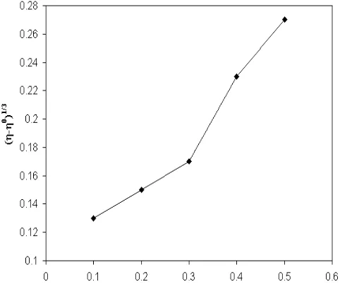
Viscosity experiments were carried on an Ostwald viscometer, immersed in a thermostated water-bath maintained at a constant temperature of 30.0 μ 0.1 C. DNA samples of approximately 0.5 mM were prepared by sonication in order to minimize complexities arising from DNA flexibility [13]. Flow time was measured with a digital stopwatch three times for each sample and an average flow time was calculated. Data were presented as (η/η0)1/3 versus the
concentration of the PP, where η is the viscosity of DNA solution in the presence of complex, and η0 is the viscosity of DNA solution in
the absence of complex. Viscosity values were calculated after correcting the flow time of buffer alone (t0), η = (t t0)/ t0 [14].
Electrochemical methods
Cyclic voltammetric study was performed on a CHI 620C electrochemical analyzer with three electrode system of glassy carbon (GC) as the working electrode, a platinum wire as auxiliary electrode and Ag/AgCl as the reference electrode. All the voltammetric experiments were carried out in single-compartment cells of volume 5-15 mL. Solutions were deoxygenated by purging with N2 prior to measurements. Increasing amounts of CT DNA
were added directly in to the cell containing the PP solution (5 X 10⁻³ M, 5 mM Tris-HCl/50 mMNaCl buffer, pH 7.1). The
concentration ranged from 0 to 400 μM for CT DNA. The solution in the cuvette was thoroughly mixed before each scan. All the experiments were carried out at room temperature.
In a typical cyclic voltammetric titration, a fixed concentration of the PP extract was taken and DNA solution in buffer was added in different ratios as done in the absorption titration, and the voltammetric response was recorded.
Results and Discussion
The interaction of small molecules with DNA plays an important role in many biological processes. These associative interactions with the DNA molecules can cause dramatic changes in the physiological functions of DNA, that might be responsible for the cytotoxic behavior of the small molecules [15]. The DNA binding capacity was evaluated based on interaction with CTDNA.
Electronic Absorption spectral studies
Absorption spectroscopy is one of the most important and useful methods to investigate the binding of any drug with DNA [16, 17]. When a small molecule interacts with DNA, it may form a complex that exhibits absorption spectrum which is different from that of the small molecule. The addition of DNA to a solution of an intercalator results in a characteristic shift of the absorption maximum to longer wavelengths (bathochromic shift or red shift) and a decrease (hypochromocity) or increase (hyperchromicity) of the absorbance [18]. The intercalation mode of micromolecule could locally elongate the DNA helix by separating the stacked base pairs and partially unwinds the DNA helix at the intercalation site [19]. Consequently, it induces the structural perturbation of the DNA duplex, which might hinder the replication of DNA and inhibit protein synthesis. These changes could cause small increase in absorption peak of double-strand DNA (dsDNA) at 260 nm, and this phenomenon proved that the opening of the dsDNA helix occurred [20].
CT-PAGE |
337
|
DNA resulted in the hypochromism and blue-shift in the UV-spectra of the PP. The hypochromism is associated with the intercalative binding of small molecules to the base pairs of DNA, because of strong stacking interactions between the aromatic chromophore of the small molecules and the base pairs of DNA [21]. After intercalating the base pairs of DNA, the * orbital of the intercalated extracts could couple with the orbital of base pairs, thus decreasing the * transition energy and further resulting in the blue-shift or red-shift. On the other hand, the coupling of orbital was partially filled by electrons, thus decreasing the transition probabilities and concomitantly resulting in the hypochromism. The other absorption peak was showed at 661.5 nm in the absence of DNA, and upon the incremental addition of DNA to extract, no significant change in the wavelength and very weak absorption change was seen shown in Fig 1. Since, only the * orbit of the extract could couple with the partly filled orbital by electrons of base pairs.
From the ultraviolet and visible absorption spectra of the extract in the absence, and presence, of DNA (Fig. 1), it was observed that the PP Extract solution exhibited peculiar hypochromic and bathochromic shifts in the absorption spectra on binding to DNA, a typical characteristic of DNA intercalation [22].
The intrinsic binding constant Kb is obtained by monitoring the
changes in the absorbance for the extracts with increasing concentration of DNA in order to compare the binding strength of the complexes with CT DNA. Kb is obtained from the ratio of slope
to the intercept from the plots of [DNA]/a f) versus [DNA]. The Kb
value is shown in Table 1. The high Kb value of 7.6 105 suggests a
stronger binding towards DNA.
Viscosity measurements
As a means to further clarify the mode of binding of the PP extract to CT DNA, viscosity measurements were carried out by varying concentration of extract. Viscosity experiment is regarded as the most critical test for the binding mode of small molecules and DNA. Generally, a classical intercalation binding causes a significant increase in the viscosity of DNA solution due to an increase in lengthening in the DNA helix to accommodate the bound ligand between the adjacent base pairs [23]. In contrast, a partial and/or nonclassical intercalation binding causes reduction in viscosity as the ligand bends (or kinks) the DNA helix and reduces its effective length, while ligands that bind exclusively in the DNA grooves typically cause less pronounced changes (positive or negative) or no changes in DNA solution viscosity under the same conditions [24,25]. Fig.2 Shows a slight increase in the flow time of the DNA with increasing concentration of extract, which is not as pronounced as those observed for the classical intercalatorethidium bromide [26]. This indicates that extract prefer to engage in DNA groove binding or surface binding with its overall size resulting in an increase in DNA viscosity, rather than an intercalative DNA interaction.
Electrochemical methods
Electrochemical methods have contributed substantially to our understanding of anticancer agents.These enable to evaluate and predict DNA interactions by DNA-binding compounds. Amongst the various electroanalytical techniques in general, cyclic voltammetry (CV) is by far the most versatile electrochemical method. The obtained results of CV from the redox properties of drugs and biomolecules might have fervent effects on interpretation of their in vivo redox behaviour or pharmaceutical activity [27].
Cyclic voltammetric experiments were performed by maintaining the concentration of PP while varying the concentration of CT DNA within 0-400 M and the voltammetric responses were recorded. The cyclic voltammogram of PP in the presence of DNA and absence of CT DNA are shown in Fig 3 and the electrochemical data are summarised in the Table 2. In the absence of CT DNA, the methanolic extract show only the oxidation peak -0.719 V (Epa) and no reduction peak in the absence of DNA. Incremental addition of DNA to PP, shows a decrease in the current intensity and negative shift of the oxidation peak potential. The resulting changes in the current and potential demonstrate interaction between the extract and DNA. This indicates that the reaction of the PP on the glassy carbon electrode surface is quasi-reversible redox process. The incremental addition of CT DNA to the PP causes only a negative potential shift and decreasing current intensity in anodic peak and no significance change of potential shift and current intensity in cathodic peak. The ipc/ipa values also
decrease in the presence of DNA.
The peak current increased initially and then decreased. The initial increase in the peak current is due to the absorption of the DNA bound complex onto the electrode surface [28]. The decrease in peak current on the addition of DNA to the complex is suggestive of an interaction between the complex and DNA [29]. A decrease in the peak-to-peak separation was observed, which is consistent with non-coordinating intercalative binding of the complexes through the planar aromatic rings between the DNA base pairs [30]. The formal potential Ef shifts slightly towards the positive side and is attributed to characteristic behavior of intercalation of the complexes into the DNA double-helix [31, 32], and suggests that PP extract bind to DNA at different rates.
PAGE |
338
|
Table 1:Absorption spectral properties of PP Extract
Extract λmax Δλ
(nm)
H%=[( r -b)/ fx
100]
Kb (M-1)
Free Bound
PP 390.0 373.0 17 32.7 7.6 X 10⁵
Figure 1: Electronic absorption spectra of PP in the absence (dash line) and presence (dark line) of increasing amounts of DNA caption.
Figure 2:The effect of PP on the relative viscosity of calf thymus DNA in 5 mMTris HCl/50 mMNaCl buffer (pH7.1).
R= [DNA]/ [PP]
Ipa x 10-5 (A) Epa (V)
0 -3.19 -0.719
0.5 -2.92 -0.723
1 -2.1 -0.732
1.5 -1.83 -0.745
2 -1.52 -0.753
2.5 -1.19 -0.762
3 -0.93 -0.783
Table 2:Electrochemical parameters of the compoundPP
Figure 3:Cyclic voltammogram of PP both in the absence (pink line) and presence (other colour line) of different concentration of DNA in 50 mMNaCl, 5 mM Tris-HCl, pH 7.2. Scane rate 100 mV s-1
Conclusion
In this work, the PP extract have been shown to possess DNA-binding abilities. The interaction of PP with calf thymus DNA was studied by absorption spectroscopy, viscometry and cyclic voltammetry. Upon binding to DNA, the absorption spectra of PP showed peculiar hypochromic effect and bathochromic shift, a slight increase in viscosity was observed in viscosity measurements, and a decrease in peak current was observed in cyclic voltammetry. The variations in the spectral characteristics, viscosity and electrochemical behavior upon binding of PP with CT- DNA indicated an intercalative mode of interaction. This study is expected to provide greater insight into the design of new anticancer drugs.
Current (A)
Potential(V)
[DNA]
PAGE |
339
|
References
[1]. Navarro JA, Salas JM, Romero MA, Vilaplana R, Gonzalez-Vilchez F, Faure R. cis-[PtCl2(4,7-H-5-methyl-7-oxo[1,2,4]triazolo[1,5-a]pyrimidine)2]: a sterically restrictive new cisplatin analogue. Reaction kinetics with model nucleobases, DNA interaction studies, antitumor activity, and structure-activity relationships. J Med Chem. 1998;41(3):332-8.
[2]. Arjmand F, Parveen S, Afzal M, Toupet L, Ben Hadda T. Molecular drug design, synthesis and crystal structure determination of CuII-SnIV heterobimetallic core: DNA binding and cleavage studies. Eur J Med Chem. 2012;49:141-50.
[3]. Zhang G, Fu P, Wang L, Hu M. Molecular spectroscopic studies of farrerol interaction with calf thymus DNA. J Agric Food Chem. 2011;59(16):8944-52.
[4]. Metcalfe C, Thomas JA. Kinetically inert transition metal complexes that reversibly bind to DNA. Chem Soc Rev. 2003;32(4):215-24.
[5]. Gilpin RK, Pachla LA. Pharmaceuticals and related drugs. Anal Chem. 1997;69(12):145R-63R.
[6]. Zhang G, Fu P, Pan J. Multispectroscopic studies of paeoniflorin binding to calf thymus DNA in vitro. J Lumin. 2013;134:303-9. [7]. Barton JK, Goldberg JM, Kumar CV,
Turro NJ. Binding modes and base specificity of tris (phenanthroline) ruthenium (II) enantiomers with nucleic acids: tuning the stereoselectivity. J Am Chem Soc. 1986;108:2081-2088. [8]. Zhao GC, Zhu JJ, Chen HY.
Spectroscopic studies of the interactive model of methylene blue with DNA by means of h-cyclodextrin. Spectrochim Acta A Mol Biomol Spectrosc. 1999;55: 1109 1117.
[9]. Sandstrom K, Warmlander S, Leijon M, Graslund A. 1H NMR studies of selective interactions of norfloxacin
with double-stranded DNA. Biochem Biophys Res Commun. 2003;304(1):55-9.
[10].Shahabadi N, Heidari L. Binding studies of the antidiabetic drug, metformin to calf thymus DNA using
multispectroscopic methods. Spectrochim ActaA Mol Biomol Spectrosc. 2012;97:406-10.
[11].Ozkan SA, Ozkan Y, Senturk Z. Electrochemical reduction of metronidazole at activated glassy carbon electrode and its determination in pharmaceutical dosage forms. J Pharm Biomed Anal. 1998;17(2):299-305.
[12].Perrin DD, Armarego WLF, Perrin DR. Purification of laboratory chemicals. 2d ed. Oxford ; New York: Pergamon Press; 1980. x, 568 p. p.
[13].Satyanarayana S, Dabrowiak JC,
Chaires JB. Tris(phenanthroline)ruthenium(II)
enantiomer interactions with DNA: mode and specificity of binding. Biochemistry. 1993;32(10):2573-84. [14].Chauhan M, Banerjee K, Arjmand F.
DNA binding studies of novel Copper(II) complexes containing L-tryptophan as chiral auxiliary: in vitro antitumor activity of Cu-Sn2 complex in human neuroblastoma cells. Inorg Chem. 2007;46(8):3072-82.
[15].Marmur J. A procedure for the isolation of deoxyribonucleic acid from micro-organism. J Mol Biol.1961; 3: 208. [16].Mansouri-Torshizi H, Saeidifar M,
Khosravi F, Divsalar A, Saboury AA, Hassani F. DNA Binding and Antitumor Activity of alpha-Diimineplatinum(II) and Palladium(II) Dithiocarbamate Complexes. Bioinorg Chem Appl. 2011;2011:394506.
[17].Kumar RS, Arunachalam S, Periasamy VS, Preethy CP, Riyasdeen A, Akbarsha MA. Surfactant-cobalt(III) complexes: synthesis, critical micelle concentration (CMC) determination,
DNA binding, antimicrobial and cytotoxicity studies. J Inorg Biochem. 2009;103(1):117-27.
[18].Kuruvilla E, Nandajan PC, Schuster GB, Ramaiah D. Acridine-viologen dyads: selective recognition of single-strand DNA through fluorescence enhancement. Org Lett. 2008;10(19):4295-8.
[19].Wilson WD, Ratmeyer L, Zhao M, Strekowski L, Boykin D. The search for structure-specific nucleic acid-interactive drugs: effects of compound structure on RNA versus DNA interaction strength. Biochemistry. 1993;32(15):4098-104.
[20].Oliveira-Brett AM, Diculescu VC. Electrochemical study of quercetin-DNA interactions: part I. Analysis in
incubated solutions. Bioelectrochemistry.
2004;64(2):133-41.
[21].Bloomfield VA, Crothers DM, Tinoco I. Physical chemistry of nucleic acids. New York,: Harper & Row; 1974. x, 517 p. p.
[22].Lafayette EA, Vitalino de Almeida SM, Pitta MG, Carneiro Beltrao EI, da Silva TG, Olimpio de Moura R, et al. Synthesis, DNA binding and topoisomerase I inhibition activity of thiazacridine and imidazacridine
derivatives. Molecules. 2013;18(12):15035-50.
[23].Shen HY, Liu YQ, Gao J, Zhen HM, Zhu N, Li J. In vitro study of DNA interaction with melamine and its related compounds. DNA Cell Biol. 2011;30(4):255-64.
[24].Arjmand F, Aziz M. Synthesis and characterization of dinuclear macrocyclic cobalt(II), copper(II) and zinc(II) complexes derived from
PAGE |
340
|
[25].Satyanarayana S, DabrowiakC, Chaires JB. (1992) Neither .DELTA.- nor .LAMBDA.-tris(phenanthroline)ruthenium(II) binds to DNA by classical intercalation . Biochemistry 31, 9319-24.
[26].Wang XL, Chao H, Li H, Hong XL, Liu YJ, Tan LF, et al. DNA interactions of cobalt(III) mixed-polypyridyl complexes containing asymmetric ligands. J Inorg Biochem. 2004;98(6):1143-50.
[27].Hart JP. Electroanalysis of biologically important compounds. New York: E. Horwood; 1990.
[28].Yang ZS, Wang YL, Zhao GC. The interaction of copper-bipyridyl complex
with DNA and cleavage to DNA. Anal Sci. 2004;20(8):1127-30.
[29].Annaraj J, Srinivasan S, Ponvel KM, Athappan P. Mixed ligand copper(II) complexes of phenanthroline/bipyridyl and curcumin diketimines as DNA intercalators and their electrochemical behavior under Nafion and clay modified electrodes. J Inorg Biochem. 2005;99(3):669-76.
[30].Lu X, Zhang M, Kang J, Wang X, Zhuo L, Liu H. Electrochemical studies of kanamycin immobilization on self-assembled monolayer and interaction with DNA. J Inorg Biochem. 2004;98(4):582-8.
[31].Srinivasan S, Annaraj J, Athappan P. Spectral and redox studies on mixed ligand complexes of cobalt(III) phenanthroline/bipyridyl and benzoylhydrazones, their DNA binding
and antimicrobial activity. J Inorg Biochem. 2005;99(3):876-82.