P
AWEŁR
EICHERT1, R
OMANR
UTOWSKI1, 2, T
OMASZG
RECZNER1, J
ERZYG
OSK1,
K
RZYSZTOFZ
IMMER1, 3, W
ITOLDW
NUKIEWICZ1Treatment of Algodystrophic Syndrome
of the Upper Extremity in Own Material
Leczenie zespołu algodystroficznego kończyny górnej
w materiale własnym
1Department of Traumatology and Hand Surgery, Silesian Piasts University of Medicine in Wrocław, Poland 2Chair and Department of Sports Medicine, Wrocław University of Physical Education, Poland
3 Unit of Sport Medicine, Silesian Piasts University of Medicine in Wrocław, Poland
Adv Clin Exp Med 2007, 16, 6, 785–791 ISSN 1230−025X
ORIGINAL PAPERS
© Copyright by Silesian Piasts University of Medicine in Wrocław
Abstract
Background.In spite of the development of new diagnostic methods and treatment possibilities, algodystrophic syndrome (Sudeck’s disease, CRPS I) still constitutes a challenge for the treating surgeon. Its etiopathogenesis is still not fully explained, diagnostic criteria are not uniform, and treatment results are unsatisfactory. Estimation of the treatment results of CRPS I of the upper extremity in own material was the purpose of this study.
Material and Methods.Between 2000 and 2005, 38 patients were treated because of algodystrophic syndrome at the Department of Trauma and Hand Surgery. Diagnosis was based on clinical examination, X−ray, and scintigra− phy. Rehabilitation and tricyclic antidepressants, anticonvulsants, vasodilators, Dexaven and Mannitol, brachial plexus blocks were used in the treatment depending on the phase of disease.
Results.The best results were achieved in patients in the first, posttraumatic phase of disease. Amelioration was achieved in most patients after use of brachial plexus blocks in the second and third phases of disease, but recur− rence of full function was achieved in less that 30% patients.
Conclusions.Effective treatment and rapid rehabilitation in the posttraumatic phase of disease prevent its further progression. Use of a brachial plexus block and then rehabilitation is an efficient method of treating patients with CRPS (Adv Clin Exp Med 2007, 16, 6, 785–901).
Key words:algodystrophic syndrome, CRPS I, scintigraphy, brachial plexus blocks.
Streszczenie
Wprowadzenie. Pomimo rozwoju nowych technik diagnostycznych i możliwości leczenia, zespół algodys− troficzny (choroba Sudecka, CRPS I) nadal jest wyzwaniem dla leczącego chirurga. Etiopatogeneza nie jest do końca wyjaśniona, kryteria diagnostyczne niejednolite, a wyniki leczenia niezadowalające.
Cel pracy. Ocena wyników leczenia zespołu CRPS I kończyny górnej w materiale własnym.
Materiał i metody.W latach 2000–2005 w Klinice Chirurgii Urazowej i Chirurgii Ręki leczono 38 chorych z po− wodu zespołu algodystroficznego. Rozpoznanie opierało się na badaniu klinicznym, radiologicznym i scyntygra− ficznym W leczeniu w zależności od okresu choroby stosowano rehabilitację oraz trójpierścieniowe leki antyde− presyjne, leki przeciwpadaczkowe, leki rozszerzające naczynia, Dexaven i Mannitol, blokady splotu ramiennego.
Wyniki.Najlepsze wyniki uzyskano u chorych w pierwszym pourazowym okresie choroby. Stosując blokady splo− tu ramiennego w II i III okresie choroby, uzyskano poprawę u większości chorych, powrót do pełnej funkcji uzy− skano jednak u niespełna 30% chorych.
Wnioski. Skuteczne leczenie i szybka rehabilitacja w pourazowym okresie choroby zapobiegają dalszej progresji choroby. Zastosowanie blokady splotu ramiennego i następnie rehabilitacja jest skuteczną metodą w leczeniu chorych z zespołem CRPS (Adv Clin Exp Med 2007, 16, 6, 785–901).
Despite great efforts all over the world, CRPS I (or reflex sympathetic dystrophy, Sudeck’s atrophy) is still an enigma. Its pathophysiology is unknown, diagnostic criteria are still debatable, and the results of treatment are poor [1, 2]. CRPS is a potentially incapacitating syndrome occurring in an extremity usually after a minor injury or operation [3–5]. Key symptoms in the acute phase include signs and symptoms of inflammation within the affected extremity and are listed in Table 1 [6]. These alter− ations are present in an area larger than and includ− ing the distal part of the extremity [7, 8]. In later stages one can observe osteoporosis, pseudomotor changes, temperature changes, vasomotor instabili− ty, palmar fascitis, and trophic changes [6].
There is no consensus on the treatment of CRPS I [4] and it depends on the stage of disease. Clinical symptoms differentiating particular stadia of disease are presented in Table 2 [6]. In the acute phase one can use anti−inflammatory agents (scav−
engers or steroids), optimize peripheral circulation (vasodilators or sympathetic blocks) if the skin
Table 1.Primary signs and symptoms of CRPS I Tabela 1.Główne objawy i symptomy zespołu CRPS I
Sign Features
(Objaw) (Opis)
Pain paramount feature, often with burn (Ból) ing, throbbing, aching, stabbing,
bursting, pressure, or crushing sensations
Swelling first physical sign: initially local (Obrzęk) soft edema, then extensive and
hard
Stiffness progressively worsens due to (Sztywność) increased fibrosis
Discoloration red, cyanotic, or pale to grayish; (Zaburzenia related to vasomotor instability koloru skóry)
Table 2. Clinical course of CRPS I
Tabela 2.Kliniczne objawy w kolejnych okresach CRPS I
Primary signs Early CRPS I: 0–3 months Intermediate CRPS I: Late CRPS I: (Pierwotne objawy) (Początkowy ostry okres: 3–9 or 12 months 9–12 months−years
0–3 m−ce) (Pośredni dystroficzny (Końcowy atroficzny okres: 3–9 do 12 m−cy) okres: 9–12 m−cy, lata) Pain paresthesia notable increase, pain with diminished pain, but severe
(Ból) motion with motion
Swelling soft local edema hard edema over extremity periarticular (minimal) (Obrzęk)
Stiffness pain−related increased stiffening due to peak stiffening, contractures
(Sztywność) fibrosis common
Discoloration red then cyanotic cyanotic, with redness over pallor
(Zaburzenia koloru joints
skóry)
Table. 3.Pharmacological targets and relate treatment of CRPS I Tabela 3.Grupy leków i ich wykorzystanie w leczeniu CRPS I
Target Therapy against target Advantages
(Cel) (Ukierunkowane leczenie) (Korzyści)
Inflammatory process naproxen p.o. [10], no effectiveness of naproxen
(Proces zapalny) prednisone p.o. [11], effective in CRPS
methyloprednisolon + lidocaine [12] good response to treatment Reactive oxygen species dimethylsulfoxid [13], recommendable in CRPS (Reaktywne formy tlenu) vitamine C [14] could be a prophylactic method Pain sensitization carbamazepine [15], useful, initial increase of pain possible (Nadwrażliwość na ból) nifedipine [16], side effects limit application
lidocaine [17] effective in early CRPS Descending control of pain amitriptyline [18] effective in neuropathic pain (Ograniczanie bólu)
Sympathetically maintained pain reserpine [19] effective in early CRPS (Nadczynność układu współczulnego)
Others calcitonin [20] improvement of pain
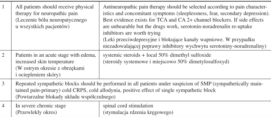
temperature of the affected extremity is lower than that of the contralateral extremity, treat all local causes of pain (trigger points), apply systemic pain medication, adapt skeletal muscle work to the lim− ited possibilities aided by a physiotherapist and/or ergotherapist, or splint the affected extremity if required [5]. In the late phase the procedures should include optimizing pain medication, pro− viding splints for comfort and protection, for the upper extremities training for one−handed activi− ties and adapting the home, and for the lower extremities providing crutches or a wheelchair and adapting the home for wheelchair use. The most often used medications and their mechanisms of action are presented in Table 3 [9]. Birklein [21] recommends the four basic steps in CRPS treat− ment presented in Table 4.
The differentiated treatment modes and the lack of satisfactory results of treatment presented by different authors encouraged the present authors to evaluate and analyze the methods of CRPS I treatment used in their clinic. Determining the results of treatment of CRPS of the upper extremity in own material was the purpose of this study.
Material and Methods
Between 2000 and 2005, 38 patients were treated because of CRPS I of the upper limb in this clinic. In the tested group, women constituted the majority (29 patients, 76%) and the patients’ ages ranged from 31 to 76 years (average: 61 years). Eighteen had ambulatory treatment and 20 were hospitalized. CRPS was a consequence of fracture of the distal radius in 26 patients, surgery of carpal
tunnel syndrome in 3, 3 patients had surgery for Dupuytren’s contracture, and there were several single cases of contusion of an upper limb, contu− sion of the wrist, and severe hand injury. Most of the patients had been treated for their trauma out− side this clinic.
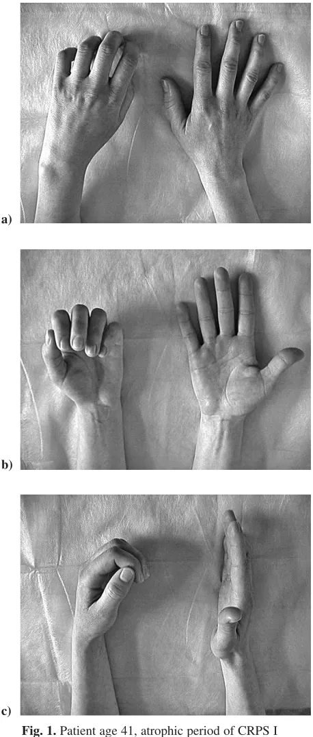
CRPS I was diagnosed when four of the five following signs and symptoms were present: pain, altered skin color, altered skin temperature, edema, and reduced range of motion, which were present in an area much larger than and also distal to the primary injury. X−ray and scintigraphy were performed as additional tests. The patients had neurological and psychiatric consultation. Figure 1 presents a clinical example of the disease in the atrophic phase. Twenty−two of the patients were treated in the first (posttraumatic, group I) period, 12 in the second (dystrophic), and 4 patients in the third (atrophic) phase. The time from fracture/ injury to starting treatment was 4–6 weeks for the patient group in the first phase (I), 12–16 weeks for the group in the second (II), and 6–9 months for those in the third phase (III). In the posttrau− matic period (I), vasodilators, tricyclic anti− depressants, tranquilizers, and non−steroid anti− inflammatory medications were used for six weeks and the so−called Szczecin method of Dexaven and Mannitol in intravenous infusion for one week. Brachial plexus blocks were used in periods II and III of disease. Brachial plexus block was estab− lished with a bolus of 15 ml of 0.25% bupivacaine and then, depending on the degree of motor block− ade, the concentration and volume of the anaes− thetic were reduced. A continuous analgesia was provided by regular application of 5–10 ml of 0.25–0.125% bupivacaine every 6 h or 12 h for one week. The aim was to achieve good sensory
Table 4.Symptom−oriented treatment options for posttraumatic CRPS I Tabela 4.Poszczególne etapy w leczeniu pourazowego CRPS I
1 All patients should receive physical Antineuropathic pain therapy should be selected according to pain character− therapy for neuropathic pain istics and concomitant symptoms (sleeplessness, fear, secondary depression). (Leczenie bólu neuropatycznego Best evidence exists for TCA and CA 2+ channel blockers. If side effects u wszystkich pacjentów) are unbearable but the drugs work, serotonin−noradrenalin re−uptake
inhibitors are worth trying
(Leki przeciwdepresyjne i blokujące kanały wapniowe. W przypadku niezadowalającej poprawy inhibitory wychwytu serotoniny−noradrenaliny) 2 Patients in an acute stage with edema, systemic steroids + local 50% dimethyl sulfoxide
increased skin temperature (steroidy systemowe i miejscowo 50% dimetylosulfoxyd) (W ostrym okresie z obrzękami
i ociepleniem skóry)
3 Repeated sympathetic blocks should be performed in all patients under suspicion of SMP (sympathetically main− tained pain−primary) cold CRPS, cold allodynia, positive effect of single sympathetic block
(Powtarzalne blokady układu współczulnego)
analgesia without motor blockade. Since motor function was unaffected, an active and painless exercise program was possible [5]. All patients had rehabilitation with calcium ionotophoresis, centrifuging massage, cryotherapy, and kinesither− apy. The follow−up time was 12 months.
Pain, mobility of the fingers and wrist, strength of the extremity, and function of the sym− pathetic nervous system were taken into account in estimating the results. Pain was measured using a 0–10 numeric rating scale (correlating with the result of the Visual Analogue Scale, VAS). Range of movement was measured in degrees (excellent; lack of extension up to 15% when the fingers are flexed the distance from the pulp to the distal pal− mar crease has to be equal to or less than 1 cm; lack of extension equal to or less than 30% with flexed fingers than can touch the palm; greater lack of extension or flexion). Strength of grip and pinch were measured with a dynamometer (excel− lent; result higher than 75%; result higher than 50%; result less than 50% of opposite hand). Autonomic (sympathetic) symptoms we deter− mined by measurement of skin temperature (dif− ference between affected and unaffected side exceeds 1.0 C°) and others autonomic testing. At the end the function of the extremity was assessed using the Quick−DASH (Disabilities of the Arm, Shoulder, and Hand) questionnaire. Excellent results meant a completely normal hand (0–15 score in Quick−DASH). Good results were pain up to a level of 2, no signs of dysfunction of the sympathetic nervous system, and limitation of range of movement (15–30 score in Quick− −DASH). A poor result was pain of more than 5 points and stiffness (> 60 score in Quick−DASH). A satisfactory result meant amelioration com− pared with the initial state (less pain, better mobil− ity of the fingers) requiring, however, further treatment.
Results
The best results were achieved in patients in the first period of disease. An essential difference was not observed in the treatment results between the group of patients treated with local anti−inflamma− tory drugs, anticonvulsants, and vasodilators and the group of patients in which the anticonvulsant was replaced by antidepressants. The effectiveness of the so−called Szczecin method (dexaven and mannitol) was similar to that of the methods pre− sented above. Treatment results in periods II and III of disease were less satisfying than in period I, but amelioration was achieved in most patients.
Discussion
When describing the results of treatment, almost all authors use similar terminology: dimin− ished pain and improved mobility. There are no Fig. 1.Patient age 41, atrophic period of CRPS I
– compulsory position of left arm: a) dorsal image of both hands, b) palmar image of both hands, c) lateral image of both hands
Ryc. 1. Chora lat 41, atroficzny okres choroby – przy− kurcz palców lewej ręki: a) strona grzbietowa obu rąk, b) strona dłoni obu rąk, c) zdjęcie boczne obu rąk
a)
b)
exact and comparable criteria in the literature. Goris [4] writes that in 90% of patients a few prob− lems may remain, such as some persistent pain, limited active range of motion, and certain decreas− es in skeletal muscle strength and endurance of the affected extremity. Compounding the significant variation due to individual differences, it is well known that these signs and symptoms also vary with the time course of the disease [22]. Stiffness is predominantly related to the pain upon movement and becomes progressively worse throughout stage I [24]. The most important principle is to start treatment as soon as possible before irreversible changes in the affected limbs occur [9, 24].
It is likely that CRPS is a disease of the central nervous system, but there are also numerous indi− cations that point to peripheral inflammatory processes, abnormal sympathetic−afferent cou− pling, and adrenoreceptor pathology [6]. A real humoral inflammation could never be proved. However, the coincidence of signs of inflammation with trophic changes and mechanical hyperalgesia in CRPS strongly resembles neurogenic inflamma− tion. Activation of primary afferent nerves leads to the release of calcitonin gene−related peptide (CGRP) and substance P (SP) from nerve endings [23]. It has been hypothesized that CRPS I may be a condition of psychogenic origin, may be psycho− logically mediated, and/or that psychological/psy− chiatric disturbances can be facilitating factors [24]. A significant number of psychiatric disorders and personality abnormalities were diagnosed in patients with CRPS I in other studies [24, 25].
The anti−inflammatory treatment of CRPS with steroids is based on controlled studies [6–8, 10–12]. Steroids have multiple effects: they inhib− it the production of inflammatory mediators, reduce the transcription rate in dorsal root ganglia cells and thereby reduce neuropeptide content of sensory neurons, and they facilitate the degrada− tion of neuropeptides [24]. Dimethyl sulfoxide 50% in a fatty cream applied four times daily is effective in reducing free oxygen−derived radicals in CRPS limbs [9, 13].
The most important class of substances being used for neuropathic pain are tricyclic antidepres− sants (TCAs). The best studies have been on amitriptyline and imipramine [14, 18]. The anal− gesic effect of TCAs is based on serotonin and noradrenaline re−uptake inhibition in the CNS and peripheral blockade of sodium channels. Newer substance classes such as combined serotonin/ noradrenaline re−uptake inhibitors may be an alter− native [26, 28].
Antiepileptic drugs are also very important in the treatment of neuropathic pain. The best evi− dence for analgesic properties are for gapentin and pregabalin, calcium−channel blocking agents [27]. There are less convincing data on carbamazepine, a sodium−channel blocker [15]. Calcitonin and diphosphonate effect bone turnover, a beneficial effect of both substances as Gobelet showed [9, 20]. Brachial plexus block as a method of treating CRPS was successfully used in some cases [28, 29]. Intermittent or continuos block of the sympathetic nervous system was successfully used on a few
Table 5.Results of treatment of algodystrophic period Tabela 5.Wyniki leczenia w zależności od okresu CRPS
Method of treatment Number of Period of Result (Wynik)
(Zastosowane leczenie) patients disease Very good Good Satisfactory Poor (Liczba (Okres (Bardzo (Dobry) (Zadowala− (Zły)
chorych) choroby) dobry) jący)
Anti−inflammatory, anti− 10 I 6 3 1
convulsant, vasodilator (Leki przeciwzapalne, przeciwdrgawkowe, rozszerzające naczynia)
Anti−inflammatory, anti− 9 I 6 2 1
depressants, vasodilator (Leki przeciwzapalne, przeciwdepresyjne, rozszerzające naczynia)
Dexaven + Mannitol 3 I 2 1
Brachial plexus blocks 12 II 2 6 4
(Blokady splotu ramiennego) Brachial plexus blocks
occasions [30]. The sensory block of the brachial plexus was maintained for 6–7 days. Then the analgesia was stopped, but the catheter was left in place in case the patient needed some more anal− gesia in the following days
Stellate ganglion blocks can be applied both therapeutically and diagnostically. Stellate blocks inhibit efferent impulses to the extremities, inter− rupting the abnormal sympathetic reflex without blocking normal somatic nerve function. Tech− nically, stellate blocks are much more demanding to perform than IV regional blocks and employ either bupivacaine (Marcaine) and lignocaine (Xylocain). Amelioration of the symptoms is usu− ally noted after about 30 min and may last up to a few hours. Three to ten sessions can be performed at 10− to 14−day intervals. The block is considered successful when a Horner’s sign develops and warming of the affected area is observed [6].
Local anaesthetic blocks of somatic nerves can be performed using lignocaine. Somatic nerve blocks are aimed at interrupting the abnormal
reflex through the somatic nerves by blocking afferent nerve impulses. Blocks can be adminis− tered two to three times per week without produc− ing local irritation.
The physical therapy management of patients with CRPS mimics most other conditions for which physical therapy interventions remain empirical and symptom based. Physical therapy appears to be a useful adjunctive therapeutic approach for patients with CRPS, particularly in an interdisciplinary setting. Physical therapy should include at least a gentle range of motion exercises, inactivation of myofascial trigger points, desensiti− zation interventions, aquatic physical therapy, pos− ture training, and movement retraining [31].
It can be concluded that correct treatment and quick rehabilitation in the posttraumatic period of disease prevent disease progression to the dys− trophic and atrophic forms and the use of a brachial plexus block and then rehabilitation is an effective method in treatment of patients with CRPS.
References
[1] Galer B, Bruehl S, Harden R.IASP diagnostic criteria for complex regional pain syndrome: a preliminary empir− ical validation study. International Association for the Study of Pain Clin J Pain 1998, 14, 48–54.
[2] Janig W, Stanton−Hicks M: Reflex Sympathetic Dystrophy: A Reappraisal. Progress in Pain Research and Management. IASP Press, Seattle 1996.
[3] Harden R:A clinical approach to complex regional pain syndrome. Clin J Pain 2000, 16, 2, 26–32.
[4] Goris R, van der Laan L:Reflex Sympathetic Dystrophy−Another View. European J Trauma 2001, 3, 99–103.
[5] Margić K, Pirc J: The treatment of complex regional pain syndrome (CRPS) involving upper extremity with con− tinuous sensory analgesia. Eur J Pain 2003, 7, 43–47.
[6] Soucacos P, Johnson E: Upper extremity reflex sympathetic dystrophy. Current Orthop 2001, 14, 356–364.
[7] Veldman P, Reynen H, Arntz I, Goris R:Signs and symptoms of reflex sympathetic: prospective study of 829 patients. Lancet 1993, 342, 1012–1016.
[8] Stanton−Hicks M:Complex regional pain syndrome (type I, RSD; type II, causalgia): controversies. Clin J Pain 2000, 16, 2, S33–40.
[9] Ludwig J, Baron R:Complex regional pain syndrome: an inflammatory pain condition? Drug Discovery Today: Disease Mechanism 2004, 4, 449–455.
[10] Kingery WS: A critical review of controlled clinical trials for peripheral neuropathic pain and complex regional pain syndrome. Pain 1997, 73, 123–139.
[11] Christensen K: The reflex dystrophy syndrome response to treatment with systemics corticosteroids. Acta Chir Scand 1982, 148, 653–655.
[12] Żyluk A: Results of the treatment of posttraumatic reflex sympathetic dystrophy of the upper extremity with regional intravenous blocks of methylprednisolone and lidocaine. Acta Orthop Belg 1998, 64, 452–456.
[13] Zuurmond W, Langendijk P, Bezemer P, Brink H, de Lange J, van Loenen AI: Treatment of acute reflex sym− pathetic dystrophy with DMSO 50% in a fatty cream. Acta Anaesthesiol Scand 1996, 40, 364–367.
[14] Cazeneuve J, Leborgne J, Kermad K, Hassan Y:Vitamin C and prevention of reflex sympathetic dystrophy fol− lowing surgical management of distal radius fractures. Acta Orthop Belg 2002, 68, 481–484.
[15] Harke H, Gretenkort P, Ladleif H, Rahman S, Harke O: The response of neuropathic pain and pain in com− plex regional pain syndrome I to carbamazepine and sustained−release morphine in patients pretreated with spinal cord stimulation: a double randomized study. Anest Analg 201, 92, 488–495.
[16] Muizelaar JP.: Complex regional pain syndrome (reflex sympathetic dystrophy and causalgia): management with the calcium channel blocker nifedypine and/or the alpha−sympathetic blocker phenoxybenzamine in 59 patients. Clin Neurol Neurosurg 1997, 99: 26–30.
[17] Wallace MS, Ridgeway B, Leung A, Gerayli A, Yaksh T:Concentration−effect relationship of intravenosus lido− caine on the allodynia of complex regional pain syndrome types I and II. Anesthesiology 2000, 92, 75–83.
[19] Chuinard R, Dabezies E, Gould J, Murphy G, Matthews R:Intravenosus reserpine for treatment of reflex sym− pathetic dystrophy. South Med J 1981, 74, 1481–1484.
[20] Gobelet C, Meier J, Schaffner W, Bischof−Delaloye A, Gerster J, Burckhardt P:Calcitonin and reflex sym− pathetic dystrophy syndrome. Clin Rheumatol 1986, 5, 382–388.
[21] Birklein F:Complex regional pain syndrome. J Neurol 2005, 252, 131–138.
[22] Soucacos P, Diznitsas L, Beris A, Xenakis T, Malizos N:Reflex sympathetic dystrophy of the upper extremity. Clinical features and response to multimodal management. Hand Clinics 1997, 13, 339–354.
[23] Birklein F, Handwerker H: Complex regional pain syndrome: how to reesolve the complexity? Pain 2001, 94, 1–6.
[24] Monti DA, Herring CL, Schwartzmann RJ, Merchese: Personality assessment of patients with complex regional pain syndrome type I. Clin J Pain 1998, 14, 295–302.
[25] Puchalski P, Żyluk A: Complex regional pain syndrome type I after fractures of the distal radius: a prospective study of the role of psychological factors. J Hand Surg 2005, 30B, 6, 574–580.
[26] Sindrup SH, Jensen TS:Efficacy of pharmacological treatments of neuropathic pain: an update and effect relat− ed to mechanism of drug action. Pain 1999, 83, 389–400.
[27] Mellick GA, Mellick LB: Reflex sympathetic dystrophy treated with gabapentin. Arch Phys Med Rehabil 1997, 78, 98–105.
[28] Ribbers GM, Geurts AC, Rijekn RA, Kerkkamp HE: Axillary brachial plexus blockade for the reflex sympa− thetic dystrophy syndrome. Int J Rahabil Res 1997, 20, 371–380.
[29] Wang L−K, Chen H−P, Chang P−J, Kang F−C, Tsai Y−C: Axillary brachial plexus block with patient controlled analgesia for complex regional pain syndrome type I: a case report. Reg Anesth Pain Med 2001, 26, 68–71.
[30] Gibbons JJ, Wilson PR, Lamer TJ, Elliot BA:Interscalene blocks for chronic upper extremity pain. Clin J Pain 1992, 8, 264–269.
[31] Dommerholt J: Complex regional pain syndrome−1: history, diagnostic criteria and etiology. J Bodywork Mov Ther 2004, 8, 167–177.
Adres do korespondencji:
Paweł Reichert
Department of Traumatology and Hand Surgery Silesian Piasts University of Medicine
R. Traugutta 57/59 50−417 Wrocław Poland
Fax: +48 71 733 27 06
E−mail: [email protected]
Conflict of interest: None declared