P
AWEŁR
EICHERT1, R
OMANR
UTOWSKI1, 2, T
OMASZG
RECZNER1, J
ERZYG
OSK1,
K
RZYSZTOFZ
IMMER1, W
ITOLDW
NUKIEWICZ1The Reasons for and Diagnostics of Algodystrophic
Syndrome of the Upper Extremity in Own Material
Przyczyny powstawania i diagnostyka zespołu algodystroficznego
kończyny górnej w materiale własnym
1Chair and Clinic of Traumatology and Hand Surgery, Silesian Piasts University of Medicine in Wrocław,
Poland
2Chair and Department of Sports Medicine, Wrocław University of Physical Education, Poland
Adv Clin Exp Med 2007, 16, 5, 669–674 ISSN 1230−025X
ORIGINAL PAPERS
© Copyright by Silesian Piasts University of Medicine in Wrocław
Abstract
Background.Despite progress in prevention and treatment, algodystrophic syndrome (reflex sympathetic dystro− phy, CRPS I), a severe complication of initially minor injury or surgery, remains a difficult diagnostic and thera− peutic problem. Analysis of events leading to CRPS I of the upper extremity and determination of the role of scintigraphy in the process of its diagnostics was the purpose of the study.
Material and Methods.Between 2000 and 2005, 38 patients were treated because of algodystrophic syndrome at the Department of Trauma and Hand Surgery, of whom 18 had ambulatory treatment and 20 were hospitalized. Diagnosis was based on clinical tests, X−rays, and scintigraphy.
Results.Patients with CRPS I as a consequence of distal radius fracture constituted the largest group of patients. Several repositions of fractures under local anesthesia and incorrect immobilization of fractures could influence the development of CRPS I. The sensitivity of scintigraphy was 85%.
Conclusions. Incorrect treatment of distal radius fractures can be a reason for CRPS I. Scintigraphy is a useful but not a determining test in the diagnostics of CRPS I (Adv Clin Exp Med 2007, 16, 5, 669–674).
Key words:algodystrophic syndrome, CRPS I, scintigraphy.
Streszczenie
Wprowadzenie.Mimo postępów w zapobieganiu i leczeniu, zespół algodystroficzny (choroba Sudecka – CRPS I) jest nadal trudnym problemem diagnostycznym i leczniczym, będącym ciężkim powikłaniem początkowo niegro− źnego urazu/operacji.
Cel pracy. Analiza przyczyn prowadzących do powstania CRPS I kończyny górnej oraz określenie roli badania scyntygraficznego w procesie diagnostyki zespołu CRPS I.
Materiał i metody.W latach 2000–2005 w Klinice Chirurgii Urazowej i Chirurgii Ręki leczono 38 chorych z po− wodu zespołu algodystroficznego. 18 chorych leczono ambulatoryjnie, 20 hospitalizowano. Rozpoznanie opierało się na badaniu klinicznym, radiologicznym i scyntygraficznym.
Wyniki.Największą grupę stanowili chorzy, u których zespół CRPS I był następstwem złamania nasady dalszej ko− ści promieniowej. Kilkakrotne repozycje złamań w znieczuleniu miejscowym oraz nieprawidłowości w unierucho− mieniu złamań mogły mieć wpływ na rozwój zespołu CRPS I. Czułość badania scyntygraficznego wynosiła 85%.
Wnioski. Nieprawidłowości w leczeniu złamań nasady dalszej kości promieniowej mogą być przyczyną powsta− nia zespołu CRPS I. Badanie scyntygraficzne jest przydatnym, ale nierozstrzygającym testem w diagnostyce ze− społu CRPS I (Adv Clin Exp Med 2007, 16, 5, 669–674).
Complex regional pain syndrome (CRPS) is a painful disorder that can develop as a dispropor− tionate consequence of a trauma affecting a limb and is characterized by pain, abnormal regulation of blood flow and sweating, edema and trophic changes of skin and subcutaneous tissues, and active and passive movement disorders [1–2]. Atkins writes that the incidence of CRPS I after a Colles’ fracture is 8% if treated conservatively and 23% if treated by an external fixator [3]. If more liberal criteria are used in diagnosis, an inci− dence of up to 37% has been reported after a Colles’ fracture [4]. Women are affected three times more often than men and the upper extremi− ties are affected twice as often as the lower extremities [5].
Doomerholt [7] writes that the first description of CRPS may date back to the 17th century when
the surgeon Ambroise Pare reported that King Charles IX suffered from persistent pain and con− tractures of the arm following a curative bloodlet− ting procedure, and then in the 19thcentury Sudeck
described complications of trauma to the limbs characterized by therapy−resistant pain, swelling, and limitations of motor function [7, 6]. For many years, the syndrome was referred to as Su− deck’s atrophy. Other names that were used in− clude minor causalgia, reflex sympathetic dystro− phy (RSD), post−traumatic pain syndrome, Su− deck’s dystrophy, algodystrophy, post−traumatic painful arthrosis, peripheral trophoneurosis and sympathalgia, and chronic trauma edema [7]. According to the new nomenclature we use CRPS, which “describes a variety of painful conditions that usually follow injury, occur regionally, have a distal predominance of abnormal findings, expected clinical course of the inciting event, often result in significant impairment of motor function, and show variable progression over time” [2]. Complex regional pain syndrome can be distin− guished into three types [7]:
1. CRPS I (RSD) follows an initiating event. It features spontaneous pain or allodynia/hyperalge− sia beyond the territory of a single peripheral nerve and is disproportionate to the inciting event. 2. CRPS II (causalgia) is similar to type I with the exception that CRPS II follows nerve injury. CRPS II is a more regionally confined presenta− tion about a joint or an area associated with a nox− ious event. Spontaneous pain is usually limited to the area involved, but may spread variably distal or proximal to the area, not in the territory of a der− matomal or peripheral nerve distribution.
3. CRPS III was created for “those difficult cases that contained pain and sensory changes, with either motor or tissue changes, but did not comply fully with the more classical forms”.
Four currently used pathophysiological theo− ries try to explain the signs and symptoms of CRPS I. Goris [4] writes about the psychosocial abnormalities theory, sympathetic dysregulation theory, causalgia theory, and the exaggerated regional inflammatory response theory. There is evidence that there are at least some families with multiple occurrences of CRPS. Recently, molecu− lar biological examination has pointed to an asso− ciation with the HLA II loci DR15 and DQ1 [9]. For diagnosis, several tests can be used that may assist in the differential diagnostic process, such as X−rays, scintigraphy, peripheral blood flow mea− surements, thermography, quantitative sensory testing, quantitative sweat testing, a sympathetic skin response test, resting sweat output (RSO) and resting skin temperature (RST), three−phase bone scintigraphy, psychological testing, and muscle strength and joint testing [2, 5, 7, 10, 11].
Knowledge about the causes leading to CRPS is essential to avoid it, and a correct choice of diagnostic tests is necessary to confirm diagnosis. Retrospective analysis of the events leading to CRPS I and determination of the role of scintigra− phy in the diagnostic process of CRPS I were the purposes of this study.
Material and Methods
Between 2000 and 2006, 38 patients were treated because of CRPS I of the upper limb in the present authors’ clinic. In the tested group, women constituted the majority (29 patients, 76%) and the average age was 61 years (range: 31–76 years). Eighteen of the patients had ambulatory treatment and 20 were hospitalized. Most patients had had prior trauma treated outside this clinic.
First the patients were questioned about the treatment the fracture or injury which led to CRPS I. By “plaster too tight” is meant immobilization which produces paresthesia, edema, and red color of the hand within a few hours of immobilization; by “immobilization too long” is meant immobi− lization longer than eight weeks after distal radius fracture. Clinical pain was essential to the diagno− sis. Intense spontaneous pain and hyperalgesia were characterized by patients as aching, burning, or pricking. By “autonomic (sympathetic) symp− toms” is meant measurement of the skin tempera− ture (the difference between the affected and unaf− fected side exceeding 1.0°C).By reduced range of motion is meant a reduction of finger flexion, weakness, tremor, exaggerated tendon reflexes, or dystonic posturing. The criteria of diagnosis are presented in Table 1.
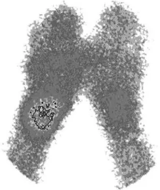
formed as additional tests. A posteroanterior radi− ograph of both hands was taken for each patient. The injuries were classified into fractures and soft− tissue injuries. The patients had neurological and psychiatric consultation and densitometry was per− formed in single cases. X−ray images are present− ed in Figure 1 and a scintigram in Figure 2.
Results
The types of injury or surgery which led to CRPS I development are collected in Figure 3. Patients with distal radius fractures treated conserv− atively definitely constituted the largest group (67%). The fact that the reported histories showed that the patients had had fractures repositioned under local anesthesia, often several times, the plaster dressing was too tight, and an extremity was often immobilized too long attracts attention (Table 2).
Table. 1.The diagnosis of CRPS I [12]
Tabela 1. Kryteria diagnostyczne CRPS I
1 Four of the five following signs and symptoms were present: pain, altered skin color, altered skin temperature, edema, reduced range of motion.
2 The symptoms were present in an area much larger than and also distal to the primary injury. 3 The symptoms were aggravated by physical activity of the affected extremity.
Fig. 1.Patient 54 years old, X−ray image: features of bone atrophy
Ryc. 1. Chory lat 54, RTG – cechy zaniku kostnego
Fig. 2. Patient 68 years old, scintigram: increased tracer uptake
Ryc. 2. Chory lat 68, badanie scyntygraficzne – wzmożony wychwyt znacznika
Fig. 3.Types of injuries and operations prior to CRPS I
Ryc. 3. Rodzaj urazu lub operacji przed rozwinięciem CRPS I
3%
67% 8%
11%
3% 8%
contusion of upper limb
fracture of distal radius
surgery for Dupuytren’s contracture
contusion of the wrist
severe hand injury
carpal Tunnel Syndrome
Table 2.CRPS risk factors
Tabela 2. Czynniki ryzyka zespołu CRPS
Several repositions 14
(Powtarzane repozycje)
Fracture immobilization in local anesthesia 14 (Nastawianie złamań w znieczuleniu
miejscowym)
Plaster too tight 15
(Za ciasne unieruchomienie)
Immobilization too long 9
(Zbyt długie unieruchomienie)
Neuro−psychic disorders 6
(Zaburzenia nerwowo−psychiczne)
Without reason 10
Pain was the basic reason for patient admis− sion. The patients were mainly in the second, dys− trophic period of disease. The percentages of occurrence of the basic symptoms and changes in X−rays and scintigrams are collected in Table 3. The sensitivity of scintigraphy was 85%; the specificity could not be determined without a con− trol group. It can be seen that only complex analy− sis, comparison of clinical images, and additional tests allows a diagnosis of CRPS.
Discussion
Soucacos reports that the primary etiological factors of CRPS I are trauma (distal radius frac− ture) (57.4%), followed by surgery (22.9%) and overuse syndromes (19.7%) [13]. Żyluk [24] writes about trauma as a reason in 68%, surgery in 14%, and others in 18%. In the present study the results are similar, the percentage of patients who had had fractures being 67%.
From the surgeon’s point of view there is the essential question whether the way of dressing a trauma influences the development of CRPS. It was documented that immobilization alone can cause all signs of CRPS [14]. Similar signs were noted in some posttraumatic conditions with spon− taneous remission [15]. Surgery on an extremity affected by CRPS is generally avoided. [16]. The present authors believe that an incorrect immobi− lizing dressing and immobilization for too long
cause venostasis on the periphery, pain, and a local inflammatory state which leads to a limitation of bone tissue rebuilding. Janig [17] reports that according to the peripheral inflammatory hypothe− sis, vasodilation might potentially be linked to the peptidergic primary afferent neurons with C−fibers and therefore to the neurogenic inflammatory component. Activation of peptidergic primary afferent neurons leads to pre−capillary vasodilation mediated by the release of calcitonin gene−related peptide and substance P and post−capillary plasma extravasation by release of substance P [17].
Repositioning a fracture with or without local anesthesia causing additional stress, pain, vaso− constriction, and worse blood flow of bone ele− ments is another factor which can lead to CRPS I. The present authors believe that this procedure can cause strong pain and limit rehabilitation. These complaints appearing in predisposed people can cause CRPS. Janig and Baron [18] report that according to the central hypothesis, the acute vasodilation might be due to inhibition of activity in cutaneous vasoconstrictor neurons, the vaso− constriction being possibly dependent on decen− tralized hypersensitivity of cutaneous blood ves− sels to impulses in cutaneous vasoconstrictor neu− rons [18].
An essential question is if the disease is pre− ventable. In a randomized, placebo−controlled study, prevention of CRPS I was shown in patients with Colles’ fractures by oral administration of vitamin C [19]. In this study the incidence of CRPS I in the control group was 22% versus 7% in the study group. Goris [4] writes that in patients with CRPS of an extremity, a new injury or opera− tion to that extremity is known to increase the chances of recurrence. The risk of developing CRPS I in the injured extremity, if not yet affected by CRPS I, is also increased [20]. In such cases it is recommended to avoid the use of a bloodless field if an operation is necessary because of the ischemia/reperfusion phenomenon and to pre− scribe prevention with scavengers or, possibly, vit− amin C for two or three days. Birklein [21] writes that the presence of patients with family histories of CRPS should trigger research on this. If one could recognize which patients are prone to devel− op CRPS, preventive treatment could be intensi− fied after trauma. Most of the patients in the pre− sent study were initially treated outside the clinic, but the authors believe that the administration of vit− amin C as a prevention of CRPS should be recom− mended particularly in patients having risk factors.
The diagnostics of CRPS is another difficult question. The present authors agree with Dommerholt [7] that there are no specific diagnos− tic tests for CRPS. The diagnosis is based on his−
Table 3.Frequency of occurrence of particular symptoms in clinical test and additional tests
Tabela 3.Częstość występowania poszczególnych objawów w badaniu klinicznym i badaniach dodatkowych
Type of symptom/additional % of patients pre−
test senting the symptom
(Objaw/badanie dodatkowe) (% pacjentów wyka− zujących objaw)
Pain 93
(Ból)
Swelling 71
(Obrzęk)
Altered skin temperature 70 (Podwyższona temperatura skóry)
Discoloration 40
(Zaburzenia zabarwienia skóry) Reduced range of motion 90 (Ograniczenie ruchomości)
X−ray 45
(Zdjęcie RTG)
Scintigraphy 85
tory and clinical examination and is not deter− mined by the results. The diagnosis is made by excluding other diagnoses that could account for the pain and other symptoms. Besides the several technical diagnostic tools that can support the diagnosis, CRPS cannot be proven by any diag− nostic measure. Birklein [21] writes that three− phase bone scintigraphy is an important diagnostic tool, although its sensitivity might be lower than has been assumed. Increased tracer uptake in the late pictures, the “mineralization phase”, is a sign of increased bone metabolism. MRI is necessary to exclude other diseases. In CRPS, edema can be seen in deep tissues and periarticularly [22]. On the other hand, Lee stated that three−phase bone scans are not reliable and can actually be confus− ing [23].
Żyluk [24] reports that the type of condition that preceded CRPS I appeared to have a signifi− cant effect on the scintigraphic image. These find− ings suggest that the fracture itself causes a signif− icant increase in the blood flow and increased fix− ation of the tracer, both in bone and in soft tissue. Thus there is the possibility of false negative inter− pretations of the scintigraphic images in patients with CRPS following soft−tissue injury, whereas false positive readings may occur in asymptomatic patients after fractures, particularly of the distal radius [24].
In the material of the present study, one−phase scintigraphy and X−rays were performed in the belief that clinical examination is basic to recog−
nizing CRPS I and scintigraphy is an additional test. The efficiency of the remaining methods is, according to literature, very differentiated. For example, Żyluk reports that on side−comparing X− rays, typically spotty osteoporosis changes can be found after 4–8 weeks and it was found that they only occur in 40% of cases [20]. In the present study this was in 50%. Driessens recommended using three−phase bone scintigraphy to differenti− ate CRPS from a pseudo−dystrophy conversion disorder based on the assumption that in CRPS the bone scan would show typical increased tracer uptake, whereas in pseudo−dystrophy there would be normal or decreased tracer uptake in the affect− ed region [25]. In the present material, scintigra− phy showed positive result in 80%, but it was not crucial because clinical symptoms of CRPS I were observed without scintigraphic changes. It should be also emphasized that increased tracer uptake is specific not only to CRPS I and it can give false positive results.
Stanton−Hinks [26] emphasize that there is no one efficient test for CRPS I and reports that we need a reliable test in the diagnosis of CRPS to discriminate between sympathetically maintained and sympathetically independent pain, and this is not, unfortunately, ensured by scintigraphy.
The authors conclude that distal radius frac− ture is the main reason of CRPS I and scintigraphy is a useful but not a determining test for the diag− nosis of CRPS I.
References
[1] Harden R:A clinical approach to complex regional pain syndrome. Clin J Pain 2000, 16, 2, 26–32.
[2] Janig W, Stanton−Hicks M:Reflex Sympathetic Dystrophy: A Reappraisal. Progress Pain Res Manag. IASP Press, Seattle.
[3] Atkins R, Duckworth T, Kanis J: Features of algodystrophy after Colles’ fracture. J Bone Joint Surg Br 1990, 72, 105–110.
[4] Goris R, van der Laan L:Reflex Sympathetic Dystrophy – Another View. Eur J Trauma 2001, 3, 99–103.
[5] Veldman P, Reynen H, Arntz I, Goris R: Signs and symptoms of reflex sympathetic: prospective study of 829 patients. Lancet 1993, 342, 1012–1016.
[6] Sudeck P: Über die akute entzündliche Knochenatrophie. Archiv Klin Chir 1900, 342, 1012–1016.
[7] Dommerholt J: Complex regional pain syndrome−1: history, diagnostic criteria and etiology. J Bodywork Mov Ther 2004, 8, 167–177.
[8] Ludwig J, Baron R: Complex regional pain syndrome: an inflammatory pain condition? Drug Discov Today: Disease Mechanism 2004, 4, 449–455.
[9] Kemler M, van de Vusse A, Berg−Loonen E, Barendse G, van Kleef M, Weber W:HLA−DQ1 associated with reflex sympathetic dystrophy. Neurology 1999, 53, 1350–1351.
[10] Cepeda M, Lau J, Carr D: Defining the therapeutic role of local anesthetic sympathetic blockade in complex regional pain syndrome: a narrative and systematic review. Clin J Pain 2002, 18, 4, 216–233.
[11] Stanton−Hicks M:Complex regional pain syndrome (type I, RSD; type II, causalgia): controversies. Clin J Pain 2000, 16, 2, S33–40.
[12] Veldman P, Goris R:Multiple reflex sympathetic dystrophy. Which patients are at risk of developing a recurrence of reflex sympathetic in the same or another limb. Pain 1996, 64, 463–466.
[13] Soucacos P, Diznitsas L, Beris A, Xenakis T, Malizos N: Reflex sympathetic dystrophy of the upper extremity. Clinical features and response to multimodal management. Hand Clinics 1997, 13, 339–354.
[15] Soucacos P, Johnson E:Upper extremity reflex sympathetic dystrophy. Curr Orthop 2001, 14, 356–364.
[16] Margić K, Pirc J:The treatment of complex regional pain syndrome (CRPS) involving upper extremity with con− tinuous sensory analgesia. Eur J Pain 2003, 7, 43–47.
[17] Janig W, Baron R: Experimental approach to CRPS. Pain 2004, 108, 307.
[18] Janig W, Baron R: Complex regional pain syndrome is a disease of the central nervous system. Clin Autonomic Res 2002, 12, 3, 150–164.
[19] Zollinger P, Tuinebreijer W, Kreis R, Breederveld R:Effect of vitamin C on frequency of reflex sympathetic dystrophy in wrist fractures: a randomised trial. Lancet 1998, 3544, 2025–2028.
[20] Puchalski P, Żyluk A: Complex regional pain syndrome type I after fractures of the distal radius: a prospective study of the role of psychological factors. J Hand Surg 2005, 30B, 6, 574–580.
[21] Birklein F: Complex regional pain syndrome. J Neurol 2005, 252, 131–138.
[22] Borre G, Borre D, Hofer B, Vogeli E: Sudeck’s dystrophy of the hand MR imaging. Clin Imaging 1995, 19, 188–192.
[23] Lee K, Kirchner J: Complex regional pain syndrome and chronic pain management in the lower extremity. Foot and Ankle Clin 2002, 7, 2, 409–419.
[24] Żyluk A:The usefulness of quantitative evaluation of three−phase scintigraphy in the diagnosis of post−traumatic reflex sympathetic dystrophy. J Hand Surg 1999, 24B, 1, 16–21.
[25] Driessens M, Block P, Geuens G:Pseudodystrophy A conversion disorder mimicking reflex sympathetic dys− trophy. Acta Orthop Belg 2002, 68, 4, 330–336.
[26] Stanton−Hicks M:Reflex sympathetic dystrophy: a sympathetically mediated pain syndrome or not? Curr Rev Pain 2000, 4, 4, 268–275.
Address for correspondence:
Paweł Reichert
Chair and Clinic of Traumatology and Hand Surgery Silesian Piasts University of Medicine
R. Traugutta 57/59 50−417 Wrocław Poland
E−mail: [email protected]
Conflict of interest: None declared