Bartosz Puła
1, A D–F,
Marta Strutyńska-Karpińska
2, A–D, F,
Alicja Markowska-Woyciechowska
3, A, B, F, Aleksandra Jethon
1, B, F,
Dariusz Wołowiec
4, A, F, Janusz Ryś
5, E, F, Marzena Podhorska-Okołów
1, E, F,
Krzysztof Grabowski
2, E, F,
Piotr Dzięgiel
1, 6, A–FExpression of Metallothionein and Ki-67 Antigen
in GISTs of Different Grade of Malignancy*
Ekspresja metalotioneiny oraz antygenu Ki-67
w guzach stromalnych o zróżnicowanym stopniu złośliwości
1 Department of Histology and Embryology, Wroclaw Medical University, Wrocław, Poland
2 Department of Gastrointestinal and General Surgery, Wroclaw Medical University, Wrocław, Poland 3 Department of Pathomorphology, Wroclaw Medical University, Wrocław, Poland
4 Clinic of Hematology, Blood Neoplasms and Bone Marrow Transplantation, Wroclaw Medical University,
Wrocław, Poland
5 Department of Tumor Pathology, Maria Sklodowska-Curie Memorial Cancer Center and Institute of Oncology,
Kraków, Poland
6 Department of Physiotherapy, Wroclaw University School of Physical Education, Wrocław, Poland
A – research concept and design; B – collection and/or assembly of data; C – data analysis and interpretation;
D – writing the article; E – critical revision of the article; F – final approval of article; G – other
Abstract
Background. Metallothioneins (MTs) are low molecular weight proteins (6–7 kDa), which have been shown to regulate zinc ion homeostasis. MTs exert anti-apoptotic and pro-proliferative effect on cancer cells. Overexpression of MT-I and MT-II isoforms has been noted in many malignant tumors, but the role of their expression in gastro-intestinal stromal tumors (GISTs) remains unclear.
Objectives. The aim of the study was to examine the relationship between expression of MT-I/II and K-67 prolif-eration antigen in a subset of GISTs presenting differential grade of malignancy.
Material and Methods. The study was conducted using immunohistochemical methods on archival paraffin sec-tions of 34 cases of GISTs. Of those, 17 tumors were classified as benign (GISTB) and 17 tumors as malignant (GISTM).
Results. The GISTM cases demonstrated higher MT-I/II expression as compared to the GISTB cases, but not sig-nificantly higher (p = 0.08). The GISTM tumors showed sigsig-nificantly higher expression of Ki-67 antigen than the GISTB cases (p = 0.01). MT-I/II and Ki-67 expression positively correlated in GISTBs (r = 0.48, p = 0.0463), but not in GISTMs.
Conclusion. The results of this study may point to a potential role of MT-I/II in the proliferation of GIST cells and disease progression (Adv Clin Exp Med 2013, 22, 4, 513–518).
Key words: GIST, metallothionein, stromal tumors.
Streszczenie
Wprowadzenie. Metalotioneiny (MT) są niskocząsteczkowymi białkami (6–7 kDa) biorącymi udział w regulacji homeostazy jonów cynku. MT wykazują działanie antyapoptotyczne oraz proproliferacyjne w komórkach nowo-tworowych. Nadekspresja izoform MT-I oraz MT-II została zaobserwowana w wielu nowotworach złośliwych, rola ich ekspresji w guzach stromalnych (GISTs) przewodu pokarmowego pozostaje jednak niejasna.
Adv Clin Exp Med 2013, 22, 4, 513–518 ISSN 1899–5276
ORIGINAl PAPERS
© Copyright by Wroclaw Medical University
Gastrointestinal stromal tumors (GISTs) are a group of mesenchymal tumors originating from precursors of Cajal cells in the gastrointestinal (GI) tract [1]. These tumors are characterized by over-expression of the c-kit gene and activation of KIT- -dependent pathways, which results in augment-ed proliferation of the tumor cells [2]. Although GISTs can occur anywhere in the GI tract, they are mainly seen in the stomach (70%) or small intes-tine (20–25%), rarely in the ileum (5%) or esopha-gus (5%) and occasionally in the abdominal cavity outside the GI tract [2–4].
GISTs vary in malignancy. Their aggressive-ness can be assessed on the basis of Fletcher’s crite-ria [5], and encompasses two parameters: the pri-mary tumor size and the number of mitotic figures observed in 50 high power fields (HPFs). Accord-ing to these criteria, tumors with a size of more than 5 cm and 5 mitoses/50HPF can be regarded as GISTs with clinically aggressive and malignant be-havior [5]. In addition, the expression of prolifer-ation markers such as Ki-67 and PCNA may be of potential value in determining the clinical aggres-siveness of GISTs [3, 6].
Metallothioneins (MTs) are intracellular, low molecular weight proteins (6–7 kDa) character-ized by a high cysteine content allowing them to bind heavy metal ions such as zinc, copper, cad-mium, lead and mercury [7]. The MT family is comprised of four main isoforms: MT-I, MT-II, III and IV, of which the I and MT-II isoforms are expressed in normal and neoplas-tic cells [8, 9]. MT-I/II are responsible for zinc and copper homeostasis, depending on the cell type. MT-I/II possess antioxidant properties, acting as scavengers of reactive oxygen species [10, 11]. Due to their properties, MT-I/II also exert anti-apop-totic and pro-proliferative effects on cancer cells in several tumor types [8, 12–17]. MT-I/II have been shown to mediate resistance to certain chemother-apeutic agents (by inactivating free radicals formed due to the metabolism of certain cytostatic drugs, e.g. anthracyclins, and binding to others, such as cisplatin) [18, 19]. The prognostic significance of
MT-I/II expression has therefore been intensively studied in numerous studies performed on other tumors, but only few analyzed the role of MTs in GISTs [8, 12, 14, 20–27].
The aim of this work was to analyze the inten-sity of MT-I/II expression in 34 GISTs of varying grade of malignancy and to correlate MT-I/II ex-pression with Ki-67 antigen exex-pression.
Material and Methods
The Patients
Archival paraffin blocks containing GISTs originating from 34 patients (19 men, 15 wom-en) operated on in the Department of Gastroin-testinal and General Surgery of Wroclaw Medical University were used in the study. The mean age of the patients was 63.8 (26–95) years. According to the criteria introduced by Miettinen and Fletch-er, the GISTs were divided into two groups: 17 benign GISTs (GISTB) and 17 malignant GISTs (GISTM) [2, 5]. All of the analyzed tumors were c-kit (CD117) positive. The primary tumor sites are shown in Table 1.
Immunohistochemistry (IHC)
The tumor samples were fixed in 10% buff-ered formalin, dehydrated and embedded in par-affin blocks. All the IHC studies were performed in 4-μm-thick paraffin sections as described [23, 24]. First, the sections were deparaffinized in xy-lene and rehydrated. For the Ki-67 IHC, the sec-tions were pre-incubated in a citrate buffer (pH 6, 10 mM) at a temperature of 95–98οC for 20
min-utes in order to reveal the epitopes. Incubation with 3% H2O2 (5 minutes) was used to block the
endogenous peroxidase. Then the sections were incubated with monoclonal antibodies direct-ed against MT-I/II (clone E9) and Ki-67 antigen (clone MIB-1) obtained from DakoCytomation (Glostrup, Denmark). The antigens were visualized
Cel pracy. Zbadanie związku między ekspresją MT-I/II oraz antygenu proliferacyjnego Ki-67 w grupie GISTs cha-rakteryzujących się zróżnicowanym stopniem złośliwości.
Materiał i metody. Badanie zostało przeprowadzone z użyciem metody immunohistochemicznej na archiwalnych skrawkach parafinowych 34 przypadków GISTs. Spośród nich 17 guzów zostało sklasyfikowanych jako łagodne (GISTB), a 17 guzów jako złośliwe (GISTM).
Wyniki. Przypadki GISTM charakteryzowały się wyższą, ale nie istotną statystycznie, ekspresją MT-I/II w porów-naniu z przypadkami GISTB (p = 0,08). Guzy GISTM wykazywały ponadto istotnie statystycznie wyższą ekspresję antygenu Ki-67 w porównaniu z guzami GISTB (p = 0,01). Zaobserwowano dodatnią korelację ekspresji MT-I/II oraz antygenu Ki-67 w przypadkach GISTB, ale nie w przypadkach GISTM.
Wnioski. Uzyskane wyniki badań mogą wskazywać na potencjalną rolę MT-I/II w procesach proliferacji GIST oraz progresji choroby (Adv Clin Exp Med 2013, 22, 4, 513–518).
using biotinylated antibodies and streptavidin conjugated with horseradish peroxidase (Dako-Cytomation lSAB+ System-HRP). Diaminoben-zidine (DAB, DakoCytomation) was used as the substrate. All the reactions were conducted using negative controls and all the slides were counter-stained with Mayer’s hematoxylin.
Evaluation of IHC Reactions
Both antigens were evaluated with previous-ly used assessment scales [22–24]. The evaluation of the MT-I/II reaction intensity was conduct-ed using Remmele’s semi-quantitative IRS meth-od [28], which is based on the intensity of the color reaction and the percentage of positive neoplastic cells in the preparation (Table 2). The scale ranges from 0–12 points (pts) and is summarized in Ta-ble 2. Ki-67 antigen expression in neoplastic cells was evaluated semiquantitatively in the whole tis-sue section, according to the percentage of posi-tive tumor cells.
Statistical Analysis
Prism 5.0 software (GraphPad, la Jolla, CA, USA) was used to analyze the data. Correlations between expression of MT-I/II, Ki-67 and the pa-tient’s age were analyzed using the Spearman cor-relation test. The Mann Whitney U-test was used to compare the intensity of MT-I/II and Ki-67 ex-pression between the GISTB and GISTM samples. The Kruskall-Wallis test was used to assess the re-lationship of the analyzed markers in relation to tumor location. The results were considered statis-tically significant at p < 0.05 in all the analyses.
Results
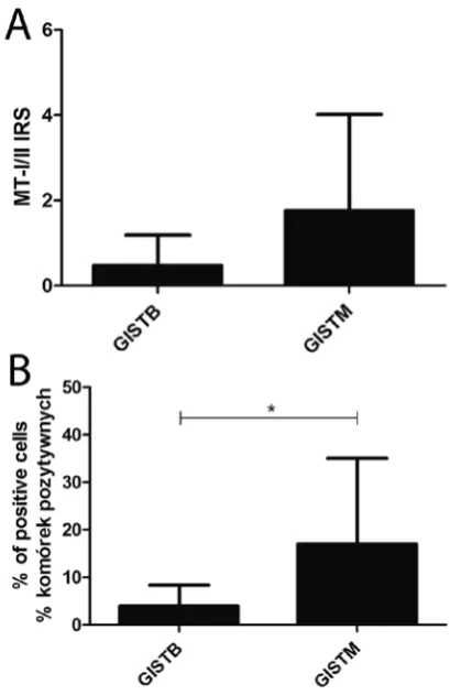
Cytoplasmic-nuclear MT-I/II reaction was noted in 16 out of the 34 analyzed cases (47.1%). MT-I/II was noted in 6/17 (35.3%) of the GISTBs and 10/17 (58.8%) of the GISTMs (Fig. 1A). The Ki-67 antigen was expressed in all the studied sam-ples (Fig. 1B). Significantly higher Ki-67 expression (p = 0.01) was noted in the GISTMs (17.0 ± 18.0%)
Table 1. localization of the GISTs in the analyzed material
Tabela 1. Umiejscowienie GIST w analizowanym materiale
localization/ /Number (Umiejscowienie/ /liczba)
Esophagus
(Przełyk) Stomach (Żołądek) Duodenum (Dwunast-nica)
Jejunum (Jelito czcze)
Ileum (Jelito kręte)
Rectum (Odbyt-nica)
Mesen-terium (Krezka)
Retroperitonal space (Przestrzeń zaotrzewnowa)
GIST 1 21 2 4 2 1 2 1
GISTB 1 13 2 1 0 0 0 0
GISTM 0 8 0 3 2 1 2 1
Table 2. Immunoreactive Scale (IRS): The percentage of positive cells (A) and the intensity of color reaction (B). The final score represents the product of these parameters (A × B)
Tabela 2. Skala IRS: procent komórek pozytywnych (A) oraz intensywność reakcji barwnej (B). Ostateczny wynik jest iloczy-nem tych parametrów (A × B)
A B
0 pts – no cells with positive reaction
0 pkt – brak komórek pozytywnych 0 pts – no staining0 pkt – brak reakcji 1 pt – to 10% cells with positive reaction
1 pkt – do 10% komórek pozytywnych 1 pt – low intensity of staining1 pkt – słaba intensywność reakcji 2 pts – 11–50% cells with positive reaction
2 pkt – 11–50% komórek pozytywnych 2 pts – moderate intensity of staining2 pkt – średnia intensywność reakcji 3 pts – 51–80% cells with positive reaction
3 pkt – 51–80% komórek pozytywnych 3 pts – high intensity of staining3 pkt – silna intensywność reakcji 4 pts – > 80% cells with positive reaction
as compared to the GISTBs (4.0 ± 4.4%). The in-tensity of MT-I/II expression, evaluated using the IRS method, demonstrated its higher expres-sion in the GISTMs (IRS 1.77 ± 2.25) as com-pared to the GISTBs (IRS 0.47 ± 0.72), but the dif-ference was not statistically significant (p = 0.08) (Fig. 2). A moderate correlation was noted be-tween MT-I/II and Ki-67 expression in the GISTBs (r = 0.48, p = 0.0463), but not in the GISTMs. No relationships were found between the intensity of expression of the analyzed markers and the pa-tients’ age, sex and tumor location.
Discussion
In several studies MTs have been shown to ex-ert anti-apoptotic and anti-oxidative propex-erties and stimulate cancer cell proliferation and metastasis in some cancer types [8–10, 13, 25]. Moreover, in some tumor types, overexpression of MT was asso-ciated with cancer cells’ resistance to cisplatin and predicted a poor outcome [18, 19]. Although in-tensive studies have been performed investigating
MT expression in different cancer types, little is known about the role of MT in GISTs [26, 27].
In studies concerning MT expression in GISTs, different assessment scales have been uti-lized to determine its expression, so the number of MT positive cases throughout the studies varied from 14.1% to 100% [26, 27]. In the current study elevated intensity of MT expression was noted in GISTM cases as compared to GISTBs. Although the differences were not significant, the results were at the threshold of significance. In a recent report by Perez-Gutierrez et al., no differences in expression of MT were shown between the low-risk and high-low-risk GISTs, but the authors took into consideration only the percentage of positive cells, while the intensity of the color reaction was omit-ted [26]. Even though the survival of the patients was not considered in the present study, the results may concur with the data of Perez-Gutierrez et al., which showed that only overexpression of MTs, but not P-glycoprotein expression, was signifi-cantly associated with poor survival rates [26]. In addition to P-glycoprotein, MT-I/II mediate che-moresitance to anthracyclines and cisplatin and
Fig. 1. Nuclear-cytoplasmic MT-I/II (A) and nuclear Ki-67 antigen expression in GISTs
Ryc. 1. Jądrowo-cytoplazmatyczna ekspresja MT-I/II (A) oraz jądrowa ekspresja antygenu Ki-67 (B) w GIST
Fig. 2. MT-I/II (A) and Ki-67 antigen (B) expression in GISTBs and GISTMs, *p < 0.05
exert anti-apoptotic and pro-proliferative proper-ties in cancer cells [8, 12–19]. That might explain why MT-I/II expression, but not P-glycoprotein expression, is a marker of poor survival rates for GIST patients. Similarly, no associations were not-ed between MT expression and the patients’ age, sex and primary tumor location.
MT overexpression has been shown to corre-late with cancer cell proliferation in several tumor types [20, 23–25, 29]. In the present study, Ki-67 antigen expression in the GISTM group was signif-icantly elevated as compared to the GISTB group. These results are in agreement with the data ob-tained by other authors, which showed that Ki-67 expression was elevated in high-risk GISTs and is associated with a poor outcome in some stud-ies [30–32]. Interestingly, MT expression corre-lated positively with Ki-67 antigen expression on-ly in the GISTBs, but not in the GISTMs, which may suggest the involvement of different path-ways in regulating cancer cell proliferation in these
two tumor groups with regard to their potential malignancy.
Soo et al. showed that MT-I/II protein ex-pression levels are lower in GISTs as compared to gastric cancer [27]. In addition, expression of the MT-2A isoform was significantly lower in GISTs than in gastric cancer [27]. In the present authors’ opinion it is difficult to compare MT expression in these two tumor types, due to their differential histogenesis [1–3, 27]. In addition, the results ob-tained by Soo et al. introduced little information concerning the role of MT in the pathogenesis of GISTs, as no comparisons with the patients’ clini-cal data were carried out [27].
In summary, MTs may be of potential interest in studies of GISTs because of its differential ex-pression in GISTB and GISTM cases. The results of the present study indicate that MT may be an additional marker of GIST malignancy, but further studies are required to analyze the utility of MT as a new potential marker in the prognosis of GISTs.
References
[1] Kindblom LG, Remotti HE, Aldenborg F, Meis-Kindblom JM: Gastrointestinal pacemaker cell tumor (GIPACT): gastrointestinal stromal tumors show phenotypic characteristics of the interstitial cells of Cajal. Am J Pathol 1998, 152, 1259–1269.
[2] Miettinen M, Lasota J: Gastrointestinal stromal tumors – definition, clinical, histological, immunohistochemical, and molecular genetic features and differential diagnosis. Virchows Arch 2001, 438, 1–12.
[3] Miettinen M, Lasota J: Gastrointestinal stromal tumors (GISTs): definition, occurrence, pathology, differential diagnosis and molecular genetics. Pol J Pathol 2000, 54, 3–24.
[4] Strutyńska-Karpińska M, Nienartowicz M, Markowska-Woyciechowska A, Rabczyński J: Analiza kliniczna i immunohistochemiczna 8 guzów stromalnych przewodu pokarmowego. Adv Clin Exp Med 2005, 14, 465–471.
[5] Fletcher CD, Berman JJ, Corless C, Gorstein F, Lasota J, Longley BJ, Miettinen M, O’Leary TJ, Remotti H, Rubin BP, Shmookler B, Sobin LH, Weiss SW: Diagnosis of gastrointestinal stromal tumors: A consensus approach. Hum Pathol 2002, 33, 459–465.
[6] Hasegawa T, Matsumo Y, Schimoda T, Hirohashi S: Gastrointestinal stromal tumor: consistent CD117 immu-nostaining for diagnosis and prognostic classification based on tumor size and MIB-1 grade. Hum Pathol 2002, 33, 669–676.
[7] Coyle P, Philcox JC, Carey LC, Rofe AM: Metallothionein: The multipurpose protein. Cell Mol life Sci 2002, 59, 627–647.
[8] Dziegiel P: Expression of metallothioneins in tumor cells. Pol J Pathol 2004, 55, 3–12.
[9] Dziegiel P, Suder E, Surowiak P, Kornafel J, Zabel M: Expression of metallothionein in synovial sarcoma cells. Appl Immunohistochem Mol Morphol 2002, 10, 357–362.
[10] De Lisle RC, Sarras MP Jr, Hidalgo J, Andrews GK: Metallothionein is component of exocrine pancreas secre-tion: implications for zinc homeostasis. Am J Physiol 1996, 271, 1103–1110.
[11] Gwinner W, Grone HJ: Role of reactive oxygen species in gromerolonephritis. Nephrol Dial Transpl 2000, 15, 1127–1132.
[12] Santon A Albergoni V, Sturniolo GC, Irato P: Evaluation of MT expression and detection of apoptotic cells in lEC rat kidney. Biochim Biophys Acta 2000, 1668, 223–231.
[13] Cherian MG, Jayasuriya A, Bay BH: Metallothioneins in human tumors and potential roles in carcinogenesis. Mutat Res 2003, 533, 201–209.
[14] Janssen AM, van Duijn W, Kubben FJ, Griffioen G, Lamers CB, van Krieken JH, van de Velde CJ, Verspaget HW:
Prognostic significance of metallothionein in human gastrointestinal cancer. Clin Cancer Res 2002, 8,1889–1896.
[15] Jin R, Chow VT, Tan PH, Dheen ST, Duan W, Bay BH: Metallothionein 2A expression is associated with cell proliferation in breast cancer. Carcinogenesis 2002, 23, 81–86.
[16] Ohshio G, Imamura T, Okada N, Wang ZH, Yamaki K, Kyogoku T, Suwa H, Yamabe H, Imamura M:
Immunohistochemical study of metallothionein in pancreatic carcinomas. J Cancer Res Clin Oncol 1996, 122, 351–355.
[18] Koropatnick J, Kloth DM, Kadhim S, Chin JL, Cherian MG: Metallothionein expression and resistance to cispla-tin in a human germ cell tumor cell line. J Pharmacol Exp Ther 1995, 275, 1681–1687.
[19] Surowiak P, Materna V, Maciejczyk A, Pudelko M, Markwitz E, Spaczyński M, Dietel M, Zabel M, Lage H:
Nuclear metallothionein expression correlates with cisplatin resistance of ovarian cancer cells and poor clinical outcome. Virchows Arch 2007, 450, 279–285.
[20] Nowak M, Madej JA, Dzięgiel P: Expression of metallothionein and its correlation with Ki-67 antigen in bitch’s mammary gland adenocarcinomas. Med Wet 2006, 62,427–431.
[21] Gomulkiewicz A, Podhorska-Okolow M, Szulc R, Smorag Z, Wojnar A, Zabel M, Dziegiel P: Correlation between metallothionein (MT) expression and selected prognostic factors in ductal breast cancers. Folia Histochem Cytobiol 2010, 48, 242–248.
[22] Królicka A, Kobierzycki C, Puła B, Podhorska-Okołów M, Piotrowska A, Rzeszutko M, Rzeszutko W, Rabczyński J, Domosławski P, Wojtczak B, Dawiskiba J, Dzięgiel P: Comparison of metallothionein (MT) and Ki-67 antigen expression in benign and malignant thyroid tumors. Anticancer Res 2010, 30, 4945–4949.
[23] Wojnar A, Pula B, Piotrowska A, Jethon A, Kujawa K, Kobierzycki C, Rys J, Podhorska-Okolow M, Dziegiel P:
Correlation of Intensity of MT-I/II Expression with Ki-67 and MCM-2 Proteins in Invasive Ductal Breast Carcinoma. Anticancer Res 2011, 31, 3027–3033.
[24] Werynska B, Pula B, Muszczynska-Bernhard B, Piotrowska A, Jethon A, Podhorska-Okolow M, Dziegiel P, Jankowska R: Correlation between Expression of Metallothionein and Expression of Ki-67 and MCM-2 Proliferation Markers in Non-Small Cell lung Cancer. Anticancer Res 2011, 31, 2833–2839.
[25] Pedersen MØ, Larsen A, Stoltenberg M, Penkowa M: The role of metallothionein in oncogenesis and cancer prognosis. Prog Histochem Cytochem 2009, 44, 29–64.
[26] Pérez-Gutiérrez S, González-Cámpora R, Amérigo-Navarro J, Beato-Moreno A, Sánchez-León M, Pareja Megía JM, Virizuela-Echaburu JA, López-Beltrán A: Expression of P-glycoprotein and metallothionein in gas-trointestinal stromal tumor and leiomyosarcomas. Clinical implications. Pathol Oncol Res 2007, 13, 203–208.
[27] Soo ET, Ng CT, Yip GW, Koo CY, Nga ME, Tan PH, Bay BH: Differential expression of metallothionein in gas-trointestinal stromal tumors and gastric carcinomas. Anat Rec (Hoboken) 2011, 294, 267–272.
[28] Remmele W, Stegner HE: Recommendation for uniform definition of an immunoreactive Score (IRS) for immu-nohistochemicalestrogen receptor detection (ER-ICA) in breast cancer. Pathologe 1987, 8, 138–140.
[29] Szelachowska J, Dziegiel P, Jelen-Krzeszewska J, Jelen M, Tarkowski R, Wlodarska I, Spytkowska B, Gisterek I, Matkowski R, Kornafel J: Prognostic significance of nuclear and cytoplasmic expression of metallothioneins as related to proliferative activity in squamous cell carcinomas of oral cavity. Histol Histopathol 2008, 23, 843–851.
[30] Vij M, Agrawal V, Kumar A, Pandey R: Gastrointestinal stromal tumors: a clinicopathological and immunohis-tochemical study of 121 cases. Indian J Gastroenterol 2010, 29, 231–236.
[31] Aoyagi K, Kouhuji K, Yano S, Miyagi M, Imaizumi T, Takeda J, Shirouzu K: Malignant potential of gastroin-testinal stromal tumor of the stomach. Int Surg 2009, 94, 1–9.
[32] Neves LR, Oshima CT, Artigiani-Neto R, Yanaguibashi G, Lourenço LG, Forones NM: Ki67 and p53 in gastro-intestinal stromal tumors – GIST. Arq Gastroenterol 2009, 46, 116–120.
Address for correspondence:
Piotr Dzięgiel
Department of Histology and Embryology Wroclaw Medical University
Chałubińskiego 6a 50-368 Wrocław Poland
Tel.: +48 71 784 13 54
E-mail: [email protected]
Conflict of interest: None declared