Olga Dzhura, Antonina Yashchenko, Vladimir Antonyuk, Alexander Lutsyk
Lectin Receptor Sites During Postnatal Osteogenesis
in Guinea Pigs
Receptory lektyn w postnatalnej osteogenezie świnek morskich
Department of Histology and Embryology, Lviv National Medical University, Lviv, UkraineAbstract
Background. Osteoporosis and its complications have become widespread, affecting large portions of the world’s population. More advanced information is needed on these pathologies to expand the possibilities for pathogenetic therapy.
Objectives. Lectin histochemistry methods offer new insights into the structure of tissue carbohydrates and their rearrangement under physiological and pathological conditions. The aim of the present investigation was to use a set of lectins with different carbohydrate affinities to study postnatal remodelling of cartilage and bone in relation to the age and sex of experimental animals.
Material and Methods. A panel of five conventional lectins – Con A, PNA, RCA, WGA, SNA, supplemented with an original fucose-specific lectin from Laburnum anagyroides bark (LABA) – was used to investigate the femoral bones of female and male guinea pigs aged 3 months, 1 year and 3 years. Tissue samples were fixed in 4% formaline, decalcified in 7% HNO3 and embedded in paraplast. The sections were subjected to a routine lectin-peroxidase-diaminobenzidine visualization technique.
Results. A pronounced labeling of growth plate chondromucoid was found with Con A, PNA and SNA. Articular cartilage showed much fainter labeling with these same lectins. Capsules of isogenic groups of chondrocytes expressed a strong affinity for SNA and WGA, encompassing a predominance of Neu5Ac/2-6Gal, DGlcNAc and NeuNAc determinants. Osseomucoid showed faint reactivity with all the lectins used, while SNA distinctly marked the line of ossification. Glycoconjugates within lacunas and osseous canaliculi, as well as cytoplasmic glycoconjugates of bone and cartilage cellular elements, expressed strong labeling with RCA and SNA. Osteoclasts reacted selectively with PNA. A comparison of lectin labeling between the experimental groups indicated that the tissue reactivity of males exceeded that of females, and that aging caused a decrease in the lectin reactivity of both genders.
Conclusions. The data extend current knowledge regarding the selective lectin labeling of osseous tissue constitu-ents, and demonstrate the applicability of lectin histochemistry methods in osteoporosis studies (Adv Clin Exp Med 2012, 21, 1, 19–26).
Key words: bone, cartilage, postnatal osteogenesis, sex-related dimorphism, lectin histochemistry.
Streszczenie
Wprowadzenie. Wzrost osteoporozy w aspekcie wiekowym stał się przyczyną przeprowadzenia bardziej dokład-nych badań patogenezy tej choroby wraz z rozszerzeniem zakresu metod badania tkanki kostnej.
Cel pracy. Przeprowadzenie eksperymentalnych badań ekspresji receptorów lektyn o różnej specyfice węglowoda-nowej w komponentach strukturalnych tkanki kostnej świnek morskich w aspekcie wiekowym, biorąc pod uwagę dymorfizm płciowy.
Materiał i metody. Zestaw, który stanowi 5 standardowych lektyn (Соn А, PNA, RCA, WGA, SNA), uzupełniony oryginalnym fukozo-specyficznym lektynowym preparatem kory Laburnum anagyroides (LAВA), został wykorzy-stany podczas przeprowadzenia badań ekspresji glikokoniugatów podczas postnatalnego remodelowania chrząstki i kości, uwzględniając płeć i wiek zwierząt doświadczalnych. Kości udowe świnek morskich, płci męskiej i żeńskiej, w wieku 3 miesięcy, 1 i 3 lat, odpowiednio zostały utrwalone w 4% neutralnej formalinie, odwapnione w 7% НNO3, a również zatopione w paraplaście. Skrawki zostały potraktowane koniugatami lektyn-peroksydazy chrzanowej, a wizualizacja receptorów została wykonana w systemie DAB – Н2О2.
Wyniki. Badania ujawniły znaczną ekspresję receptorów lektyny Соn А, PNA oraz SNA w składzie chondromukoidu nasadowych płytek wzrostu, w porównaniu ze słabszym wiązaniem tych lektyn z chrząstka powierzchni stawowej. Otoczki izogenicznych grup chondrocytów wykazywały dużą przynależność do lektyny SNA i WGA, co świadczy
Adv Clin Exp Med 2012, 21, 1, 19–26 ISSN 1899-5276
ORIGINAL PAPERS
o zwiększonej ekspresji w ich składzie Neu5Ac/2-6Gаl, DG1cNAc i NeuNAc determinantę. Oseomukoid wykazał słabą reaktywność ze wszystkimi wykorzystanymi lektynami, podczas gdy lektyna SNA konturowała linie kostnienia. Glikokoniugaty zatok i kanalików kostnych, jak również cytoplazmatyczne glikokoniugaty elementów komórkowych chrząstki oraz kości, demonstrowały znaczną przynależność do lektyny RCA i SNA. Osteoklasty sporadycznie reago-wały z lektyną PNA. Porównywanie wiązania lektyn między grupami zwierząt doświadczalnych ujawniło, że reaktyw-ność komponentów tkankowych samców była wyższa niż u samic, biorąc pod uwagę te same struktury. W starszych grupach wiekowych reaktywność tkanek u obu płci była ograniczona.
Wnioski. Otrzymane wyniki pozwalają poszerzyć wiadomości o możliwościach wykorzystania lektyn jako selek-tywnych markerów komórek oraz ich subpopulacji, jak również można je wykorzystać jako podstawę badań doty-czących patologicznych zmian węglowodanów w tkance kostnej (Adv Clin Exp Med 2012, 21, 1, 19–26).
Słowa kluczowe: kość, chrząstka, postnatalna osteogeneza, płciowo uwarunkowany dymorfizm, histochemia lektyn.
Current knowledge about chondrogenesis and osteogenesis during normal development as well as under different physiological and pathological con-ditions is far from complete. The frequency of skel-etal disorders, particularly osteoporosis, has been constantly increasing during past decades [1, 2], and various new approaches are being used for their in-vestigation and treatment. Among these are lectin histochemistry methods for detecting the carbohy-drate determinants of glycosaminoglycans, proteo-glycans, glycoproteins and glycolipids [3–5, 8].
Lectin labeling in situ and in vitro has provided new insights into the early stages of osteogenesis and revealed the important role of glycocojugates in the mechanisms of growth and metabolism in cartilage and bone [6, 7]. However, the current au-thors found no references in the available literature to the late stages of postnatal development, or to sex-related differences in glycoconjugate expres-sion during endochondral ossification. The goal of the present investigation was to study the redis-tribution of lectin receptor sites in the chondro-osseous junction at subsequent stages of postnatal development, particularly in relation to the sex of the experimental animals, and to compare these changes with parathormone levels and bone min-eralization profiles.
Material and Methods
Male and female guinea pigs aged 3 months, 1 year and 3 years were divided into six experimen-tal groups. Plasma parathormone levels were de-termined using an immunoradiological test system (BioSource hPTH-IRMA, Belgium). Serum levels of calcium, magnesium and phosphorus were de-tected with a standard kit (LaChema, Czech Re-public). The calcium and magnesium content in bone tissue were detected using atomic absorption spectrometry on an ААС-115 spectrometer (LO-MO, St Petersburg, Russia), following the standard protocol at wavelengths 422.7 nm for Ca and 285.2 nm for Mg. Phosphorus content was determined
using an SF-46 spectrophotometer (LOMO, St. Pe-tersburg, Russia) at 360–370 nm.
For the lectin histochemistry, femoral bones were removed after administering an ether anes-thesia overdose and decapitating the animals. Tis-sue samples were fixed in 4% neutral formaline for 24 hours; decalcified in several changes of 7% HNO3, using a preparatory needle to assess the de-gree of decalcification; incubated for 48 hours in a 5% solution of aluminum–potassium–phosphate salts; washed for 24 hours in tap water and embed-ded in paraplast. Sections 7 µm thick were labeled with lectin-peroxidase conjugates with subsequent visualization in 3,3’-diaminobenzidine tetrahydro-chloride-H2O2 as described in an earlier article [8].
A routine set of five lectins (Con A, PNA, RCA, WGA, SNA) was supplemented with an original fucose-specific lectin from Laburnum anagyroides
bark (LABA) [12] (Table 1). The lectins were puri-fied and labeled with horseradish peroxidase at the Lectinotest Laboratory (Lviv, Ukraine; http://www. meduniv.lviv.ua/depart/ lectinotest.html). In the control experiments, the lectins were omitted from the incubation medium. For general morphology, the sections were stained with hematoxylin and eosin. Morhological analysis was conducted with a Zeiss KM 470600-9901 microscope (Germany), and photographs were taken using an AVerMedia video system (Taiwan). The statistical analysis was performed using Microsft Office Excel 2003 soft-ware (USA).
Results
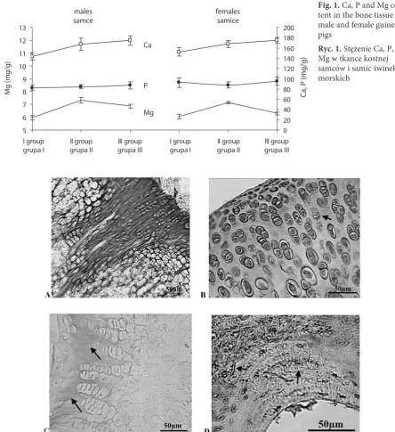
An age-dependent elevation of parathormone levels was observed in males, compared to a de-crease in the same parameter in females. In both sexes, aging caused an increase in Ca and P con-tent in bone tissue. In blood serum these indices remained at approximately same levels in males, and decreased slightly in females (Table 2, Fig. 1).
aged 1 year there were epiphyseal growth plates in the knee and hip-joint cartilage, whereas in females of the same age group, growth plates had disap-peared and the bones were completely ossified.
Intercellular spaces within the growth plates of the experimental animals expressed strong re-activity with Con A, PNA and WGA; moderate reactivity with SNA and RCA; and faint-to-neg-ative reactivity with LABA (Figures 2A and 2C). In comparison to the growth plates, the articular cartilage expressed much fainter labeling with the lectins (Tables 3 and 4).
Chondrocytes from different locations were la-beled with RCA and SNA, the lectins used to visu-alize different phases of the mitotic cycle (Fig. 2B).
Glycoconjugates in the perinuclear space of the developing chondrocytes expressed intense WGA affinity, whereas isogenic group capsules demon-strated strong reactivity with WGA and SNA (Fig-ures 2B and 3A).
Osseomucoid demonstrated a rather faint affin-ity for all the lectins used except for SNA, which dis-tinctly labeled the cement line (tidemark) (Fig. 3A). Islets of cartilage within the developing osseous tissue reacted to Con A and SNA; aging caused a decrease in the number of these islets.
Glycoconjugates within lacunas and osseous canaliculi, as well as cytoplasmic glycoconjugates of osteoblasts and osteocytes, reacted strongly to RCA and SNA (Figures 2D and 3B). Osteoclasts
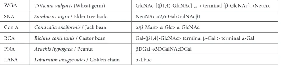
Table 1. Panel of lectins used, their sources and specificities
Tabela 1. Lista wykorzystanych lektyn, ich źródła i specyfika
WGA Triticum vulgaris (Wheat germ) GlcNAc-[(β1,4)-GlcNAc]1–2 > terminal [β-GlcNAc]n>NeuAc
SNA Sambucus nigra / Elder tree bark NeuNAc α2,6-Gal/GalNAcβ1 Con A Canavalia ensiformis / Jack bean α/β-Man> α-Glc> α-GlcNAc
RCA Ricinus communis / Castor bean Gal-(β1,4)-GlcNAc> terminal β-Gal > terminal α-Gal PNA Arachis hypogaea / Peanut βDGal →3DGalNAcDGal
LABA Laburnum anagyroides / Golden chain α-LFuc
Table 2. Concentration of parathormone in blood, Ca, P, Mg in blood and bone of male and female guinea pigs during postnatal life
Tabela 2. Stężenie parathormonu we krwi, Ca, P, Mg we krwi oraz w tkance kostnej samców i samic świnek morskich w postnatalnej osteogenezie
Groups,
n = 5 Biochemical investigation (Badanie biochemiczne) parathyroid
hormone, pg/ml M ± m
mineral components in blood serum,
mM/l mineral components in bone, mg/g total Ca
level, M ± m inorganic P, M ± m total Mg level, M ± m
Ca,
M ± m P, M ± m Мg, M ± m
Males
І 10.02 ± 0.21 2.32 ± 0.11 1.93 ± 0.09 0.96 ± 0.04 143.45 ± 7.31 82.45 ± 4.51 5.96 ± 0.17 ІІ 11.54 ± 0.47* 2.48 ± 0.16 1.89 ± 0.06 0.83 ± 0.05 167.2 ± 12.16 84.56 ± 3.70 7.31 ± 0.21** ІІІ 11.32 ± 0.54 2.43 ± 0.12 1.9 ± 0.11 0.95 ± 0.05 174.4 ± 9.25* 87.27 ± 7.14 6.87 ± 0.16*
Сr – 0.9825 –0.9928 – 0.9403 0.75 –
Females
I 11.06 ± 0.34 2.56 ± 0.12 2.02 ± 0.04 1.12 ± 0.07 152.25 ± 8.31 92.65 ± 9.32 6.05 ± 0.18 ІІ 10.81 ± 0.26 2.24 ± 0.17 2.04 ± 0.08 0.99 ± 0.03 167.8 ± 6.64 87.72 ±5.82 7.16 ± 0.29* ІІІ 9.94 ± 0.13* 2.16 ± 0.05* 1.95 ± 0.13 1.15 ± 0.02 175.05 ± 5.34 95.43 ± 7.91 6.30 ± 0.21
Сr – 0.774 0.9269 – –0.846 –0.659 –
were selectively labeled with PNA and SNA (Fig-ures 3C and 3D).
A comparison among the groups of animals revealed that the lectin reactivity of the males’ bone and cartilage exceeded that of the females, and that aging caused a reduction in lectin labeling in the tissues of both genders.
Discussion
A differential mode of parathormone secre-tion was detected, and reference levels of this hor-mone in male and female guinea pigs during their postnatal development were established. The main physiological action of parathormone is directed
5 6 7 8 9 10 11 12 13
I group
grupa I grupa IIII group grupa IIIIII group I groupgrupa I grupa IIII group grupa IIIIII group 0 20 40 60 80 100 120 140 160 180 200 males
samce femalessamice
M
g
(m
g/
g)
Ca
, P
(m
g/
g)
Ca
P
Mg
Fig. 1. Са, Р and Mg con-tent in the bone tissue of male and female guinea pigs
Ryc. 1. Stężenie Ca, P, Mg w tkance kostnej samców i samic świnek morskich
Fig. 2. Lectin labeling of the bone and cartilage of male guinea pigs aged 3 months. A) Expression of Con A receptor sites within the metaepiphyseal plate chondromucoid; B) Mitotic figures within isogenic cell groups of the articular cartilage revealed with SNA; C) Faint LABA reactivity of the metaepiphyseal growth plate; D) Expression of SNA receptor sites in bony lacunas and canaliculi during osteon formation
at stimulating the resorptive activity of osteoclasts, and at inhibiting the reabsorption of phosphate ions by nephrons. Both of these mechanisms cause an elevation of Ca levels in blood plasma [9].
In the current study, the data on plasma Ca and P levels in male and female guinea pigs corre-late positively with parathormone levels, whereas the intensity of these ions’ deposition in bone tis-sue is apparently influenced by other biologically active substances, i.e. estrogen and testosterone. The increased ossification rate of growth plates in
females in comparison with males is probably due to the stimulatory effect of somatotrophins and androgens on chondrocyte proliferation and the elongation of bones in males, and on the inhibi-tory effect of estrogens in females [9].
Large amounts of mannosyl and sialic acid residues were detected in the growth plates of all the investigated groups of animals. Cartilage os-sification inhibited lectin reactivity and decreased the carbohydrate content of the mineralized bone. These results are in good agreement with the
find-Table 3. Lectin receptor sites in bone tissue of male guinea pigs during postnatal life
Tabela 3. Receptory lektyn w tkance kostnej samców świnek morskich podczas postnatalnej ontogenezy Group
(Grupa) Elements of cartilage and bone (Tkanka chrzęstna i kostna) Lectins (Lektyny)
WGA LABA Con A PNA RCA SNA
I
Distant zone AC Chondromucoid AC Cells AC Epiphyseal plate – ZUC – ZCC – ZBC – ZC Periosteum Osseomucoid Osteocytes Osteoblasts Osteoclasts Endosteum ++ + + +/± ++ ++ ++ + ± ± + – + + – – – +/± + ±/– + – – – – + ++ + ± + +++ +++ ++ + ± ± ± – + ++ ++ ± ± +++ ++ ++ + ± ± ± ++ + + + + + ++ ++ + + + + + – + + + ++ ± +++ +++ ++ ++ ± ++ ± – + ІІ
Distant zone AC Chondromucoid AC Cells AC Epiphyseal plate – ZUC – ZCC – ZBC – ZC Periosteum Osseomucoid Osteocytes Osteoblasts Osteoclasts Endosteum + ± ± ± ++ ++/+ ++/+ + ± ± ± – + – – – – ± ± – + – – – – ± ± ±/+ ± + ++/+ ++/+ + + ± ± ± – + ± ± + – + + ± +++ ± ± ± – + + ± + ± ++ ++ + + ± + + – + ± ± ++ ± ++ ++ + ++ ± ++ + ++ + ІІІ
Distant zone AC Chondromucoid AC Cells AC Periosteum Osseomucoid Osteocytes Osteoblasts Osteoclasts Endosteum ± ± ± + – – – – + – – – ± – – – – – ± ± ± + ± ± ± – + ± ± + ++ ± ± ± + + ± ± + ++ ± ± ± – + ± ± ++ + ± + + – + AC – articular cartilage; ZUC – zone unaltered cartilage; ZCC – zone columnar cartilage; ZBC – zone bubbles cartilage; ZC – zone calcification. Reactivity: – unreactive, ± weak, + moderate, ++ strong, +++ intense or very strong.
ings of other researchers [10, 11], in spite of the use of different species, different fixatives and different lectin labeling protocols.
The strong Con A reactivity of developing car-tilage and bone may be due partly to high mannosyl residue content in type II collagen molecules in the cartilage, and partly to the presence (reported by other researchers) of D-mannopyranosyl, D-glu-copyranosyl and D-fructopyranosyl residues on the surface of osteoprogenitor cells [7, 12] or elevated expression of a-D-mannose-rich glycoproteins by apoptotic cells [13]. In this context mannoso-glycans apparently play an important role during cartilage growth and development. This notion is confirmed by the phenomenon of the stimulation of osteogenesis by subcutaneous injection of Con A [6]. It is also noteworthy that mannosoglycans (Con A receptor sites) are expressed in the early stages of chondrohistogenesis [14]; this reactivity decreases in the ossification centers [5, 15].
Sex-related differences in lectin binding were also observed: Con A, PNA and SNA labeling in the growth plate of males exceeded that of females, espe-cially when comparing 1-year-old animals. The
ex-pression of Con A receptor sites in the growth plates was higher than in the articular cartilage. Similar enhancement of Con A reactivity was described by Zschabitz et al. [12]in developing rat cartilage.
The large amounts of WGA and SNA receptor sites detected in the isogenic group capsules is in agreement with Cormack’s report of high glyco-conjugate content in these elements [16], and with the observations of Farnum and Wilsman, who described strong WGA binding to the intercellular septa in growth plates and pericellular glycocon-jugates around chondrocytes. The low content of fucosoglycans in cartilage and bone noted in the current study is in agreement with the observa-tions of Zschabitz et al. [12].
In the current investigation, osteoclasts were intensely labeled by PNA and SNA, this observa-tion is in agreement with other researchers’ re-ports of a high content of DGal(b1-3)DGlcNAc
determinants (receptor sites for Maclura pomifera
lectin) in these cells [10, 11], and with Howard and Batsakis’sobservations of PNA strong affin-ity for macrophages-histiocytes [17], which are closely related to osteoclasts.
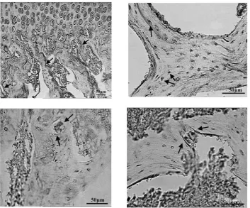
Fig. 3. A) Expression of SNA receptor sites in the chondro-osseous junction of a male guinea pig 3 months of age; B) RCA labeling of osteocytes within bony lacunas in a male guinea pig 3 years of age; C) PNA receptor sites within the cytoplasm of an osteoclast in Howship’s lacuna in a female guinea pig 3 years of age; D) SNA binding to osteo-clasts in a male guinea pig aged 1 year
In the current study a decrease was observed in the expression of carbohydrate determinants during postnatal development of cartilage and bone. In guinea pigs both tissues demonstrated high content of Con A and SNA receptor sites (mannosoglycans and sialoglucans), with faint LA-BA reactivity (low fucosyl content). The findings regarding the lectin reactivity of guinea pig carti-lage and bone are in agreement with the results of other researchers data on rat and human tissues,
Table 4. Lectin receptor sites in bone tissue of female guinea pigs during postnatal life
Tabela 4. Receptory lektyn w tkance kostnej samic świnek morskich podczas postnatalnej ontogenezy Group
(Grupa) Elements of cartilage and bone (Tkanka chrzęstna i kostna) Lectins (Lektyny)
WGA LABA Con A PNA RCA SNA
I
Distant zone AC Chondromucoid AC Cells AC Epiphyseal plate – ZUC – ZCC – ZBC – ZC Periosteum Osseomucoid Osteocytes Osteoblasts Osteoclasts Endosteum ++ ++ ± + ++ ++ ++ + ± ± ± – + – ± – – ± ± – + – – – – + ++ + ± + ++ ++ ++ + ± ± ± – + ++ ++ ± ± +++ ++/+ ++/+ + ± ± ± ++ + + + + ± ++ + + + + + + – + + + ++ ± + ++ + ++ ± ++ ± – + ІІ
Distant zone AC Chondromucoid AC Cells AC Periosteum Osseomucoid Osteocytes Osteoblasts Osteoclasts Endosteum + ±/+ ± + ± – – – + – – – + – – – – ± ± ± ± + ± ± ± – + ± ± + ++ ± ± ± – + + ± + ++ ± ± ± – + ± ± ++ ++ ± ++ + ++ + ІІІ
Distant zone AC Chondromucoid AC Cells AC Periosteum Osseomucoid Osteocytes Osteoblasts Osteoclasts Endosteum ± ± ± + – – – – + – – – ± – – – – ± ± ± ± + ± ± ± – ± ± ± ± ++ ± ± ± + + ± ± + ++ ± ± ± – + ± ± ++ + ±/+ + + – + AC – articular cartilage; ZUC – zone unaltered cartilage; ZCC – zone columnar cartilage; ZBC – zone bubbles cartilage; ZC – zone calcification. Reactivity: – unreactive, ± weak, + moderate, ++ strong, +++ intense or very strong.
AC – chrząstka stawowa; ZRC – strefa chrząstki rezerwowej; ZP – strefa proliferacji; ZMH – strefa dojrzewania i hipertrofii; ZC – strefa kalcynacji. Poziom zabarwienia: – niski, nieaktywny, ± słaby, + umiarkowany, ++ silny, +++ intensywny lub bardzo silny.
References
[1] Davidson M, De Simone ME: Osteoporosis update. Clin Rev 2002, 12, 75–82.
[2] Karsh J: Diagnostic challenges in osteoporosis. Indication for bone densitometry and establishing secondary causes. Can Fam Physician 2001, 47, 1244–1250.
[3] Farnum CE: Binding of lectin-fluorescein conjugates to intracellular compartments of growth-plate chondrocytes in situ. Am J Anat 1985, 174, 419–435.
[4] Lyons TJ, Stoddart RW, McClure SF, McClure J: The tidemark of the chondro-osseous junction of the normal human knee joint. J Mol Hist 2005, 36, 207–215.
[5] Zschabitz A, Gabius H, Krahn V, Michiels I, Schmidt W, Koepp H, Stofft E: Distribution patterns of neoglyco-protein-binding sites (endogenous lectins) and lectin-reactive glycoconjugates during cartilage and bone forma-tion in human finger. Acta Anat (Basel) 1995, 154, 272–282.
[6] Izbicka E, Dunstan C, Horn D, Adams R: Mitogenic lectin concanavalin A induces calvarial bone formation in vivo via indomethacin-sensitive pathway. Calcif Tissue Int 1997, 60, 204–209.
[7] Wlodarski K, Ostrowski K, Chlopkiewicz B, Koziorowska I: Correlation between the agglutinability of living cells by concanavalin A and their ability to induce cartilage and bone formation. Calcif Tissue Int 1974, 16, 251–255. [8] Lutsyk AD, Yashchenko AM, Detiuk ES, Lutsyk MD: Lectin receptors in salivary glands of a rat during postnatal
development. Arch Anat Histol Embryol (Leningrad) 1986, 91, 27–35 (in Russian).
[9] Ganong WF: Review of medical physiology. McGraw-Hill, New-York 2001, 20th ed., 351– 362.
[10] Kagayama M, Sasano Y, Akita H: Lectin binding in bone matrix of adult rats with special referense to cement lines. Tohoku J Exp Med 1993, 170, 81–91.
[11] Nakamura M, Akita H, Mizoguchi I, Kagayama M: A histochemical localization on Maclura pomifera lectin dur-ing osteogenesis. Histochemistry 1989, 92, 225–230.
[12] Zschabitz A, Krahn V, Gabius H, Weiser H, Khaw A, Biesalski H, Stofft E: Glycoconjugate expression of chon-drocytes and perichondrium during hyaline cartilage development in the rat. J Anat 1995, 187, 67–83.
[13] Bilyy RO, Antonyuk VO, Stoika RS: Cytochemical study of role of α-D-mannose and β-D-galactose-containing glycoproteins in apoptosis. J Mol Histol 2004, 35, 829–838.
[14] Farnum CE, Wilsman NJ: Lectin-binding histochemistry of non-decalcified growth plate cartilage: a postembed-ment method for light microscopy of epon-embedded tissue. J Histochem Cytochem 1984, 32, 593–607.
[15] Lyons TJ, Stoddart RW, McClure SF, McClure J: Lectin and other histochemical studies of the articular cartilage and the chondro-osseous junction of the normal human knee joint. J Mol Hist 2007, 38, 13–23.
[16] Cormack DH: Ham’s histology. Lippincott, Philadelphia 1987, 9-th ed., 279–307.
[17] Howard DR, Batsakis JG: Peanut agglutinin: a new marker for tissue histiocytes. Am J Clin Path 1982, 77, 401–408.
Address for correspondence:
Olga DzhuraDepartment of Histology and Embryology, Lviv National Medical University 69 Pekarska St.
Lviv 79010 Ukraine
Tel.: (032) 786 444 E-mail: [email protected]
Conflict of interest: None declared