Magdalena Beśka
1, Izabela Walawender
1, Jacek Kasperski
2, Dariusz Skaba
3,
Przemysław Nowak
1, Edyta Reichman-Warmusz
4, Ryszard Szkilnik
1Biological Peculiarities
of the Analgesic Drug Nefopam in Rats*
Biologiczne właściwości nefopamu – leku przeciwbólowego u szczurów
1 Department of Pharmacology, Medical University of Silesia, Zabrze, Poland 2 Department of Prosthetic Dentistry, Medical University of Silesia, Zabrze, Poland
3 Department of Conservative Dentistry and Endodontics Division of Dental Propedeutics, Medical University
of Silesia, Zabrze, Poland
4 Department of Histology, Medical University of Silesia, Zabrze, Poland
Abstract
Background. It is well known that besides the typical symptoms of Parkinson’s disease, non-motor disabilities were found to be major contributing factors to impairments in disease-related quality of life. The scope of the non-motor manifestations of Parkinson’s disease is broad and includes depression, pain, disturbances in mood, cognition, autonomic function, sleep, perceptual changes, and impulse control. Pain as a primary symptom is usually located on the side of the body that is most compromised by the disease. The treatment always demands a great adjustment of dopamine agonist, local injections of steroids, massages, physiotherapy, and analgesic therapy, which improve the life quality of patients.
Objectives. The aim of this study was to examine the biological effects of nefopam, a non-opioid analgesic drug, in rats.
Material and Methods. Locomotor activity, stereotypy, catalepsy, depressive behavior, and motor coordination were examined in adult male Wistar rats after nefopam administration. Furthermore, the dopamine (DA) synthesis rate in the frontal cortex, nucleus accumbens, and striatum after nefopam challenge was determined and microdi-alysis of the striatum was performed.
Results. Nefopam administered in doses of 1, 5, 10, 20, and 40 mg/kg i.p. was without effect on locomotor activity in the rats, although higher doses (20 or 40 mg/kg i.p.) evoked behavioral stereotypy. Nefopam ameliorated motor coordination (assessed with the rotarod test) and diminished the cataleptogenic effect of SCH 23390. In biochemi-cal studies it was shown that nefopam reduced the DA synthesis rate in the frontal cortex, nucleus accumbens and striatum and augmented DA release in the striatum in rats.
Conclusions. These data lead to the proposal that the “behavioral-biochemical profile” of this analgesic justifies its use in patients with motor abnormalities, for example in Parkinson’s disease (Adv Clin Exp Med 2010, 19, 1, 21–31).
Key words: nefopam, locomotor activity, motor coordination, catalepsy, dopamine, rats.
Streszczenie
Wprowadzenie. Wiadomo, że typowym motorycznym objawom choroby Parkinsona towarzyszą inne zaburzenia, które znacznie pogarszają jakość życia pacjentów z tym schorzeniem. Zalicza się do nich depresję, ból, pogorszenie funkcji kognitywnych, zaburzenia snu oraz objawy pochodzące z układu autonomicznego. Spośród wymienionych, ból jest jedną z najczęstszych skarg tych pacjentów. Leczenie wymaga najczęściej korekty dawek stosowanych już agonistów dopaminowych, miejscowych iniekcji kortykosteroidów, masaży, fizykoterapii oraz podawania leków przeciwbólowych.
Cel pracy. Uzyskanie odpowiedzi, czy nefopam – nieopioidowy analgetyk wykazuje inne, oprócz przeciwbólowych, właściwości biologiczne u szczurów.
Adv Clin Exp Med 2010, 19, 1, 21–31 ISSN 1230-025X
oRIgINAL PAPERS
© Copyright by Wroclaw Medical University
Nefopam (3,4,5,6-tetrahydro-5-methyl-1-phe-nyl-1H-2,5-benzoxazocinehydrochloride) is a cen-trally acting non-opioid analgesic agentwith anti-shivering effects that is structurally related to antihistamines and antiparkinson drugs. Nefopam inhibits the synaptic uptake of dopamine (DA), norepinephrine (NE), and serotonin (5-HT) in an amphetamine-like fashion. It has also been found that this drug has affinity to serotonergic 5-HT2A,
5-HT2C, 5-HT3, α1-adrenergic, and dopaminergic
D1 receptors [1]. However, it neither binds to
opi-ate receptors nor inhibits prostaglandin synthesis. Some data suggest that descending serotonergic pathways are involved in nefopam-induced anti-nociception, but the detailed mechanism remains unclear [2]. The pharmacological characteristics of its antishivering properties resemble those of other drugs, particularly methylphenidate hydrochlo-ride, clonidine, and orphenadrine, which decrease muscle rigidity and postoperative shivering.
Furthermore, gomaa et al. [3] reported that nefopam stimulated immune functions and improved the defense mechanism, which may be of future therapeutic value in diseases that need immunological enhancement. others demon-strated that nefopam was more effective than car-bamazepine, a reference antiepileptic drug, in its ability to protect cerebellar neuronal cultures from the neurodegeneration induced by veratridine [4]. The same group found that nefopam prevented N-methyl-D-aspartate (NMDA)-mediated exci-totoxicity following stimulation of L-type voltage-sensitive calcium channels by the specific agonist BayK8644. They concluded that the novel action of nefopam may be important both for its central analgesic effects and for its potential therapeutic use in neurological and neuropsychiatric disor-ders involving excessive glutamate release [5]. As has been noted, excessive activation of glutamate receptors under conditions of oxidative and meta-bolic stress may contribute to neuronal
dysfunc-tion and degeneradysfunc-tion in diseases ranging from stroke, Alzheimer’s disease, and Parkinson’s dis-ease to psychiatric disorders [6].
Dopaminergic neurons in the substantia nigra, which control body movements, are among the most prominent populations of neurons to degenerate in Parkinson’s disease. oxidative stress due to aging, DA oxidation, and mitochondrial dysfunction may render dopaminergic neurons vulnerable to excito-toxicity. In fact, activation of glutamate receptors is required for the neurotoxic actions of mitochon-drial complex I inhibitors towards dopaminergic neurons [7]. Moreover, an ongoing debate over the past decade also relates to the concern of whether L-dihydroxyphenylalanine (L-DoPA) or other dopaminomimetics promote or reduce reactive oxygen species formation in the brain, thereby possibly accelerating or decelerating the progres-sion of Parkinson’s disease [8, 9]. Furthermore, the involvement of other system (e.g. the serotoninergic or histaminergic) in regulating receptor sensitivity status as well as in modulating DA release and reac-tive oxygen species production in DA-denervated striatum have been investigated [10–13].
one must recognize that besides the typical symptoms of Parkinson disease (tremor, rigid-ity, etc.), non-motor disabilities were found to be major contributing factors to impairments in disease-related quality of life. The scope of the non-motor manifestations of Parkinson disease is broad and includes depression, pain, disturbances in mood, cognition, autonomic function, sleep, perceptual changes, and impulse control [14]. Pain as a primary symptom is usually located on the side of the body that is most compromised by the disease. The main motor symptom related to pain is rigidity, which occurs frequently during the “off” episodes, i.e. during the period in which the antiparkinson medication loses its effect until the next administration of the dose. The treatment always demands a great adjustment of DA
ago-Materiał i metody. U dorosłych szczurów samców szczepu Wistar zbadano aktywność lokomotoryczną z oceną stereotypii, katalepsję, badanie działania przeciwdepresyjnego oraz koordynację ruchową po podaniu nefopa-mu w różnych dawkach. Dokonano ponadto oceny szybkości syntezy dopaminy (DA) w korze czołowej, jądrze półleżącym przegrody i prążkowiu po podaniu badanego leku oraz wykonano badanie mikrodializy prążkowia.
Wyniki. Nefopam stosowany w dawkach 1,0; 5,0; 10; 20 oraz 40 mg/kg i.p. (dootrzewnowo) nie wpływał na aktywność lokomotoryczną badanych szczurów. Po podaniu większych dawek (20 lub 40 mg/kg i.p.) obserwowa-no u zwierząt zachowanie stereotypowe. Nefopam poprawiał natomiast kordynację ruchową (w teście rota-rod) oraz osłabiał kataleptogenne działanie SCH 23390. W badaniach biochemicznych wykazano, że badany lek zmniej-szał szybkość syntezy DA w korze czołowej, jądrze półleżącym przegrody oraz prążkowiu, nasilał także uwalnianie DA w prążkowiu u szczurów.
Wnioski. Na podstawie przeprowadzonych badań należy stwierdzić, że „behawioralno-biochemiczny profil” bada-nego analgetyku przemawia za tym, że może on być z dużym bezpieczeństwem stosowany u osób z zaburzeniami funkcji motorycznych, np. w chorobie Parkinsona (Adv Clin Exp Med 2010, 19, 1, 21–31).
Biological Peculiarities of Nefopam in Rats 23
nist, local injections of steroids, massages, phys-iotherapy, and analgesic therapy, which improve the life quality of patients [15]. Considering the above, the aim of the present study was to analyze the “behavioral-biochemical profile” of nefopam to address the possible non-analgesic effects of this drug.
Material and Methods
Subjects
All subjects were male Wistar rats (200–250 g) obtained from the University Animal Department (Katowice, Poland) housed in plastic cages with free access to food and water in a well-ventilated room at 22 ± 2°C. The animals were maintained on a 7:00 a.m. to 7.00 p.m. light-dark cycle. All experimental procedures were approved by the Local Bioethics Committee for Animal Care and were performed in accordance with the Principles of Laboratory Animal Care.
Open Field
The locomotor activity and stereotypy of the rats were recorded individually for each animal [16]. Rats were placed in an open-field chamber that consisted of a transparent Plexiglas box (40 × 40 × 20 cm) with an automated behavioral monitor equipped with an X–Y–Z arrangement of infrared photoreceptor beams (16 × 16 × 8 per side) (opto-Varimex, Columbus Instruments, Columbus, oH, USA). Each apparatus was connected to a com-puter with operating software that recorded all horizontal and vertical beam breaks. The rats were brought into the laboratory one day before experi-ment for adaptation. on the day of the experiexperi-ment the rats were injected with saline (1.0 ml/kg i.p.) or nefopam (1.0, 5.0, 10, 20, or 40 mg/kg i.p.) and placed in a chamber. The software enabled deter-mining the distance traveled (a measure of loco-motor activity) and repetitive loco-motor movements (a measure of stereotypy). The position of each rat was determined every 100 msec. The total num-ber of beam breaks was cumulated every 25 min for a period of 75 min. Each group consisted of 10 rats.
Locomotor Coordination
(Rotarod Test)
The rotarod test [17] has been used to assess motor coordination and balance alterations in rodents. Ten minutes after saline pretreatment (1.0 ml/kg), the rats were individually placed
on a wooden bar 3 cm in diameter. The bar cir-culated longitudinally four times per minute and the length of time (in sec) on the rotating bar was recorded. Any rat remaining on the bar for 300 sec was placed back in its cage. This test was carried out twice more on each rat, with 10 min intervals between tests. The mean time was cal-culated for each rat. Then the rats were injected with nefopam (1.0, 5.0, 10, 20, or 40 mg/kg). After 15 min, 30 min, 1 h, 2 h, and 3 h, motor coordination was examined according to the methods described above. Each group consisted of 10 rats.
Catalepsy
The inclined screen procedure of Iorio et al. [18] was used to assess SCH 23390-induced cata-lepsy. Saline vehicle (1.0 ml/kg) or nefopam (1.0, 5.0, 10, 20, or 40 mg/kg) were administered before SCH 23390 HC1 (0.5 mg/kg i.p.) injection. Thirty minutes later the rats were placed in the center of a 25 × 50 cm wire mesh screen (1.0 cm squares) inclined 60’ from the horizontal. The time (in s) taken for each rat to move any paw at least one screen division was recorded (up to 60 s). Each group consisted of 10 rats.
Porsolt Forced Swim Test
Depressive behavior was tested in the Porsolt forced swim test [19], a standard animal test of depression often used to show the efficacy of antidepressants. Immobility during a forced swim can be reduced by a range of clinically active antidepressant drugs. Rats were individu-ally placed in a narrow cylinder of water from which there is no escape. After an initial period of vigorous activity, the rats adopt a character-istic immobile posture, which has come to be known as behavioral despair or learned helpless-ness. The rats in this study were habituated to the forced swim cylinder. Each animal was placed for 10 minutes in a cylindrical tank (50 cm high, 22 cm in diameter) filled with water (25°C) to 25 cm. No scoring of immobility was performed during habituation. The rats were then returned to their home cages.
HPLC for Electrochemical
Detection of L-DOPA
(Indirect Method
for DA Synthesis Rate)
The synthesis rate of DA was measured by the indirect method described by Carlsson et al. [20] based on L-DoPA tissue accumulation after challenge with the aromatic amino-acid inhibitor hydroxybenzylhydrazine (NSD-1015). Rats were administered saline (1.0 ml/kg) or nefopam (1.0, 5.0, 10, 20, or 40 mg/kg) and 30 min later NSD 1015 (100 mg/kg i.p.). Thirty minutes after the second injection the animals were sacrificed by decapitation and their brains immediately excised. The corpus striatum and frontal cortex were sepa-rated and placed on dry ice. Then the tissues were weighed and stored at –70°C pending assay. The frozen tissue were homogenized for 15–20 sec in 0.5 ml of ice-cold trichloracetic acid (0.1 M) con-taining 0.05 mM ascorbic acid.
After centrifugation (5 min, 5000 × g), the supernatants were filtered through 0.2-µm cellulose membranes (Titan MSF Microspin filters, Scientific Resources Inc., Eatontown, gB) and stored at –80°C until analysis. L-DoPA and 5-HTP levels were mea-sured in 20-μl aliquots of the supernatants injected onto an HPLC Rusing gilson instrument (gilson, France) including a model 141 electrochemical detector with flow cell, a model 302 piston pump with a 5SC head, a model 802 manometric module, thermostat, Hypersil BDS C18, 10 × 4 mm, 3 µm pre-column, and Hypersil BDS C18, 250 × 4.6 mm, 3 µm chromatographic column (ThermoQuest gB). The working potential was +700 mV and sensitivity was 10 nA/V. The mobil phase consisted of 75 mM NaH2Po4, 1.7 mM 1-octanesulphonic acid, 5 µM
EDTA (all Avocado Research Chemicals Ltd.), 100 µl triethylamine (Sigma), and 9.5% acetonitrile (Lab-Scan), adjusted to pH 3 with phosphoric acid (Fluka). The flow rate was maintained at 0.7 ml/min at a temperature of 22°C. Peaks were automatically integrated by a UCI-100 universal chromatographic interface (Dionex, germany). Each group consisted of 6–7 rats.
Microdialysis Procedure
Rats were anesthetized with relanium (Polfa) (10 mg/kg i.p.) and ketamine (Parke-Davis) (80 mg/ kg i.p.) and placed in a stereotaxic frame. The der-mis overlying the skull was incised and retracted to expose the skull plate. A small burr hole was drilled to allow implantation of a dialysis probe with a 4-mm active membrane (ID 75 µm, oD 150 µm; Polymicron Technologies Inc., USA) into
the right striatum (A +0.7, L +3.0, V –7.0 accord-ing to the Paxinos and Watson stereotaxis atlas). Two stainless steel screws were mounted near the probe and fastened to the skull with dental cement (Duracryl Plus, Spofa, Prague, Czech Republic).
on the following day the free ends of the probe were connected to Teflon tubes and continuously per-fused with artificial cerebrospinal fluid (145 mM Na+,
2.7 mM K+, 1.2 mM Ca2+,151.7 Cl–) at a flow rate of
2.0 µl/min (Micodialysis pump, Harvard Apparatus Model 22, gB). Samples were collected every 20 min and injected directly onto a 3 µm 150 × 3 mm column (MD 150/RP-18, ESA, USA) using a mobile phase consisting of 1.7 mM 1-octanesulfonic acid, 25 µM EDTA, 100 µl triethylamine/1000 ml, and 10% ace-tonitrile in 75 mM phosphate buffer at pH 3 and a flow rate of 0.6 ml/min. A guard cell (+250 mV), and flow-through electrochemical cell (E1 +250, E2 –175) were
used for analysis with a Coulochem (ESA, USA) data analysis system to integrate the peak areas of NE, DA, 3,4-dihydroxyphenylacetic acid (DoPAC), and homovanillic acid (HVA) [21–23].
When the dialysate DA levels were constant (at about 1.5 h from the beginning of perfusion), the rats were injected with nefopam (10 mg/kg
i.p.). The observation was continued for 180 min. Seven rats were used in this experiment.
Data Analysis
group differences were analyzed by Student’s
t-test. A p value < 0.05 was taken as the level of significant difference.
Results
Locomotor Activity
and Stereotype Behavior
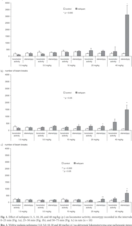
Nefopam administered in doses of 1.0, 5.0, 10, 20, and 40 mg/kg did not affect locomotor activity compared with the saline-treated rats in any of the examined periods of time (0–25 min, 25–50 min, and 50–75 min) (Figs. 1a–c). Rats challenged with nefopam at the higher doses (20 and 40 mg/kg) exhibited stereotypic reactions (e.g. stereotyped walking, sniffing) compared with the control group. In the first period (0–25 min) the differ-ences were statistically significant for the dose of 40 mg/kg (p < 0.05), in the second period for the doses of 10, 20, and 40 mg/kg (p < 0.05), and in the third for 20 (p < 0.005) and 40 mg/kg (p < 0.05).
0 500 1000 1500 2000 2500 3000 3500 4000
locomotor
activity stereotypy locomotoractivity stereotypy locomotoractivity stereotypy locomotoractivity stereotypy locomotoractivity stereotypy 1.0 mg/kg 5.0 mg/kg 10 mg/kg 20 mg/kg 40 mg/kg
control nefopam
number of beam breaks
* p < 0.005
*
Fig. 1. Effect of nefopam (1, 5, 10, 20, and 40 mg/kg i.p.) on locomotor activity, stereotypy recorded in the intervals 0–25 min (Fig. 1a), 25–50 min (Fig. 1b), and 50–75 min (Fig. 1c) in rats (n = 10)
Ryc. 1. Wpływ podania nefopamu (1,0; 5,0; 10; 20 and 40 mg/kg i.p.) na aktywność lokomotoryczną oraz zachowanie stereoty-powe oceniane w przedziałach: 0–25 min (ryc. 1a) 25–50 min (ryc. 1b) oraz 50–75 min (ryc. 1c) u szczurów (n = 10)
0 500 1000 1500 2000 2500 3000 3500 4000
locomotor
activity stereotypy locomotoractivity stereotypy locomotoractivity stereotypy locomotoractivity stereotypy locomotoractivity stereotypy 1.0 mg/kg 5.0 mg/kg 10 mg/kg 20 mg/kg 40 mg/kg
control nefopam
*
* p < 0.05
*
*
number of beam breaks
0 500 1000 1500 2000 2500 3000 3500 4000
locomotor
activity stereotypy locomotoractivity stereotypy locomotoractivity stereotypy locomotoractivity stereotypy locomotoractivity stereotypy 1.0 mg/kg 5.0 mg/kg 10 mg/kg 20 mg/kg 40 mg/kg
control nefopam
**
** p < 0.005 * p < 0.05
*
number of beam breaks
a)
b)
the amount of climbing (number of rearings) in the second period of testing (35–50 min, p < 0.05) compared with the saline-treated rats (Fig. 2).
Motor Coordination (Rotarod
Test)
Nefopam at all doses prolonged the time on the rod compared with the controls. Significant changes were observed at 15 (p < 0.05), 30 (p < 0.05), 60 (p < 0.005), and 120 min (p < 0.05) after its administration at a dose of 1.0 mg/kg); 15 (p < 0.005), 30 (p < 0.05), and 60 min (p < 0.005) at 5.0 mg/kg ip); 15 (p < 0.05), 30 (p < 0.05), and 60 min (p < 0.005) at 10 mg/kg, 30 (p < 0.05), 60 (p < 0.005), 120 (p < 0.05), and 180 min (p < 0.05) at 20 mg/kg; and 15 (p < 0.05), 30 (p < 0.005), and 60 min (p < 0.05) at 40 mg/kg (Tab. 1).
Catalepsy
Nefopam injected at a dose of 10 mg/kg i.p.
30 min before dopamine D1/D5 receptor antagonist
SCH 23390 challenge (0.5 mg/kg i.p.) significantly diminished the cataleptic response at 15, 30, and 45 min (p < 0.05) of observation compared with the control group (SCH 23390 alone) (Fig. 3).
Porsolt Forced Swim Test
Nefopam administered at a dose of 10 mg/kg did not affect immobility time compared with the saline-treated rats (Fig. 4).
L-DOPA Assay
Nefopam administered at all the tested doses (1.0, 5.0, 10, 20, and 40 mg/kg) reduced L-DoPA content in the frontal cortex, nucleus accumbens, and striatum. Significant changes in the frontal cortex were observed after nefo-pam injected at doses of 5.0, 10, 20 and mg/kg (p < 0.05) and 40 mg/kg (p < 0.005) and in the nucleus accumbens and striatum at all the tested doses (1.0 mg/kg p < 0.05 and 5.0, 10, 20, and 40 mg/kg p < 0.005) (Fig. 5).
In vivo
Microdialysis Study
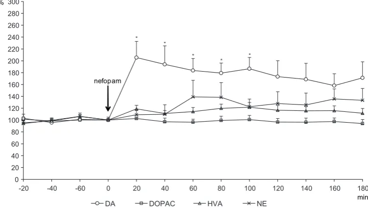
Nefopam (10 mg/kg) acutely produced a sharp increase in the striatal microdialysate DA concen-tration of up to 206% compared with the base-line (at 20 min), then a gradual decrease in DA release was seen (data significant between 20–100 min, p < 0.05). In contrast, there was no difference in DA metabolite (DoPAC and HVA) microdi-alysate compared with the baseline data (before nefopam challenge). Nefopam also had no effect on NE release (Fig. 6).
Discussion
The present study demonstrated that nefopam, besides its analgesic effect, possesses some other biological properties. The most important finding
Fig. 2. Effect of nefopam (1, 5, 10, 20, and 40 mg/kg i.p.) on the number of rearings recorded in the following inter-vals: 0–25 min, 25–50 min, and 50–75 min in rats (n = 10). Explanations as in Fig. 1.* p < 0.05; ** p < 0.005
Ryc. 2. Wpływ podania nefopamu (1,0; 5,0; 10; 20 and 40 mg/kg i.p.) na liczbę wspięć ocenianą w przedziałach: 0–25 min; 25–50 min oraz 50–75 min u szczurów (n = 10). objaśnienia jak na ryc. 1. * p < 0,05; ** p < 0,005
0 20 40 60 80 100 120 140 160
180 1 mg/kg 5 mg/kg 10 mg/kg 20 mg/kg 40 mg/kg
number of rearings
control nefopam
*
Biological Peculiarities of Nefopam in Rats 27
is that its having no effect on general locomotor activity significantly improved motor coordina-tion tested on the rotarod apparatus. As has been noted, nefopam augmented DA release in the stri-atum. A study by Rosland and Hole [24] showed that nefopam inhibits DA, NE, and 5-HT reuptake in synaptosomal preparations from rat forebrain, hippocampus, and striatum. Amphetamine and its derivatives also promote DA efflux, mainly by reverse transport through monoamine uptake transporter [25]. Both in vivo voltametry and brain microdialysis studies demonstrated an increase in DA efflux after amphetamine, which attained its maximum response 20–40 min after injection with concomitant reduction in DoPAC and HVA efflux [26, 27]. The excess of DA in the synaptic cleft produces increased locomotor activation in laboratory animals [28].
In the present study the DA release evoked by nefopam (10 mg/kg) was not accompanied by locomotor activity changes; only higher doses (20 and 40 mg/kg) produced behavioral stereotypy, as is also observed after high-dose amphetamine challenge [29]. Nefopam is not an exception in this case because several drugs that inhibit DA reuptake do not affect locomotor activity (e.g. bupropion) [30]. More interesting is that nefopam
significantly improved motor coordination. In this case it somewhat resembles amphetamine deriva-tives, which to some extent ameliorate both motor and cognitive performance. Low doses of meth-ylphenidate, which increase catecholamine release in the prefrontal cortex, have recently been shown to improve delayed alternation performance in rats. Low doses of methylphenidate also improved performance of a sustained attention task, the five-choice attention task that similarly depends upon the prefrontal cortex [31]. It has also been shown that another DA agonist improved performance and motor coordination in laboratory animals [32]. Bergquist et al. [33] found that in unilaterally 6-hydroxydopamine-lesioned rats, a dose-depen-dent improvement in rod performance during perfusion of the substantia nigra with the non-selective DA agonist apomorphine was observed. In contrast, perfusion of the substantia nigra with the DA D1/D5 antagonist SCH 23390
dose-depend-ently impaired rod performance. Furthermore, the results of a study by Andersson et al. [34] indicate that partial depletion of nigral DA stores can significantly impair motor functions and that increased nigral DA release can counteract minor impairments of striataldopaminergic neurotrans-mission.
Table 1. Effect of nefopam (1, 5, 10, 20, and 40 mg/kg i.p.) on motor coordination in rats (n = 10)
Tabela 1. Wpływ nefopamu (1,0; 5,0; 10; 20 and 40 mg/kg i.p.) na koordynację ruchową u szczurów (n = 10) groups
(grupy) 15’ 30’ 60’ 120’ 180’
Control
(Kontrolna) 19.2 ± 5.6 26.2 ± 6.0 31.3 ± 8.5 73.5 ± 20.6 121.9 ± 39.7 Nefopam (1.0 mg/kg) 137.4 ± 39.3
*
138.6 ± 40.4*
198.1 ± 37.4**
172.5 ± 35.1*
186.2 ± 32.9 Control(Kontrolna) 16.3 ± 5.1 56.3 ± 25.1 84.7 ± 37.4 177.7 ± 41.3 262.2 ± 28.6 Nefopam (5.0 mg/kg) 171.5 ± 40.0
**
179.5 ± 36.8*
176.9 ± 26.6**
226.8 ± 25.3 229.1 ± 31.9 Control(Kontrolna) 18.2 ± 4.6 40.9 ± 9.1 27.8 ± 8.3 91.3 ± 38.1 78.4 ± 37.5 Nefopam (10 mg/kg) 42.3 ± 8.0
*
80.1 ± 16.8*
131.5 ± 31.6**
102.3 ± 42.3 157.8 ± 44.2 Control(Kontrolna) 83.7 ± 36.5 104.4 ± 38.7 83.8 ± 29.3 120.1 ± 38.5 80.0 ± 27.4 Nefopam (20 mg/kg) 127.6 ± 32.8 226.0 ± 37.0
*
243.5 ± 26.9**
249.3 ± 34.1*
252.7 ± 32.6**
Control(Kontrolna) 55.3 ± 29.1 147.9 ± 41.2 164.2 ± 40.2 198.4 ± 42.1 245.9 ± 33.7 Nefopam (40 mg/kg) 177.3 ± 40.9
*
259.6 ± 29.9**
249.5 ± 36.1*
250.7 ± 28.8 230.7 ± 33.0 * p < 0.05; ** p < 0.005.Fig. 3. Effect of nefopam (10 mg/kg i.p.) on catalepsy after SCH 23390 (0.5 mg/kg i.p.) administration in rats (n = 10). Explanations as in Fig. 1. * p < 0.05
Ryc. 3. Wpływ nefopamu (10 mg/kg i.p.) na nasilenie katalepsji po podaniu SCH 23390 (0,5 mg/kg i.p.) u szczurów (n = 10). objaśnienia jak na ryc. 1. * p < 0,05
0 10 20 30 40 50 60
15' 30' 45' 60' 90' 120' seconds
cataleps
y
control nefopam
* *
*
* p < 0.005
Fig. 4. Effect of nefopam (10 mg/kg i.p.) on immobility time in rats (n = 10). Explanations as in Fig. 1
Ryc. 4. Wpływ nefopamu (10 mg/kg i.p.) na czas bezruchu u szczurów (n = 10). objaśnienia jak na ryc. 1
0 50 100 150 200 250 300
immobility time
control nefopam
The present study also showed that nefopam injected at a dose of 10 mg/kg 30 min before the DA D1/D5 receptor antagonist SCH 23390
chal-lenge (0.5 mg/kg) significantly diminished the cataleptic response compared with the control group (SCH 23390 alone). It is worth knowing that catalepsy is regarded as a relevant animal model of Parkinson’s disease [35]. It has been shown that drugs that reduce the cataleptogenic response of SCH 23390 or the haloperidol ameliorate motor deficits in Parkinson’s disease. As mentioned in the introduction, the incidence of severe depression is very common in Parkinson’s disease. The use of
Biological Peculiarities of Nefopam in Rats 29
blockers, are frequently used for treating depres-sion and they showed antidepressant-like action in the forced swimming test [36]. Neurochemical studies have demonstrated that 5-HT reuptake blockers (SSRI, e.g. paroxetine, fluoxetine) mark-edly increase 5-HT concentration in the vicinity of 5-HT-containing cells in the midbrain raphe nuclei, which leads to activation of 5-HT autore-ceptors and consequently results in inhibition of
cell firing and reduction of 5-HT release in the forebrain. Terminal autoreceptors further limit the increase in the level of synaptic 5-HT produced by SSRI [37]. Another consequence of SSRI challenge is the reduction of the rate 5-HT and DA synthesis [38, 39]. The present study also found decreased DA synthesis (measured by L-DoPA accumula-tion) after nefopam administration.
In conclusion, besides some similarities in
bio-Fig. 5. Effect of nefopam (1, 5, 10, 20, and 40 mg/kg i.p.) on L-DoPA content in the frontal cortex, nucleus accum-bens, and striatum in rats (n = 6–7)
Ryc. 5. Wpływ nefopamu (1,0; 5,0; 10; 20 and 40 mg/kg i.p.) na zawartość L-DoPA w korze czołowej, jądrze półleżącym przegrody oraz prążkowiu u szczurów (n = 6–7)
0 500 1000 1500 2000 2500 3000 3500
frontal cortex nucleus accumbens striatum ng/g wet tissue
saline nefopam 1.0 mg/kg nefopam 5.0 mg/kg nefopam 10 mg/kg nefopam 20 mg/kg nefopam 40 mg/kg
**
*
** ** * p < 0.05
** p < 0.005
** * *
** * *
**
** ** **
Fig. 6. Effect of nefopam (10 mg/kg i.p.) on the microdialysate DA, DoPAC, HVA, and NE concentration in the stria-tum of rats (n = 7). * p < 0.05
Ryc. 6. Wpływ nefopamu (10 mg/kg i.p.) na zawartość DA, DoPAC, HVA i NE w mikrodializatach prążkowia u szczurów (n = 7). *p < 0,05
0 20 40 60 80 100 120 140 160 180 200 220 240 260 280 300
-20 -40 -60 0 20 40 60 80 100 120 140 160 180
DA DOPAC HVA NE
* *
*
* *
nefopam
chemical profile between nefopam and antidepres-sants, the present study showed no antidepressant-like action of the drug (in the forced swimming test). Summing up, the data of this study lead to
the proposal that the “behavioral-biochemical profile” of the analgesic nefopam justifies its use in patients with motor abnormalities, for example in Parkinson’s disease.
References
Girard P
[1] , Coppé MC, Verniers D, Pansart Y, Gillardin JM: Role of catecholamines and serotonin receptor sub-types in nefopam-induced antinociception. Pharmacol Res 2006, 54, 195–202.
Hunskaar S
[2] , Fasmer OB, Broch OJ, Hole K: Involvement of central serotonergic pathways in nefopam-induced antinociception. Eur J Pharmacol 1987, 138, 77–82.
Gomaa AA
[3] , Aly SA, Badary MS, Ahmed EA: The immunopotentiator effects of nefopam. Int Immunopharmacol 2007, 7, 266–271.
Novelli A
[4] , Groppetti A, Rossoni G, Manfredi B, Ferrero-Gutiérrez A, Pérez-Gómez A, Desogus CM, Fernández--Sánchez MT: Nefopam is more potent than carbamazepine for neuroprotection against veratridine in vitro and has anticonvulsant properties against both electrical and chemical stimulation. Amino Acids 2007, 32, 323–332.
Novelli A
[5] , Díaz-Trelles R, Groppetti A, Fernández-Sánchez MT: Nefopam inhibits calcium influx, cgMP for-mation, and NMDA receptor-dependent neurotoxicity following activation of voltage sensitive calcium channels. Amino Acids 2005, 28, 183–191.
Mattson MP
[6] : glutamate and neurotrophic factors in neuronal plasticity and disease. Ann N Y Acad Sci 2008, 1144, 97–112.
Büeler H
[7] : Impaired mitochondrial dynamics and function in the pathogenesis of Parkinson’s disease. Exp Neurol 2009, 218, 235–246.
Kostrzewa RM, Kostrzewa JP, Brown RW, Nowak P, Brus R:
[8] Dopamine receptor supersensitivity: development, mechanisms, presentation, and clinical applicability. Neurotox Res 2008, 14, 121–128.
Nowak P, Kostrzewa RA, Skaba D, Kostrzewa RM:
[9] Acute L: -DoPA Effect on Hydroxyl Radical- and DoPAC- -Levels in Striatal Microdialysates of Parkinsonian Rats. Neurotox Res 2010, 17, 299–304.
Nowak P, Bortel A, Dabrowska J, Biedka I, Slomian G, Roczniak W, Kostrzewa RM, Brus R:
[10] Histamine H(3)
receptor ligands modulate L-dopa-evoked behavioral responses and L-dopa derived extracellular dopamine in dopamine-denervated rat striatum. Neurotox Res 2008, 13, 231–240.
Nowak P, Jochem J, Zwirska-Korczala K, Josko J, Noras L, Kostrzewa RM, Brus R:
[11] ontogenetic noradrenergic
lesion alters histaminergic activity in adult rats. Neurotox Res 2008, 13, 79–83.
Nowak P, Nitka D, Kwieciński A, Jośko J, Drab J, Pojda-Wilczek D, Kasperski J, Kostrzewa RM, Brus R: [12]
Neonatal co-lesion by DSP-4 and 5,7-DHT produces adulthood behavioral sensitization to dopamine D(2) recep-tor agonists. Pharmacol Rep 2009, 61, 311–318.
Nowak P, Noras L, Jochem J, Szkilnik R, Brus H, Körossy E, Drab J, Kostrzewa RM, Brus R:
[13] Histaminergic
activity in a rodent model of Parkinson’s disease. Neurotox Res 2009, 15, 246–251.
Chaudhuri KR
[14] , Schapira AH: Non-motor symptoms of Parkinson’s disease: dopaminergic pathophysiology and treatment. Lancet Neurol 2009, 8, 464–474.
Beiske AG
[15] , Loge JH, Rønningen A, Svensson E: Pain in Parkinson’s disease: Prevalence and characteristics. Pain 2009, 141, 173–177.
Przegaliñski E, Filip M, Papla I, Siwanowicz J:
[16] Effect of serotonin (5-HT)1B receptor ligands on cocaine sensiti-zation in rats. Behav Pharmacol 2001, 12, 109–116.
Jones BJ, Roberts DJ:
[17] The quantitative measurement of motor incoordination in naive mice using an accelerating rota-rod. J Pharm Pharmacol 1968, 20, 302–304.
Iorio LC, Barnett A, Billard W, Gold EH:
[18] Benzodiazepines: Structure-activity relationships between Dl receptor blockade and selected pharmacological effects. Adv Exp Med Biol 1986, 204, 1–14.
Porsolt RD, Anton G, Blavet N, Jalfre M:
[19] Behavioural despair in rats: a new model sensitive to antidepressant treatments. Eur J Pharmacol 1978, 47, 379–391.
Carlsson A, Davis JN, Kher W, Lindqvist M, Atack CV:
[20] Simultaneous measurement of tyrosine and trypto-phan hydroxylase activities in brain in vivo using an inhibitor of the aromatic amino acid decarboxylase. Naunyn- -Schmiedeberg’s Arch Pharmacol 1972, 275, 153–168.
Takeda H
[21] , Matsumiya T, Shibuya T: Detection and identification modes for the highly sensitive and simultaneous determination of various biogenic amines by coulometric high-performance liquid chromatography. J Chromatogr 1990, 515, 265–278.
Brus R, Nowak P, Sokoła A, Kotrzewa RM, Shani J:
[22] Behavioral and biochemical effects of new central dopamine D3 and D4 receptor antagonists in rats. Pharmacol Rev Comm 2002, 12, 39–59.
Nowak P, Brus R, Oświęcimska J, Sokoła A, Kostrzewa RM:
[23] 7-Nitroindazole enhances amphetamine-evoked
dopamine release in rat striatum. An in vivo microdialysis and voltammetric study. J Physiol Pharmacol 2002, 53, 251–263.
Rosland JH
[24] , Hole K: The effect of nefopam and its enantiomers on the uptake of 5-hydroxytryptamine, nor-adrenaline and dopamine in crude rat brain synaptosomal preparations. J Pharm Pharmacol 1990, 42, 437–438.
Khoshbouei H
Biological Peculiarities of Nefopam in Rats 31
Kasperska A, Brus R, Sokoła A, Kostrzewa RM, Shani J:
[26] Sexual differentiation in the central dopaminergic effect of nitric oxide donors and inhibitor on stereotyped behavior changes induced by amphetamine, but not by apo-morphine. Pharmacol Rev Comm 1999, 10, 329–339.
Nowak P
[27] , Bortel A, Dabrowska J, Oswiecimska J, Drosik M, Kwiecinski A, Opara J, Kostrzewa RM, Brus R:
Amphetamine and mCPP effects on dopamine and serotonin striatal in vivo microdialysates in an animal model of hyperactivity. Neurotox Res 2007, 11, 131–144.
Brus R
[28] , Nowak P, Szkilnik R, Mikolajun U, Kostrzewa RM: Serotoninergics attenuate hyperlocomotor activity in rats. Potential new therapeutic strategy for hyperactivity. Neurotox Res 2004, 6, 317–325.
Milesi-Hallé
[29] A, McMillan DE, Laurenzana EM, Byrnes-Blake KA, Owens SM: Sex differences in (+)-amphet-amine- and (+)-methamphet(+)-amphet-amine-induced behavioral response in male and female Sprague-Dawley rats. Pharmacol Biochem Behav 2007, 86, 140–149.
Sitges M
[30] , Reyes A, Chiu LM: Dopamine transporter mediated release of dopamine: role of chloride. J Neurosci Res 1994, 39, 11–22.
Berridge CW
[31] , Devilbiss DM, Andrzejewski ME, Arnsten AF, Kelley AE, Schmeichel B, Hamilton C, Spencer RC:
Methylphenidate preferentially increases catecholamine neurotransmission within the prefrontal cortex at low doses that enhance cognitive function. Biol Psychiatry 2006, 60, 1111–1120.
Thullier F
[32] , Lalonde R, Lestienne F: Effects of dopaminergic agents and of an NMDA receptor antagonist on motor coordination in Lurcher mutant mice. Pharmacol Biochem Behav 1999, 63, 213–219.
Bergquist F
[33] , Shahabi HN, Nissbrandt H: Somatodendritic dopamine release in rat substantia nigra influences motor performance on the accelerating rod. Brain Res 2003, 973, 81–91.
Andersson DR
[34] , Nissbrandt H, Bergquist F: Partial depletion of dopamine in substantia nigra impairs motor performance without altering striatal dopamine neurotransmission. Eur J Neurosci 2006, 24, 617–624.
Khisti RT
[35] , Mandhane SN, Chopde CT: Haloperidol-induced catalepsy: a model for screening antidepressants effective in treatment of depression with Parkinson’s disease. Indian J Exp Biol 1997, 35, 1297–1301.
Castagné V
[36] , Porsolt RD, Moser P: Use of latency to immobility improves detection of antidepressant-like activity in the behavioral despair test in the mouse. Eur J Pharmacol 2009, 616, 128–133.
Hervas Queiroz CMT, Adell A, Artigas F:
[37] Role of uptake inhibition and autoreceptor activation in the control of 5-HT release in the frontal cortex and dorsal hippocampus of the rat. Brit J Pharmacol 2000, 130, 160–166.
Stenfors C
[38] , Yu H, Ross SB: Pharmacological characterisation of the decrease in 5-HT synthesis in the mouse brain evoked by the selective serotonin re-uptake inhibitor citalopram. Naunyn Schmiedebergs Arch Pharmacol 2001, 363, 222–232.
Dremencov E
[39] , El Mansari M, Blier P: Effects of sustained serotonin reuptake inhibition on the firing of dopamine neurons in the rat ventral tegmental area. J Psychiatry Neurosci 2009, 34, 223–229.
Address for correspondence:
Przemysław Nowak
Department of Pharmacology Medical University of Silesia H. Jordana 38
41-808 Zabrze Poland
Tel./fax: +48 32 272 26 83 E-mail: [email protected]
Conflict of interest: None declared