Distribution of insertion sequences associated with Tn1546 and clonal
diversity of vancomycin-resistant enterococci isolated from patients in
Tehran, Iran
Oskoui M 1*, Farrokh P1
1Department of Bacteriology, Pasteur Institute of Iran, Tehran, Iran.
Received: December 2009, Accepted: February 2010.
ABSTRACT
Background and objectives: Infection with vancomycin-resistant enterococci (VRE) has caused a therapeutic problem. VanA and VanB resistant types are the predominant phenotypes among vancomycin resistant enetrococci. Transposon 1546 (Tn1546) harboring the vanA gene cluster, plays an important role in the horizontal transfer of vanA gene. In this study, we examined the phenotypic and genotypic diversity of a number of clinical VRE.
Materials and Methods: Twenty-four clinical VRE isolated from two university hospitals in Tehran were examined based on their antimicrobial susceptibility, Tn1546 related element organization and pulsed-field gel electrophoresis (PFGE) patterns. Integration of well-studied insertion sequence elements IS1216V, IS1542 and IS1251 was examined by PCR mapping and sequencing.
Results: From 24 isolates, 15 isolates with VanA phenotype and 9 isolates with VanB phenotype were identified which both groups interestingly possessed the vanA gene.According to PCR mapping, our isolates were assigned to 6 main groups. In 14 (58.3%) isolates, IS1216V was inserted in vanX-vanY region and/or in truncated left-hand of Tn1546-like elements. In 11 (45.8%) isolates, both IS1216V and IS1542 were inserted in vanX-vanY and orf2-vanR regions, respectively and none of them harbored IS1251. Interestingly, PFGE of the isolates showed a high degree of diversity.
Conclusion: PCR mapping revealed that VanA elements in our isolates were highly heterogeneous. Overall, we found no correlation between transposon type and PFGE pattern. Genetic diversity of VRE provides practical information for epidemiological studies and our data showed horizontal transfer of VRE in this region.
Keywords: Vancomycin- resistant enterococci, Tn1546, insertion sequences, Iran.
INTRODUCTION
Enterococci are part of the normal flora in the gastrointestinal tracts of humans (1, 2); however, they can also be the main cause of nosocomial infection especially in immunocompromised patients (3, 4). Vancomycin-resistant enterococci (VRE) have presented a global problem for treatment (3, 5, and 6). VRE are
phenotypically and genotypically heterogenous (7) and there are six types of glycopeptide resistance in enterococci (VanA to VanE and VanG) (8, 9). VanA phenotype with acquired inducible resistance to both vancomycin and teicoplanin, and VanB phenotype with variable level of resistance to vancomycin but susceptibility to teicoplanin are the most predominant ones (10, 11). Recently, VRE with vanA genotype, susceptible to teicoplanin (VanB phenotype-vanA genotype), has become increasingly prevalent in Asia (11).
VanA gene cluster is carried on transposon Tn1546 or closely related elements (5, 12) consisting of vanR, vanS, vanH, vanA, vanX, vanY, vanZ genes (3). orf1 and orf2 with transposition function are also * Corresponding author: Mahvash Oskoui, Ph.D.
Address: Pasteur Institute of Iran. 69, Pasteur Ave.Tehran-13164, Iran.
Tel: +98-21-66405535 Fax: +98-2166405535 E-mail: [email protected]
16 OSKOUI ET AL . IRAN. J. MICROBIOL. 2 (1) : 15-22
present in the left end of Tn1546. Due to integration of insertion sequences (ISs), point mutation, and deletions in nonessential genes or integration regions, there are considerable variations among Tn1546
elements (4, 5, 12).
Investigation of IS elements in VanA gene cluster provides useful information for epidemiological studies of the dissemination of VRE due to Reference Amplicon size (bp)
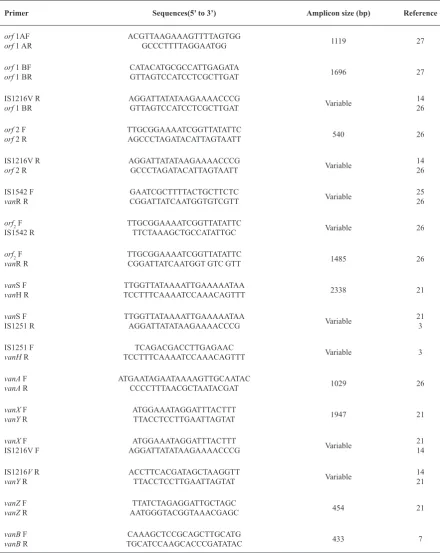
Sequences(5’ to 3’) Primer
27 1119
ACGTTAAGAAAGTTTTAGTGG GCCCTTTTAGGAATGG
orf 1AF orf 1 AR
27 1696
CATACATGCGCCATTGAGATA GTTAGTCCATCCTCGCTTGAT orf 1 BF
orf 1 BR
14 26 Variable
AGGATTATATAAGAAAACCCG GTTAGTCCATCCTCGCTTGAT IS1216V R
orf 1 BR
26 540
TTGCGGAAAATCGGTTATATTC AGCCCTAGATACATTAGTAATT
orf 2 F orf 2 R
14 26 Variable
AGGATTATATAAGAAAACCCG GCCCTAGATACATTAGTAATT IS1216V R
orf 2 R
25 26 Variable
GAATCGCTTTTACTGCTTCTC CGGATTATCAATGGTGTCGTT IS1542 F
vanR R
26 Variable
TTGCGGAAAATCGGTTATATTC TTCTAAAGCTGCCATATTGC orf2 F
IS1542 R
26 1485
TTGCGGAAAATCGGTTATATTC CGGATTATCAATGGT GTC GTT orf2 F
vanR R
21 2338
TTGGTTATAAAATTGAAAAATAA TCCTTTCAAAATCCAAACAGTTT vanS F
vanH R
21 3 Variable
TTGGTTATAAAATTGAAAAATAA AGGATTATATAAGAAAACCCG vanS F
IS1251 R
3 Variable
TCAGACGACCTTGAGAAC TCCTTTCAAAATCCAAACAGTTT IS1251 F
vanH R
26 1029
ATGAATAGAATAAAAGTTGCAATAC CCCCTTTAACGCTAATACGAT vanA F
vanA R
21 1947
ATGGAAATAGGATTTACTTT TTACCTCCTTGAATTAGTAT vanX F
vanY R
21 14 Variable
ATGGAAATAGGATTTACTTT AGGATTATATAAGAAAACCCG vanX F
IS1216V F
14 21 Variable
ACCTTCACGATAGCTAAGGTT TTACCTCCTTGAATTAGTAT IS1216V R
vanY R
21 454
TTATCTAGAGGATTGCTAGC AATGGGTACGGTAAACGAGC vanZ F
vanZ R
7 433
CAAAGCTCCGCAGCTTGCATG TGCATCCAAGCACCCGATATAC vanB F
vanB R
horizontal transfer of Tn1546-like elements (3, 13). The most common insertion sequences reported in vanA gene cluster are IS1216V, IS1542, IS1251, and IS1476 (14). Although IS1216V is known in most of the vanA elements, the other IS elements appear to be geographically restricted (14). Beside horizontal transfer of resistance gene cluster, clonal dissemination of VRE was determined in various studies (14, 15).
Although vancomycin-resistant enterococci have been reported worldwide, investigation of resistant isolates from different geographic locations provides useful information (16, 17). In this study, our goal is determination of the phenotypic as well as genotypic diversity of clinical VRE in this region with using antimicrobial susceptibility, PCR mapping and Pulse-field gel electerophoresis.
MATERIALS AND METHODS
Bacterial isolates and identification. A total of 24 VRE clinical isolates, collected between May 2006 and May 2007 from Milad and Sina University Hospitals in Tehran, were studied. These VRE were isolated from nearly 500 enterococci. With the exception of isolates from blood samples, all enterococci were isolated from urine specimens. These isolates were identified by conventional biochemical reactions (18). E. faecium BM4147, E. faecalis ATCC 29212, and E. faecalis V583 were used as control strains.
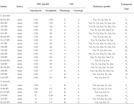
Antimicrobial agents and MIC determination. Antimicrobial susceptibility of the isolates were tested by the disc diffusion method and interpreted according to the Kerby-Bauer method (19). The antibiotics (MAST Diagnostics Ltd. Merseyside, Table 2. The phenotypic and genetic characteristics of vancomycin resistant isolates of enterococci isolated from patients in Tehran.
‡ Vm, Vancomycin; Tc, teicoplanin; Am, ampicillin; Cm, chloramphenicol; Cp, ciprofloxacin; Em, erythromycin; Te,
tetracycline
Isolate Source MIC (μg/ml) Van
†
Resistance profile‡ Transposon
Type
Vancomycin Teicoplanin Phenotype Genotype
E. faecium
B-201 IPI urine >256 >256 A A Vm, Tc, Cp, Em, Te A1
495 IPI urine >256 128 A A Vm, Tc, Cp, Em, Te, Am, Cm B3
105 IPI urine >256 128 A A Vm, Tc, Cp, Em, Te, Am, Cm A1
107 IPI urine >256 96 A A Vm, Tc, Cp, Em, Am, Cm B1
431 IPI urine >256 64 A A Vm, Tc, Cp, Em, Te, Am, Cm F7
B-364 IPI urine >256 64 A A Vm, Tc, Cp, Em, Te F6
N-117 IPI urine >256 64 A A Vm, Tc, Cp, Em, Te, Am C
557 IPI urine >256 64 A A Vm, Tc, Cp, Em, Te, Am, Cm F1
390 IPI urine >256 64 A A Vm, Tc, Cp, Em, Te, Am, Cm B2
110 IPI urine >256 64 A A Vm, Tc, Cp, Em, Am B1
436 IPI urine >256 64 A A Vm, Tc, Cp, Em, Te, Am, Cm E1
108 IPI urine >256 64 A A Vm, Tc, Cp, Em, Am, Cm B1
B-269 IPI urine >256 64 A A Vm, Tc, Cp, Em E2
102 IPI urine >256 48 A A Vm, Tc, Cp, Em, Te, Am A3
B-148 IPI blood >256 32 A A Vm, Tc, Cp, Em, Te, Am A2
106 IPI urine >256 1.5 B A Vm, Cp, Em, Te, Am, Cm B1
109 IPI urine >256 1 B A Vm, Cp, Em, Te, Am D
E-83 IPI urine >256 0.5 B A Vm, Cp, Em, Te F5
E. faecalis
523 IPI urine >256 1 B A Vm, Cp, Em, Te, Am F3
N-89 IPI urine >256 0.5 B A Vm, Cp, Em, Te, Cm F2
E-125 IPI urine 128 0.5 B A Vm, Cp, Em, Te F8
578 IPI urine 32 0.38 B A Vm, Cp, Em F4
524 IPI urine 16 1.5 B A Vm, Cp, Em, Te, Am F10
18 OSKOUI ET AL . IRAN. J. MICROBIOL. 2 (1) : 15-22
England) used for disc diffusion assays included vancomycin (30μg), teicoplanin (30μg), tetracycline (30μg), erythromycin (15μg), chloramphenicol (30μg), ampicillin (10μg), and ciprofloxacin (5μg). Minimum inhibitory concentration (MIC) of vancomycin (SERVA FEINBIOCHEMICA GmbH & Co., Germany) and teicoplanin were determined using broth microdilution (19) and Etest (AB Biodisk, Solna, Sweden), respectively.
Characterization of Tn1546-like elements. Extraction of chromosomal and plasmid DNA were performed using Bacterial Genomic and Plasmid Miniprep kits (Metabion, Martinsried, Germany). VanA and VanB-type enterococci were examined by PCR with primers specific for the vanA and vanB genes.
For structural analysis of Tn1546-like elements,
PCR was carried out with previously published primers for orf1, orf 2, orf 2-vanR, vanS-vanH, vanA, vanX-vanY, and vanZ (Table 1). Presence of well-studied IS elements IS1216V, IS1542 and IS1251 in the orf 2-vanR , vanS-vanH and vanX-vanY intergenic regions were confirmed with additional PCR by one primer in the published sequence of Tn1546 and one in the published IS sequence (Table 1). In order to determine precise left ends of the Tn1546, DNA fragments were amplified with a combination of Tn1546-derived primers and primers based on the IS1216V which was inserted at the left end of the transposon. The primer sequences and amplicon size of products are listed in Table 1.
PCR amplification was carried out on a Eppendorf thermal cycler with the following protocol: initial denaturation at 95°C for 4 min; this was followed by 30 cycle of DNA denaturation at 95°C for 30 S,
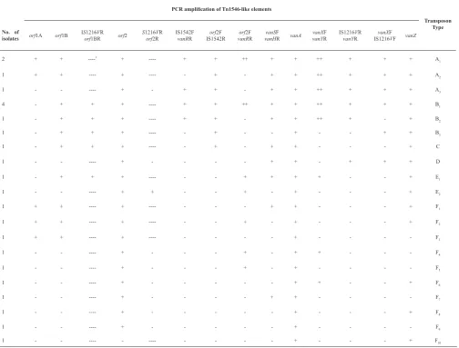
Transposon Type PCR amplification of Tn1546-like elements
vanZ vanXF IS1216VF IS1216VR
vanYR vanXF vanYR vanA vanSF vanHR orf2F vanRR orf2F IS1542R IS1542F vanRR
S1216VR
orf2R
orf2 IS1216VR
orf1BR
orf1B
orf1A No. of
isolates A1 + + + ++ + + ++ + + ----+ ----* + + 2 A2 + + + ++ + + -+ - ----+ ----+ + 1 A3 + + + ++ + + -+ + -+ -1 B1 + + + ++ + + ++ + + ----+ + + -4 B2 + -+ ++ + + -+ + ----+ + + -1 B3 + + -+ -+ - ----+ + + -1 C + -+ + -+ - ----+ + + -1 D + + + -+ + -+ -1 E1 + -+ + + + - ----+ + + -1 E2 + -+ -+ -+ + -1 F1 + -+ + - ----+ ----+ + 1 F2 + -+ -+ - ----+ ----+ + 1 F3 -+ - ----+ ----+ + 1 F4 -+ + -+ -+ -1 F5 -+ -+ -+ -1 F6 + -+ + -+ -1 F7 -+ + -+ -1 F8 + -+ -+ -1 F9 -+ -+ -1 F10 + -+ - - -1
primer annealing at 55-56°C for 1 min and DNA extension at 72°C for 1 min; and final extension at 72°C for 10 min.
DNA sequence analysis. PCR amplicons of vanX-vanY and orf2-vanR regions which were larger than those of the prototype vanA gene cluster were sequenced with IS1216V and IS1542 primers. To determine the DNA sequence of the left end of the VanA transposon derivatives, PCR products of this region were sequenced with IS1216V primers (Macrogen Research, Seoul, Korea).
Pulse-field gel electerophoresis (PFGE). PFGE was performed as described previously (20). Genomic DNA was digested with SmaI (Fermentas, Vilnius, Lithuania), and separated on a 1% agarose gel using a contour-clamped homogeneous-field apparatus (CHEF DR III system; Bio-Rad Laboratories, Richmond, CA). Salmonella Braenderup H9812 was used as molecular weight marker after XbaI (Fermentas, Vilnius, Lithuania) digestion. The agarose gels were run at 14°C and 6 V/cm for 21 h, with a linear pulse time of 5 to 40 sec at an angle of 120 degrees. The banding patterns were analyzed using Gelcompar II version 4.0 (Applied Maths Sint-Matens-Latem, Belgium).
RESULTS
Bacterial isolates and antibiotic resistance. Eighteen Out of 24 VRE were E. faecium and the remaining were E. faecalis. All the isolates were resistant to vancomycin, ciprofloxacin and erythromycin discs. Furthermore, 79.2%, 66.7%, 62.5% and 41.7% of the isolates were resistant to tetracycline, ampicillin, teicoplanin and chloramphenicol, respectively (Table 2). The MIC of vancomycin and teicoplanin of the isolates are given in Table 2. Fifteen out of 18 E. faecium showed the VanA phenotype, while all the E. faecalis and 3 of the E. faecium displayed VanB phenotype.
Structural analysis of Tn1546 element by PCR mapping. When PCR was carried out with the vanA specific primer, PCR product of expected size was obtained from 15 isolates with VanA phenotype. None of the 9 isolates with VanB phenotype possessed the vanB gene, but all of them harbored the vanA gene.
PCR mapping of Tn1546-like elements of 24 isolates revealed 6 main different transposon types (A-F) according to the patterns of ISs inserted into Tn1546 (Table 3). Type A was characterized by an IS1216V insertion in the vanX-vanY intergenic region Lithuania) digestion. The agarose gels were run at
14°C and 6 V/cm for 21 h, with a linear pulse time of 5 to 40 sec at an angle of 120 degrees. The banding patterns were analyzed using Gelcompar II version 4.0 (Applied Maths Sint-Matens-Latem, Belgium).
RESULTS
Bacterial isolates and antibiotic resistance. Eighteen Out of 24 VRE were E. faecium and the remaining were E. faecalis. All the isolates were resistant
to vancomycin, ciprofloxacin and erythromycin discs.
Furthermore, 79.2%, 66.7%, 62.5% and 41.7% of the isolates were resistant to tetracycline, ampicillin, teicoplanin and chloramphenicol, respectively (Table 2). The MIC of vancomycin and teicoplanin of the isolates are given in Table 2. Fifteen out of 18 E. faecium showed the VanA phenotype, while all the E. faecalis and 3 of the E. faecium displayed VanB phenotype.
Structural analysis of Tn1546 element by PCR mapping. When PCR was carried out with the vanA
specific primer, PCR product of expected size was
obtained from 15 isolates with VanA phenotype. None of the 9 isolates with VanB phenotype possessed the
vanB gene, but all of them harbored the vanA gene. PCR mapping of Tn1546-like elements of 24 isolates revealed 6 main different transposon types (A-F) according to the patterns of ISs inserted into Tn1546 (Table 3). Type A was characterized by an IS1216V insertion in the vanX-vanY intergenic region and an IS1542 insertion in the orf2-vanR region. Type B was
specified by the presence of two copies of IS1216V at the left-hand of Tn1546 and the vanX-vanY region and an IS1542 insertion in the orf2-vanR intergenic region. Type C was indicated with one copy of IS1216V in the left-hand of Tn1546 and the presence of IS1542 in the orf2-vanR intergenic region. Type D and E were characterized with one copy of IS1216V in the vanX -vanY region and one copy in the left-hand of Tn1546, respectively. Group F had no insertion sequences (Table 3).
Through amplification of intergenic regions, only
9 and 6 isolates showed larger size of amplicon, approximately 3000bp, in the vanX-vanY and orf 2-vanR regions, respectively (Table 3).
Sequence analysis of the VanA transposons. Sequencing of vanX-vanY and orf2-vanR amplicons larger than those of the prototype VanA gene cluster (larger than 3000bp) showed that IS1216V and IS1542
17
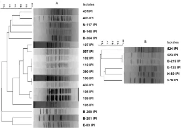
Fig. 1. PFGE analysis of vancomycin resistant E. faecium (A) and E. faecalis
(B). The phylogenetic tree is based on cluster analysis of the unweighted pair group method with average linkages (UPGAMA) and Dice analysis.
Isolates
431IPI 495 IPI N-117 IPI B-148 IPI B-364 IPI 107 IPI 557 IPI 102 IPI 110 IPI 390 IPI 106 IPI 436 IPI 108 IPI 109 IPI 105 IPI B-269 IPI B-201 IPI E-83 IPI
A
A
B Isolates
524 IPI 523 IPI B-219 IPI E-125 IPI N-89 IPI 578 IPI
20 OSKOUI ET AL . IRAN. J. MICROBIOL. 2 (1) : 15-22
and an IS1542 insertion in the orf2-vanR region. Type B was specified by the presence of two copies of IS1216V at the left-hand of Tn1546 and the vanX -vanY region and an IS1542 insertion in the orf 2-vanR intergenic region. Type C was indicated with one copy of IS1216V in the left-hand of Tn1546 and the presence of IS1542 in the orf2-vanR intergenic region. Type D and E were characterized with one copy of IS1216V in the vanX-vanY region and one copy in the left-hand of Tn1546, respectively. Group F had no insertion sequences (Table 3).
Through amplification of intergenic regions, only 9 and 6 isolates showed larger size of amplicon, approximately 3000bp, in the vanX-vanY and orf 2-vanR regions, respectively (Table 3).
Sequence analysis of the VanA transposons. Sequencing of vanX-vanY and orf2-vanR amplicons larger than those of the prototype VanA gene cluster (larger than 3000bp) showed that IS1216V and IS1542 were inserted in these regions, respectively. We published partial sequence of IS1216V and IS1542 with access numbers of FJ416860, FJ416861, GQ273971 and GQ273972 in GenBank (www.ncbi. nlm.nlh.gov/Genbank/submit.html). Sequencing of amplicons of the left end of Tn1546-like elements confirmed insertion of IS1216V in this region.
PFGE profiles. Analysis of the 18 E. faecium and 6 E. faecalis banding patterns differentiated 9 and 5 PFGE t
ypes respectively, with a similarity value of 0.7 (fig.1). So, the VRE isolates showed a high degree of heterogeneity. Among vancomycin resistant E. faecium, 9 isolates, with more than 78% similarity in their banding patterns, were the prevalent type.
DISCUSSION
Up to now, phenotypic and genotypic varieties of the vancomycin-resistant enterococci have been investigated in several previous studies (13, 21). In the present study, we compared the phenotypic (antibiotic resistant patterns) as well as genotypic traits of 24 VRE, isolated from patients admitted in two major hospitals of Tehran. The predominant species in this study were E. faecium (75%) with E. faecalis accounting for 25% of the remaining isolates. Overall, the VRE isolates in spite of resistant to vancomycin, were 100% resistant to erythromycin and
ciprofloxacin; furthermore, ampicillin showed a high frequency of resistance among E. faecium (94.5%). Similar to the previous study (22), chloramphenicol was the only drug that showed a lower rate of resistance among our isolates. Since consumption of antibiotics and the related selective pressures causes antibiotic resistance in community, it seems that high level of resistance to these antibiotics is related to their high consumption (1).
All the VRE harbored the vanA gene; however, 9 (37.5%) of them exhibited the VanB phenotype-vanA genotype. Incongruence of the VanB phenotype-vanA genotype has been found in Japan, Taiwan, and Korea (23), but to our knowledge, this is the first report of VanB-vanA VRE from humans in Iran.
According to PCR mapping, VanA elements were highly heterogeneous and none of them were completely similar to the prototype BM4147. Similar results were also obtained with previous studies (13, 14).
Based on PCR and sequencing results, IS1216V was found in 14 isolates (58.3%) in vanX-vanY and/or in the left-hand of Tn1546-like elements and had higher frequency among VRE. In 11 isolates (45.8%), both IS1216V and IS1542 were inserted in vanX-vanY and orf 2-vanR regions. IS1251 was identified neither with vanS F-vanH R primers nor with published primers of IS1251 and vanS/vanH through PCR reaction. Thus, the distribution of ISs in Tn1546-like elements of our isolates is similar to those among Europeans and Koreans (2, 21, 24) but not to American VRE isolates (1, 12, 13).
vanZ, as a nonessential gene in Tn1546, was detected in 19 (79.2%) of our isolates using PCR. The vanR, vanS, vanH and vanX genes are essential for the expression of vanA resistance, therefore, it was surprising that some isolates lacked these amplicons after PCR with intergenic region primers. PCR results obtained from each single gene showed that in 100%, 87.5%, 79.2% and 62.5% of the isolates vanR, vanS, vanH and vanX were amplified, respectively (data was not shown). In this study, negative PCR reactions were repeated several times, but absence of PCR products may indicate disruptions or insertions in these regions and these kinds of difficulties with amplifying these regions of some VanA elements have been reported previously (25).
findings were reported in Europe and Korea (14, 25). Based on some reports, point mutation, insertion of IS elements and deletion of intergenic region can be responsible for the VanB phenotype-vanA genotype (2, 11, 23). It seems that loss of some Tn1546 parts can be responsible for this change in our isolates.
Using PFGE we found high degree of diversity among the isolates. While 6 E. faecium with more than 78% similarity had both IS1216V and IS1542 in vanX-vanY and orf 2-vanR regions, we generally found no correlation between the positions of Insertion sequences in the Tn1546-like elements and the isolates PFGE types. The genetic diversity among Tn1546 elements was shown in several previous studies (14, 24, and 25). The variability in transposon type in various countries may be explained by the different antibiotic selective pressures against glycopeptides (2, 25), as well as the geographic differences in the transposon distribution (14) and movement of ISs in Tn1546 during outbreak (25).
In conclusion, our results revealed high degree of diversity and unique characterization among VRE in clinical specimens in this region. Thus, the genetic diversity indicated horizontal transfer of VRE rather than their clonal dissemination in this region and offered useful information for further epidemiological studies.
ACKNOWLEDGMENT
We are grateful to Dr. Mohammad Mehdi Feizabadi for providing the control isolates used in this study and also we thank Dr. Ali Nojoumi for carefully reading of the manuscript.
REFERENCES
Mendez-Alvarez S, Perez-Hernaadez X, Claveria-Mar-1.
tin F. Glycopeptide resistance in Enterococci. Internatl. Microbiol. 2000; 3: 71-80.
Oh JY, An S, Jin JS, Lee YC, Cho DT, Lee JC. Pheno-2.
typic and genotypic differences of the vancomycin-re-sistant Enterococcus faecium isolates from humans and poultry in Korea. J. Microbiol. 2007; 45: 466-472. Simjee S, White DG, McDermott PF, Wagner DD, 3.
Zervos MJ, Donabedian SM, et al. Characterization of
Tn1546 in vancomycin-resistant enterococcus faecium
isolated from canine urinary tract infections: evidence of gene exchange between human and animal entero-cocci. J. Clin. Microbiol. 2002; 40: 4659-4665. Biavasco F, Foglia G, Paoletti GZ, Magi G, Guagli-4.
anone E, Sundsfjord A, et al. VanA-type enterococci
from humans, animals, and food: species distribution, population structure, Tn1546 typing and location, and virulence determinants. Appl. Environ. Microbiol. 2007; 73: 3307-3319.
Henrique PM, Palazzo ICV, Zanella RC, Darini ALC. 5.
Molecular characterization of Enterococci harboring genotype and phenotype incongruence related to glyco-peptide resistance isolated ib Brazilian hospitals. Mem. Inst. Oswaldo. Cruz. 2008; 103: 301-305.
Gu L, Cao B, Liu Y, Guo P, Song S, Li R,
6. et al. A
new Tn1546 type of VanB phenotype-vanA genotype vancomycin-resistant Enterococcus faecium isolates in mainland China. Diagn. Microbiol. Infect. Dis. 2009; 63: 70-75.
Dahl KH, Simonsen GS, Olsvik Ø, Sundffjord A.
7.
Het-erogeneity in the vanB gene cluster of genomically di-verse clinical strains of vancomycin-resistant Enterococci.
Antimicrob. Agents Chemother. 1999; 43: 1105-1110. Guardabassi
8. L, Dalsgaard A. Occurrence, structure, and
mobility of Tn1546-like element in environmental iso-lates of vancomycin-resistant Enterococci. Appl. Envi-ron. Microbiol. 2004; 70: 984-990.
Naas
9. T, Fortineau N, Snanoudj R, Spicq C, Durrbach A,
Nordmann P. First nosocomial outbreak of vancomycin-resistant enterococcus faecium expressing a vanD-like phenotype associated with a vanA genotype. J. Clin. Mi-crobiol. 2005; 43: 3642-3649.
Camargo ILBC, Del Peloso PF, Da Costa Leite CF, 10.
Goldman GH, Darini ALC. Identification of an unusual VanA element in glycopeptides-resistant Enterococ-cus faecium in Brazil following international transfer of a bone marrow transplant patient. Can. J. Microbiol. 2004; 50: 767-770.
Park IJ, Lee WG, Shin JH, Lee KW, Woo GJ. VanB 11.
phenotype-vanA genotype Enterococcus faecium with heterogeneous expression of teicoplanin resistance. J. Clin. Microbiol. 2008; 46: 3091-3093.
Jung WK, Hong SK, Koo HC, Kwon NH, Park Y.H. 12.
Nucleotide sequence of IS1678, an insertion sequence in the vanA cluster of enterococci. Antimicrob. Agents Chemother. 2005; 49: 1666-1667.
Jensen LB, Ahrns P, Dons L, Jones RN, Hammerum 13.
AM, Aarestrup FM. Molecular analysis of Tn1546 in
Enterococcus faecium isolated from animals and hu-mans. J. Clin. Microbiol. 1998; 36: 437-442.
Huh JY, Lee WG, Lee K, Shin WS, Yoo JH. Distribu-14.
tion of insertion sequences associated with Tn1546-like elements among Enterococcus faecium isolates from pa-tients in Korea. J. Clin. Microbiol. 2004; 42: 1897-1902. Donabedian S, Hershberger E, Thal LA, Chow JW, 15.
Clewell DB, Robinson-Dunn B, et al. PCR fragment
length polymorphism analysis of vancomycin-resistant
Enterococcus faecium. J. Clin. Microbiol. 2000; 38: 2885-2888.
Novais
16. C, Coque TM, Sousa JC, Baquero F, Peixe L,
and The portuguese resistance study group. Local ge-netic patterns within a vancomycin-resistant enterococ-cus faecalis clone isolated in three hospitals in Portugal.
22 OSKOUI ET AL . IRAN. J. MICROBIOL. 2 (1) : 15-22
Novais C, Sousa JC, Coque TM, Peixe L, and The por-17.
tuguese resistance study group. Molecular character-ization of glycopeptide-resistant Eterococcus faecium
isolates from Portuguese hospitals. Antimicrob. Agents
Chemother. 2005; 49: 3073-3079.
Manero A, Blanch AR. Identification of Enterococcus 18.
spp. with a biochemical key. Appl. Environ. Microbiol. 1999; 65: 4425-4430.
Clinical and Laboratory Standards Institute. Perfor-19.
mance standards for antimicrobial susceptibility testing, 16th informational supplement, M100-S16. Clinical and Laboratory Standards Institute, Wayne, Pa, USA. 2006. McEllistrem MC, Stout JE, Harrison LH. Simplified 20.
protocol for Pulsed-Field Gel Electrophoresis analysis of Streptococcus pneumonia. J. Clin. Microbiol. 2000; 38(1): 351-353.
Jung WK, Hong SK, Lim JY, Lim SK, Kwon NH, Kim 21.
JM, et al. Phenotypic and genetic characterization of
vancomycin-resistant enterococci from hospitalized hu-mans and from poultry in Korea. FEMS Microbiol. Lett. 2006; 260: 193-200.
Busani L, Grosso MD, Paladini C, Graziani C, Pantosti 22.
A, Biavasco F, et al. Antimicrobial susceptibility of van-comycin-susceptible and –resistant enterococci isolated in
Italy from raw meat products, farm animals, and human infections. Int. J. Food Microbiol. 2004; 97: 17-22. Ko KS, Baek JJ, Lee J, Oh WS, Peck KR, Lee NY,
23. et
al. Molecular characterization of vancomycin-resistant
Enterococcus faecium isolates from Korea. J Clin Mi-crobiol. 2005; 43: 2303-2306.
Willems RJL., Top J, Braak NVD, Belkum AV, Mevius 24.
DJ, Hendriks G, et al. Molecular diversity and evolu-tionary relationships of Tn1546-like elements in entero-cocci from humans and animals. Antimicrob. Agents Chemother. 1999; 43: 483-491.
Woodford
25. N, Adebiyi AA, Palepou MI, Cookson BD.
Diversity of VanA glycopeptide resistance elements in enterococci from humans and nonhuman sources. Anti-microb. Agents Chemother. 1998; 42: 502-508. Miele A, Bandera M, Goldstein BP. Use of primers se-26.
lective for vancomycin resistance gene to determine van
genotype in enterococci and to study gene organization in VanA isolates. Antimicrob. Agents Chemother. 1995; 39: 1772-1778.
Yu H, Seol S, Cho D. Diversity of Tn
27. 1546-like elements