*Corresponding author: Mojgan Bandehpour PhD. Department of Biotechnology, School of Advanced Technologies in Medicine, Shahid Beheshti Univer-sity of Medical Sciences, Tehran, Iran
Tel: +98-21-22439957 Fax: +98-21-22439956
E-mail: [email protected]
Cloning, expression and purification of the factor H binding protein and
its interaction with factor H
Fatemeh Yarian1, Mojgan Bandehpour2,3*, Negar Seyed4, Bahram Kazemi1,2,3
1Department of Biotechnology, School of Medicine, Shahid Beheshti University of Medical Sciences, Tehran, Iran
2Department of Biotechnology, School of Advanced Technologies in Medicine, Shahid Beheshti University of Medical Sciences, Tehran, Iran
3Cellular and Molecular Biology Research Center, Shahid Beheshti University of Medical Sciences, Tehran, Iran
4Department of Immunotherapy and Leishmania Vaccine Research, Pasteur Institute of Iran, Tehran, Iran
Received: July 2015, Accepted: November 2015
ABSTRACT
Background and Objective: Neisseria meningitidis is a leading cause of meningitis and sepsis worldwide. The factor H binding protein (fHBP) is a key virulence factor of Neisseria meningitidis that is able to selectively bind to human factor H, the key regulator of the alternative complement pathway, which it has important implications for meningococcal pathogene-sis and vaccine design. The aims of present research were cloning, expression, purification of fHbp and confirmation of the interaction between serum factor H (fH) and produced factor H binding protein.
Materials and Methods: A 820 base pairs fhbp gene fragment was amplified by PCR and cloned into expression vector pE -T28a (+) in Bam HI and SalI restriction enzymes sites. Recombinant DNA was expressed in BL21 (DE3) cell. fHBP protein was purified by Ni-NTA agarose resin. Coupling of recombinant protein into CNBr activated Sepharose 4B resin was carried out for application in serum fH protein purification. (fH-fHBP) interaction was confirmed by SDS-PAGE and far-western blotting.
Results and Conclusions: SDS-PAGE results showed a 35 kDa protein band. 150 kDa fH protein was purified by designed Sepharose 4B resin. Far-western blotting confirmed (fH-fHBP) interaction and proper folding of factor H binding protein.
Keywords: N. meningitidis, FHBP, Cloning, FH protein, Far-western blotting.
croorganism and an exclusive human pathogen that usually exists in an asymptomatic nasopharyngeal carriage state. However, N. meningitidis can cause devastating invasive disease, such as septicemia or meningitis, following penetration of the mucosal tissue, invasion of the bloodstream, and colonization of the meninges. The virulence of N. meningitidis
is influenced by multiple factors: capsule polysac
-charide and surface adhesive proteins expression (outer membrane proteins including pili, porins PorA and B, adhesion molecules Opa and Opc), iron sequestration mechanisms, and endotoxin
(lipooli-ORIGINAL
AR
TICLE
INTRODUCTION
-gosaccharide, LOS) (1). fHBP was initially identi -fied as a surface-exposed lipoprotein with unknown
function named GNA1870 (genome-derived Neisse
-ria antigen 1870) (2). The complement system plays an important role in innate immune defense against pathogenic microbes. Factor H is a 150-kDa soluble protein that is the main regulator of the alternative pathway (AP) which it is secreted in the nasophar-nyx (3). Some microbes and viral pathogens such as Echinococcus granulosus, Onchocerca volvulus and HIV, avoid complement-mediated killing sys-tem by recruiting fH to their surfaces (4). Pioneer-ing study demonstrated that the interaction of fH with the meningococcus GNA1870 (fHBP) is the principal of fH-binding meningococcal protein (5). Most (fH-fHBP) interaction studies have been done in vivo (5). Therefore we attempted to produce re-combinant fHBP and investigate it in vitro. All to-gether the production of recombinant proteins are one of the biotechnology special skills for drug or vaccine and nutrition science researches. Currently, recombinant proteins are found fundamentally in ev-ery medical testing and biological research laborato-ry (6). So the recombinant fHBP will be applied in medical sciences like the design of diagnostic kit or meningococcus vaccine preparation.
MATERIALS AND METHODS
Cloning of fHBP gene into pET 28a (+). The fhbp gene was selected based on the fhbp gene sequence in the GenBank accession nos. ACA52540.1. It was synthesized into pGH plasmid (Bioneer, Korea).
The fhbp gene was amplified from pGH plasmid by
specific primers. Amplification was carried out in 30 cycles of 40 sec at 94 °C, 40 sec at 64 °C and 1 min
at 72 °C. The PCR product was confirmed by nucle
-ic acid sequencing. Following the initial confirma
-tion, PCR product and pET28a (+) (Novagen, USA) were digested with BamHI and SalI (Fermentas, Lithuania). Ligation was carried out with T4 DNA Ligase (Fermentas, Lithuania). The ligation reaction was transformed into E. coli Top10 competent cell (7). The recombinant plasmids were confirmed by colony PCR and restriction enzyme analysis.
Expression of the recombinant fhbp gene.E. coli BL21 (DE3) was used for protein expression as host
with 50µg/µl kanamycin (Merck,Germany) in LB
medium for selection, 0.5 mM IPTG (Isopropyl-be
-ta-D-thiogalacto-pyranoside) (Merck,Germany) as inducer. Samples were collected 3, 5, 7, and 9 hours after induction. The cells were harvested, treated with lysis buffer (50 mM Tris base, 10% glycerol, 0.1%Triton X-100) (Merck, Germany) and the lysate was analyzed by SDS-PAGE and the quantity of the expressed protein was estimated by comparing the intensity of the protein bands.
Western blot analysis. Proteins resolved by
SDS-PAGE were electrophoretically transferred to a ni
-trocellulose membrane (Wathman, UK). TBS buffer (Tris-Buffered Saline containing 3% BSA (Bovine Serum Albumin) (Sigma, USA) was used for block-ing the membrane. The membrane immersed in
1:2000 dilution of ALP (alkaline phosphatase) con
-jugated anti His-tag monoclonal antibody (Abcam, UK) 2 hours at room temperature. Subsequently, it was visualized for color after development in NBT/ BCIP substrate solution (Roche, Germany).
Purification of the protein. The recombinant
protein purification was carried out by Ni-NTA col
-umn as specified by the manufacturer's instructions
(Novagen, USA). The purified protein was subse
-quently analyzed by SDS-PAGE and western blotting using anti His-tag antibody.
Coupling of recombinant protein into CNBr activated Sepharose 4B and purification of fH serum protein. For studying of (fH-fHBP) inter-action, purified fHbp protein was coupled to CNBr
activated Sepharose 4B by the manufacturer's in
-structions (Novagen,USA). 500 µl of human serum was dialyzed by coupling buffer (NaHCO3 0.1 M, NaCl 0.5 M, PH 8). The affinity chromatography was performed for isolation of fH protein from se-rum sample. It was eluted by two different pH buffer (Glycin,NaCl, pH:2 and Diethanolamin,NaCl pH:11) (Merck,Germany).
milk). The eluted factor H was analyzed by western
blotting using an ALP-conjugated anti-His tag anti
-body (Abcam, UK) (8).
RESULTS
The preparation of gene fragment. The fhbp gene was amplified with specific primers as shown in Fig. 1. The pET28a/fHBP plasmid was confirmed
by specific PCR, universal PCR and restriction anal
-ysis (Fig. 2).
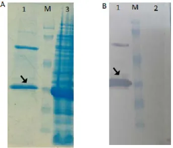
Expression of fHBP recombinant protein. Re -combinant protein expression was confirmed by western blotting (Fig. 3A). The optimum incubation time after induction was considered 3 hours (Fig. 3B).
Purification of the recombinant protein. Puri-fication of the recombinant protein was carried out
using Ni-NTA affinity column (Fig. 4A). The expect
-ed protein band was obtain-ed in elut-ed fraction and confirmed by western blotting (Fig. 4B).
Purification of serum fH protein. Serum factor
H protein was purified using CNBr activated Sep
-harose 4B coupled to fHBP. It was analyzed by SDS-PAGE and a ͠~150 KD protein was detected (Fig. 5)
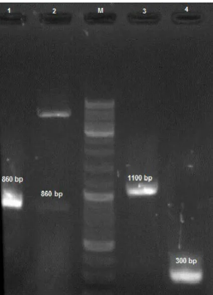
Fig. 1. Specific PCR product of fhbp gene: Lane M, 100bp DNA ladder ; Lane 1, 860bp PCR product.
Fig. 3. SDS-PAGE and Western blot analysis of the recombinant protein expression at different times: (A) Lane M, molecular weight marker,Lane1, BL 21; Lane 2, 0 time; Lane 3, 3h after induction. (B) Lane 1,3h ; Lane 2, 5h ; Lane 3; 7h after induction with 0.5 mM IPTG. Lane 4, BL 21 cell; Lane 5, 0 time.
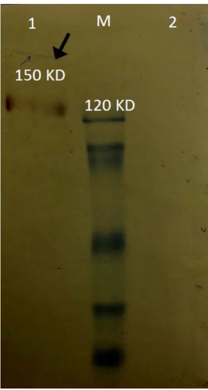
Far Western Blot analysis. fH-fHBP interaction was determined by anti-His tag antibody. A band with approximate size of 150 KD appeared on the membrane (Fig. 6).
DISCUSSION
Neisseria meningitidis is an obligate human patho-gen and important cause of septicemia and meningi-tis that it is known a major cause of morbidity and mortality worldwide. To cause disease, the bacterium must successfully survive in the bloodstream where it has to avoid being killed by host innate immune mechanisms, particularly the complement system (9). Most N. meningitidis strains studied to date, express fHBP, with levels of expression that vary significantly among isolates (2). Humans clear meningococci via both the classical and alternative complement path-ways (10). Factor H is a 150-kDa glycoprotein typi-cally presents in human plasma at concentrations of 300 to 500 μg/ml (11). fH is the main inhibitor of the
alternative complement pathway and a key discrimi-nator between host and pathogen cells. Many patho-gens, however, have evolved to evade this alternative complement pathway by binding fH on the bacteri-al surface. Although fH binding proteins have been
identified in many pathogens, the sequence and struc
-ture of fHBP are unique to N. meningitidis and a few other neisserial species (12). Interestingly, fHBP only binds human fH, which may be one explanation of the species-specificity of meningococci for the human host (13). The ability of fHBP to bind human fH with high affinity has implications not only for its role as a Fig. 5. Western blot analysis of the purified fH protein by
affinity chromatography: Lane M, molecular weight marker, Lane 1, purified factor H protein
Fig. 6. fH-fHBP binding confirmation by Far western blot analysis:
virulence factor in vivo, but also it can be as a target antigen for vaccination against meningococcus. An-ti-fHBP antibodies can elicit protection by two mech-anisms: direct complement-mediated killing of the bacterium and blocking fH binding to the bacteria to increase the susceptibility of the bacterium to be killed by the alternative complement pathway (5). Several studies have shown that inhibition of fH binding to fHBP results in increased susceptibility of the bacte-ria to complement-mediated bactericidal activity (14). The main objective of present study was producing of recombinant fHBP and fH-fHBP interaction study in vitro. Confirmation of this step, can be used in the field of vaccine and specific antibodies production. Prokaryotic systems are powerful tools for production of this recombinant protein. Escherichia coli is one of the best micro organisms of choice in this field. It is a factory and well-established micro organism for the most popular expression platform. Recombinant DNA is extensively used in biotechnology, medicine and researches (15). For this reason, pET expression system has been developed for the cloning and ex-pression of recombinant proteins in E. coli based on the T7 promoter (16). So, successful sub cloning of 820 bp fHBP gene into pET 28a (+) expression vec-tor was carried out by enzymatic digestion. Western blotting technique using Anti-6X His-tag antibody
confirmed 35 kDa molecular mass recombinant pro
-tein expression. Purified pro-tein by Ni-NTA agarose resin showed two distinct monomer and dimer bands on gel and western blotting. There were two ways to check (fH-fHbp) interaction. One of them was design-ing of new affinity chromatography which resin binds to recombinant fHBP. CNBr activated Sepharose 4B is able to coupling with some proteins. Therefore, by activation of resin according to manufacturer’s pro-tocol, fHbp was coupled into resin. fH is a ligand for fHBP (5) and it has typically 150-kDa glycoprotein presents in human serum at concentrations of 300 to 500 μg/ml (11, 17). Because of unique structure of
this recombinant protein and specific coupling con
-dition, its purification will be very specific. Rickard Nilsson in 2013 described an affinity chromatography system, based on the HVR of the M5 protein, allowing efficient, single-step purification of fH from human serum (8). Another way to check fH-fHBP interaction was far-western blotting. The binding of fH to surface of sero groups A, B, and C of N. meningitidis strains were detected by FACS and Far-Western blot analysis (18). Far-western blotting (WB) was derived from the
standard WB method to detect protein-protein inter-actions in vitro. In Far WB, proteins in a cell lysate containing prey proteins are firstly separated by SDS or native PAGE, and transferred to a membrane, as in a standard WB. The proteins in the membrane are then denatured and renatured. The membrane is then
blocked and probed, usually with purified bait pro
-tein (s). The bait pro-teins are detected on spots in the membrane where a prey protein is located, if the bait proteins and the prey protein together form a complex. Compared with other biochemical binding assays, Far WB allows prey proteins to be endogenously ex-pressed without purification, and determines whether two proteins bind to each other directly (19, 20). So, to demonstrate fH-fHBP interaction, we successfully used Far-Western blotting. In present study, human se-rum fH protein was used as a prey protein and purified recombinant fHBP as a bait one. Recombinant fHBP consists of 6X-His tag peptid, which is detectable with conjugated Anti-6X His-tag antibody.
Based on the results of this study, considerable amount of recombinant fHbp protein was expressed in E. coli BL21 (DE3). It was used for designing a prop -er chromatography column to purify a large amount of the factor H protein.
ACKNOWLEDGEMENT
This article was extracted from Fatemeh Yarian’s thesis and was carried out in Cellular and Molecular
Biology Research Center of Shahid Beheshti Univer
-sity of Medical Sciences. We thanks Iran National Sci-ence Fundation for supporting of this research.
REFERENCES
1. Stephens DS. Biology and pathogenesis of the evolu-tionarily successful, obligate human bacterium Neisse-ria meningitidis. Vaccine 2009;27 Suppl 2:B71-7. 2. Masignani V, Comanducci M, Giuliani MM, Bambi
-ni S, Adu-Bobie J, Arico B, et al. Vaccination against Neisseria meningitidis using three variants of the lipo-protein GNA1870. J Exp Med 2003;197:789-99. 3. Zipfel PF, Jokiranta TS, Hellwage J, Koistinen V, Meri
S. The factor H protein family. Immunopharmacology 1999;42(1-3):53-60.
4. Diaz A, Ferreira A, Sim RB. Complement evasion by
H in the hydatid cyst wall. J Immunol 1997;158:3779-3786.
5. Madico G, Welsch JA, Lewis LA, McNaughton A, Per -lman DH, Costello CE, et al. The meningococcal vac-cine candidate GNA1870 binds the complement regu -latory protein factor H and enhances serum resistance. J Immunol 2006;177:501-510.
6. Sreenivas S, Krishnaiah SM, Govindappa N, Basavara -ju Y, Kanojia K, Mallikar-jun N, et al. Enhancement in production of recombinant two-chain Insulin Glargine by over-expression of Kex2 protease in Pichia pastoris.
Appl Microbiol Biotechnol 2015;99:327-336.
7. Ghodsi S, Gharavi S, Ghadam P. Cloning the hbs gene from Bacillus subtilis and expression of the HBsu protein in Escherichia coli. Iran J Microbiol 2010;2(3):152-6.
8. Nilsson OR, Lannergard J, Morgan BP, Lindahl G, Gustafsson MC. Affinity purification of human fac -tor H on polypeptides derived from streptococcal m protein: enrichment of the Y402 variant. PloS one 2013;8(11):e81303.
9. Stephens DS, Greenwood B, Brandtzaeg P. Epidemic meningitis, meningococcaemia, and Neisseria menin-gitidis. Lancet 2007;369(9580):2196-2210.
10. Wright V, Hibberd M, Levin M. Genetic polymor -phisms in host response to meningococcal infection: the role of susceptibility and severity genes. Vaccine 2009;27 Suppl 2:B90-102.
11. Haralambous E, Dolly SO, Hibberd ML, Litt DJ, Udalova IA, O'Dwyer C, et al. Factor H, a regulator of complement activity, is a major determinant of me-ningococcal disease susceptibility in UK Caucasian patients. Scand J Infect Dis 2006;38:764-771.
12. Lambris JD, Ricklin D, Geisbrecht BV. Complement evasion by human pathogens. Nat Rev Microbiol 2008;6:132-142.
13. Granoff DM, Welsch JA, Ram S. Binding of comple -ment factor H (fH) to Neisseria meningitidis is specific for human fH and inhibits complement activation by rat and rabbit sera. Infect Immun 2009;77:764-769. 14. Giuntini S, Beernink PT, Reason DC, Granoff DM.
Monoclonal antibodies to meningococcal factor H binding protein with overlapping epitopes and discor-dant functional activity. PloS one 2012;7(3):e34272. 15. Chou CP. Engineering cell physiology to enhance
re-combinant protein production in Escherichia coli. Appl Microbiol Biotechnol. 2007;76:521-32.
16. Studier FW, Moffatt BA. Use of bacteriophage T7 RNA polymerase to direct selective high-level expres -sion of cloned genes. J Mol Biol 1986;189:113-130. 17. Pio R, Elsasser TH, Martinez A, Cuttitta F. Identifi
-cation, characterization, and physiological actions of factor H as an adrenomedullin binding protein present in human plasma. Microsc Res Tech 2002;57:23-27. 18. Schneider MC, Exley RM, Chan H, Feavers I, Kang
YH, Sim RB, et al. Functional significance of fac -tor H binding to Neisseria meningitidis. J Immunol 2006;176:7566-775.
19. Berggard T, Linse S, James P. Methods for the detec -tion and analysis of protein-protein interac-tions. Pro-teomics 2007;7:2833-2842.