BERTRAM FLESHLER
Case Western Reserve University School of Medicine, Cleveland, Ohio 44106
Most, if not all, ingested pro-tein is transported across the gut as its constituent amino acids. Thus, the way the gut handles amino acids is an important physiologic problem, and their mishandling by the gut in disease states represents an important threat to general body activities. I propose to dis-cuss some of the knowledge that has been gathered over the past 10 to 15 years concerning the mechanisms of amino acid ab-sorption. The data to be pre-sented have been gathered from three major kinds of studies: the studies of in vitro model systems, physiologic studies of humans, and studies of patients, with syndromes involving specific amino absorption abnormalities. Some of these data have been accumulated in our own laboratory; much of what I have to present is the work of other investigators.
In Vitro Studies on Gut
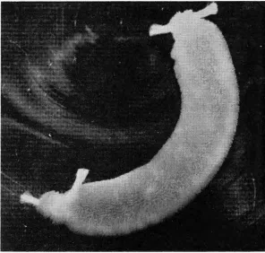
Figure 1 shows an experimental technique which has become a classic model for the study of transport of various substances, not only amino acids. This is the everted gut sac. The technique in-volves making everted gut seg-ments from the intestines of small animals. The animal's gut is re-moved as quickly as possible, cleaned, turned inside out so that the mucosa is on the outside, and small sacs made of the segments
*
Presented at the Thirty-Ninth Annual McGuire Lecture Series, No-vember 9-10, 1967, Medical College of Virginia, Richmond.MCV QUARTERLY 4(1): 21·31, 1968
of the gut. The sacs are then placed in various bathing solutions con-taining the substances to be tested.
Figure 2 shows the kinds of re-sults one can obtain with this kind of method. As indicated by the dotted line, the concentration of the substance tested, in this case the amino acid, L-tryptophan, was the same on both the mucosa! and the serosal sides. The measurement of the concentration changes at various time intervals following the placing of the substance in the sac is an indication of what movement is going on. Under aerobic con-ditions (the box on the left), the mucosa! concentration of trypto-phan (black column) decreased after an hour's incubation, and the serosal concentration increased. Remember that the initial concen-tration was the same on both sides. Anaerobically, when oxygen was not allowed in the flask, concen-tration against a gradient no longer occurred.
The importance of aerobic, energy-dependent systems for transport of L-tryptophan from the mucosa! surface to the serosal side is shown in Figure 3. In this study, tryptophan initially was placed only on the mucosa! side, in con-trast to the previous study. None was on the serosal side. After an hour's incubation aerobically, not only had some tryptophan been transported from the mucosa! to the serosal side, but there also now existed a concentration gradi-ent between these two sides. By contrast, anaerobically the trans-fer from the mucosa! to serosal side was very much diminished,
and no concentration gradient had been developed.
Figure 4 shows an experiment with a substance for which there is no active transport mechanism. The test substance was pyridoxine. In the upper bar is shown the situation when pyridoxine was placed at equal concentration on both sides of the everted gut sac. There was no concentration gradi-ent developed from the mucosa to serosa either aerobically or anaero-bically. The bottom set shows the situation when the pyridoxine was placed only on the mucosa! side. Transfer was slow; no concentra-tion gradient developed, and it did not make any difference whether the environment was anaerobic or aerobic. This further confirms the idea that energy-dependent systems are of significance in transporting tryptophan, whereas they are not of significance in transporting pyri-doxine.
By the use of this in vitro tech-nique, unphysiologic as it may sound, much information has been gathered as to the transport of amino acids. Table 1 shows some data obtained with a series of amino acids using the everted gut sac technique. A concentration gradient
>
1.00, as indicated in the third column, indicates that there was accumulation of these amino acids on the serosal side as com-pared to the mucosa!· side, the amino acids having been in each instance placed in equal concen-trations on both sides of the mem-brane at the start of the experiment. It is obvious that different amino acids behave in somewhat differentmanners. In addition to testing single amino acids, mixtures of amino acids and amino acids in which parts of the amino acid mol-ecules have been modified have also been studied.
On the basis of such studies in the everted gut sac, it appears that certain types of amino acids are transported by specific mecha-nisms. It is presently thought that there exist separate specific trans-port mechanisms for the mono-carboxylic neutral amino acids; for dibasic amino acids, such as lysine, arginine, and ornithine along with the neutral amino acid, cystine; for the dicarboxylic amino acids; and, possibly, for proline, hydroxy-proline and, questionably, glycine.
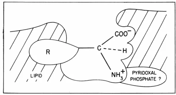
Figure 5 shows a sketch of how amino acids might interact with a membrane carrier. This scheme applies, most likely, to the neutral amino acids. The carboxyl, the hydrogen, and the alpha amino groups all are quite specific, since substitutions at any one of these sites decrease the transport rate of the amino acid. The question mark next to the pyridoxal phosphate in-dicates that certain studies suggest that this substance may be an im-portant co-factor in neutral amino acid transport. The longer the side group, indicated here as the R, the more lipophilic the substance is and the greater the affinity of the amino acid for the lipid carrier or the lipid membrane. It has been shown in the group of neutral amino acids that those with a longer side chain, i.e., those with a more lipophilic side chain, com-pete preferentially for transport with those with shorter side chains. Thus, on the basis of these ani-mal in vitro experiments, of which this has been only a brief resume, there has evolved a concept of amino acid absorption as being one in which there are several specific carrier mechanisms in-volved.
Studies on Human Gut
In order to study amino acid ab-sorption in humans, different
tech-AMINO-ACID ABSORPTION
Fig. 1-Everted sac method of Wilson and Wiseman. Tied sac of everted hamster jejunum containing 1 ml of fluid. X 2.64 (Reprinted with permission from T. H. Wilson, Intestinal Absorption. Philadelphia: W. B. Saunders, 1962, p. 33.)
AE.ROBIC ANAEROBIC
40
E 20 - - CONCENTRATION INITIAL
Ol BOTH SJDE.S
~
0
D
Sero so I • Mucosol Time: I hourTABLE 1
Rates of Transference of Amino -Acids an{ Concentration Gradients Developed by Sacs of Everted Intestine
Rate of transference Concentration gradient Amino Acid (µl./mg dry wt./hr) developed
Praline* 14.0± 3.2 2.08 ± 0.18
Threonine 12.0 ± 2.7 1.90 ± 0.28
Alanine 11.5 ± 3.5 1.82 ± 0.32
Glycine* 10.1 ± 2.4 1.65 ± 0.19
Serine 8.8 ± 2.2 1.61 ± 0.19
Valine 8.2 ± 2.7 1.42 ± 0.19
Histidine* 5.3 ± 2.8 l.42 ± 0.22
Hydroxyproline 6.3 ± 1.8 1.36 ± 0.14
Phenylalanine 5.4 ± 2.2 l.24 ± 0.11
lsoleucine 4.0 ± 1.2 1.19 ± 0.09
* These results are taken from Wiseman (1955) and are included in this table for the sake of completeness. The experiments were performed under identical conditions.
Adapted with permission from J. Physiol. 133: 628, 1956.
AEROBIC ANAEROBIC
40
E INITIAL
..__
20-
MUCOSALCl CONCE.NTRATION
::i.
0
D
Serosol • Mucosa! Time ' I hourFig. 3- Typical everted loop experiments using "H-labelled L-tryptophan initially on the _outside of the sac (mucosa! side) only. Aerobically active transport r:sult~ m the development of a concentration gradient. Anaerobically only d1ffus1on occurs. (Reprinted with permission from The Scientific Basis of
Medi-cine Annual Reviews. London: The Athlone Press, 1963, p. 173.)
niques must be developed. Aside from the difficulty of obtaining pieces of human gut to make everted gut sacs, such sacs could not be used in the same way as sacs from hamster gut. Besides pos-sible species differences in trans-port, from the practical standpoint there is the fact that, in the larger species, such as dog, the muscle wall is so thick that penetration of substance from mucosa through to the serosal side is ineffective. In hu-mans two major kinds of approaches have been used. One method is to take pieces of gut at operations, or by biopsy, and perform in vitro studies. I shall discuss these studies later, mainly in regard to certain disease states. Another method is to perfuse the gut under more or less physiologic conditions. Per-fusion experiments . to study in-testinal absorption of amino acids and other substances in the human have been widely used. In general the method is as follows: A sub-ject is intubated with a tube which most often contains two holes, a perfusing hole and an aspirating hole, some distance down the gut. The test substance is then per-fused along with a non-absorbable reference marker, polyethylene glycol being one that is commonly and popularly used today. The dis-appearance from the gut lumen of the substance to be studied in re-lation to· changes in marker con-centration as a measure of water absorption has been used as a test for absorption of the specific sub-stance being studied. Thus, if the concentration of amino acids drops during the test, in relation to the concentration polyethylene glycol, some of the amino acid has been absorbed during the period of per-fusion.
Table 2 shows the kind of data obtained when the absorption of glycine was studied by the perfu-sion method. As the glycine con-centration in the perfusion fluid was increased, the amount absorbed,
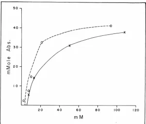
pressed as millimoles per 15-cen-timeter gut segment perfused, also increased. The increase in the amount absorbed, however, was not linearly related to the increase in the glycine concentration of the perfusion fluid. The rate of glycine absorption reached a limiting value. This is illustrated in Figure 6, which shows the results of
studies of L-isoleucine absorption in two subjects. Plotted are
milli-moles absorbed against the
con-centration of isoleucine perfused. As the concentration increased, the
amount absorbed also increased, but the absorption rate tailed off as the concentration increased. If simple diffusion were the process involved, one would expect a straight-line relationship between
the concentration perfused and the amount absorbed. Instead, this
curve suggests saturation kinetics
and is, thus, consistent with the
idea that some sort of active trans-port process is taking place.
Figure 7 shows the results of transport fluids in which the gut
was perfused with solution
con-taining both glycine and L-alanine. At the bottom of Figure 7 are in-dicated the concentrations of each
substance for any one study. For example, "fifty" means that a 50
mM solution of glycine and a 50 mM solution of L-alanine were
per-fused together. Three patients were
TABLE 2
Absorption of Glycine
Perfused (mM)
10
25
50
75
100 150
300
Absorbed (mmoles/15
min/15-cm gut segment)
1 ± 0
3± 1 3 ± 1
6 ± 1
7 ± 1 10 ± 3
15 ± 4
Adapted with permission from J. Clin. Inv. 45: 1435, 1966.
AMINO-ACID ABSORPTION
AEROBIC ANAEROBIC SYSTEM
100
E
EQUAL0;50 BOTH SIDES
~
100
E OUTSIDE
"ii
50 ONLY~
0
0
Serosal • Mucosa! Time : I hourFig. 4-Everted loop experiments using 0H-labelled pyridoxine HCI. The
interrupted line indicates the initial concentrations. With equal concentrations on both sides of the sac initially (upper figure), there is no active transport for no concentration gradient develops. When pyridoxine is only present on the outside (lower figure), it diffuses across into the serosal fluid equally well whether incubation is aerobic or anaerobic. (Reprinted with permission from The Scientific Basis of Medicine Annual Reviews. London: The Athlone Press, 1963, p. 174.)
studied, and on each several
ex-periments were performed. It can
be seen that, at every concentration
studied, the absorption of alanine,
shown by clear bars, was always
greater than the absorption of
gly-cine. If passive diffusion process
alone were involved, the two sub-stances would not be expected to so drastically influence each oth-er's absorption.
Another way of testing whether
or not substances share a common
site for absorption involves hold-ing the concentration of one sub-stance constant but changing the concentration of the other
sub-stance. The first is then called the
inhibitor; the second, the test
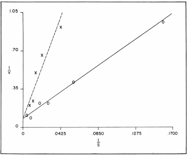
sub-stance. Figure 8 shows a
Line-weaver-Burk plot, a double
recip-rocal plot in which l/S is the
reciprocal of the concentration
per-fused, and 1/V represents the
re-ciprocal of the amount absorbed.
The solid line connecting these
open circles indicates the a
bsorp-tion of glycine at various concen-trations when perfused alone. The dotted line indicates the absorption of glycine in the same subject when perfused at various concentrations
together with 150 mM L-alanine as
the inhibitor. This line is displaced
upwards and to the left; in other
words, the absorption of glycine
is decreased in the presence of
the inhibitor, L-alanine. However,
as the concentration of glycine is increased, the line tends to meet the line representing absorption of glycine alone; it intercepts the ver-tical axis at the same place. The
fact that the lines under these two
conditions arrive at the same point is consistent with a form of
in-hibition called competitive
inhibi-tion, that is, where two substances
compete for the same transport mechanism or for the same
bind-ing site. If the inhibition were not
competitive, that is, if the binding site were being poisoned in some
way by the inhibitor, then the
in-tercepts would not be expected to
be at the same place. These studies in humans with glycine and alanine
50
40
(f)
..0 30 <(
Q) 0 ~ 20
E
10 I I I
/o
II
I I I I
o:
I I
I I I
Cl.-I I
I
20
--
...
---
---0
40 60 80 100 120
mM
Fig. 6-Plots of L-isoleucine absorption (mmole/15 min/ 15-cm gut segment)
against concentration of isoleucine perfused. The results obtained on two human
subjects with intubation tubes for perfusion are shown.
D
15w
c:
z Q)
E z o>
<C Q) _J
"'
<C-s
.!.i o> 10
a:: E
0 (..)
ml
LO
...
"'
w ~
z :::> c: 0
·e
~ 5
<.!) ~
LL.. ...
"'
0 ~ 0 z E
0 E
I-ll..
a:: 0
0
({)
m 50 75 150 150 90 ·150 300
<C
CONCENTRATIONS OF GLYCINE AND L-ALANINE
IN THE PERFUSATES (mM)
Fig. 7- Absorption of glycine and L-alanine when perfused together at
equimolar concentrations. The first three studies (starting at the left) were
performed in Subject 1, the fourth in Subject 2, and the last three in Subject 3.
In the first study, perfusate osmolality was 245 mOsm per kg and in the last
two studies, 396 and 620 mOsm per kg, respectively. The height of the column
indicates mean absorption. (Reprinted with permission from J. Clin. Inv.
are, thus, consistent with the studies in everted gut sacs in in
vitro preparations. They indicate
that these particular amino acids are absorbed by active transport mechanisms.
The influence of the lipophilic side chain, suggested by the everted gut sac experiments, can also be
seen when three amino acids are
tested together. Figure 9 shows the results of perfusing together at
equimolar concentrations:
isoleu-cine, alanine, and glycine.
Isoleu-cine was always absorbed faster than alanine, which, in turn, was
always absorbed faster than the
glycine.
On the basis of these studies and the everted gut sac studies, one would then predict that L-isoleucine would be a very effective inhibitor of glycine absorption. Surprisingly, however, this was not found. Figure 10 shows Lineweaver-Burk plots of glycine absorption. The solid line
indicates glycine absorption when
glycine alone was perfused. The
small-dash line indicates the effect
of 150 mM alanine; the large-dash line is the effect of isoleucine as an inhibitor at 150 mM. No inhibitory effect by the L-isoleucine on gly
-cine absorption was seen. Isoleu-cine, as indicated previously, cer-tainly is transported more quickly than is glycine. These studies show that isoleucine is not, however, as effective an inhibitor of glycine
ab-sorption as is alanine. This might
well indicate that there are two
steps in amino acid transport. One
would be a postulated entry step
in which the length of the lipo-philic chain is important and in which the longer side chain of isoleucine produces greater affinity
with the lipid membrane, enabling
it to be transported more quickly.
The second step, perhaps an exit
step, may be the specific carrier
mechanism and, indeed, may be the rate-limiting step for the proc-ess. Isoleucine and glycine do not
appear to share this later step.
AMINO-ACID ABSORPTION
1.05
I I
I
I IX I I
I I
I
.70 I I
X I I I
I
I
v
x/
I
I I
.35 I
I I
/x
IX 0 0 I
I
0
0
0 .0425 .0850 .1275 .1700
.!.. s
Fig. 8-Results of perfusing glycine alone (straight line). Results of perfusing
glycine in the presence of 150 mM L-alanine (dash line). (Reprinted with
permission from J. Clin. Inv. 45: 1437, 1966.)
7.
5.
0
~
u"'
~ 3.
E
.
.
1.
.
/
..
/
./
.)
/
..
·
/
/
.
/ '
/ /
16 24 3 2 mM
Fig. 9-Results of perfusing isoleucine (dotted line), alanine (dash line), and
In Vitro Studies on Gut Mucosa
In vitro studies on the distribu -tion ratios of amino acids between
gut mucosa and bathing solution
may shed additional light on
spe-cific transport mechanisms for cer-tain amino acids. Results obtained from such studies are shown in Table 3. By distribution ratio is
meant the concentration of amino acid taken up by the mucosa in
relation to the concentration in
the bathing fluid. A distribution
ratio
>
1.00 indicates that tissueaccumulated amino acids. For
in-stance, when lysine alone was in-cubated with the tissue, the distri-bution ratio was 4.57 ± 1.84. When
lysine was incubated together with either arginine or cystine, the distri-bution ratios found were con sider-ably and statistically decreased as
compared to the situation when ly
-sine alone was incubated. Glycine incubated together with lysine had
no effect on lysine uptake by the tissue, indicating that lysine,
argi-nine, and cystine very likely form one family, glycine not being part of that family. Cystine incubated alone gives a distribution ratio of
3.61 ± 0.85. When incubated to-gether with lysine as the inhibitor, the distribution ratio is less. Glycine
again has no effect on cystine
dis-tribution in the tissue.
Thus, many interesting results have already been obtained from in vitro studies and studies on normal humans in regard to amino acid absorption of the gut. Much remains to be learned about the normal processes by which amino
acids are handled by the gut. The
fact that various mixtures of amino
acids may be handled differently can be of considerable metabolic
significance, since it has been
shown that lack of certain amino
acids in the diet results in poor
growth rates and poor wound
heal-ing rates. It appears that the body
has to be presented with certain
optimum mixtures of amino acids for optimum function. With this,
then, as some indication of what
.H
.3 6
.30
. 2 4 I
v . I 8
. I 2
.0 6
, ,
,'
.04
,'
,
, ,
,
0
, , , ,
. 0 8
I
s
, ,
, ,
,•
. I 2
.
//
/0
.16
Fig. IO-Results of perfusing glycine alone (straight line). Results of perfusing
glycine in the presence of 150 mM alanine (dotted line). Results of perfusing
glycine in the presence of 150 mM isoleucine (dash line).
TABLE 3
Inhibition of Normal Gut Mucosa! Uptake of L-Lysine-C14 and L-Cystine-S3'
by a Second Amino Acid
Distribution Significance of differ-Substrate Inhibitor No. of Tests Ratio ence from control (p)
L-lysine None 12 4.57 ± 1.84
L-lysine L-arginine 4 1.13 ± 0.22 <0.001 L-lysine L-cystine 4 1.77 ± 0.26 <0.001 L-lysine Glycine 3 4.81 ± 0.40 >0.6
L-cystine None 10 3.61 ± 0.85
L-cystine L-lysine 4 2.31 ± 0.35 <0.01 L-cystine Glycine 3 3.63 ± 0.96 >0.9
Adapted with permission from Science 143: 483, 1964.
Copyright by the American Association for the Advancement of Science.
AMINO-ACID ABSORPTION
TABLE 4
Uptake of L-Lysine-cu and L-Cystine-S36 by Gut Mucosa from Normal Subjects
and Patients with Cystinuria
Normal subjects Cystinuric subjects
Distribution Ratio (Mean ± SD)
L-lysine
13.4 ± 1.91
1.4 ± 0.26
(p
<
0.001)L-cystine
4.5 ± 1.26
1.2 ± 0.28 (p
<
0.001)Adapted with permission from Science 143: 483, 1964.
Copyright by the American Association for the Advancement of Science.
TABLE 5
Evidence Favoring a Jejuna! Transport Defect for Tryptophan in Hartnup Disease
1. Ingested L-tryptophan can be recovered in feces of patients but not of
normal subjects.
2. Plasma levels of tryptophan after ingestion of the amino acid rise more
slowly and remain high for a longer time in cases of Hartnup disease than
in normal subjects.
3. Bacterial breakdown products of tryptophan are excreted in greater amounts
and for longer periods after ingestion of the amino acid in Hartnup disease
patients than in normal subjects.
Adapted with permission from Quart. J. Med. 29: 415, 1960.
TABLE 6
Evidence Concerning a Defect of Tryptophan Absorption in the Blue Diaper Syndrome
1. Tryptophan content of stools, before and after loading with tryptophan,
higher in patients than controls.
2. Excess urinary excretion of indolic metabolites, especially after tryptophan feeding, formed in lower gut by action of bacteria on unabsorbed tryptophan.
3. I. V. tryptophan in patient increased metabolites of kynurenine pathway
several times as compared to similar amounts of oral tryptophan. No such
discrepancy between oral and I. V. doses found in controls.
4. Plasma tryptophan levels following oral loading low in patients compared
with controls.
Compiled with permission from Quart. J. Med. 29: 407-421, 1960.
28
the state of the art is at the moment insofar as amino acid un-der normal conditions is
con-cerned, let us turn briefly to some
abnormalities of amino acid ab-sorption.
Abnormalities of Amino Acid
Absorption
Cystinuria
The first entity to be discussed is cystinuria. This is a genetically
determined disorder in which the
outstanding clinical manifestation is the formation of cystine renal
calculi. It has been known that
aminoaciduria occurs in cystinuric patients. Only over the last few years, however, has it been deter-mined that a specific amino acid
transport defect can also be seen
in the gut. The results of in vitro
studies with isotopically labeled
lysine and cystine incubated with
gut mucosa obtained from
cysti-nuric and normal subjects are shown
in Table 4. The amino acid
distri-bution ratios in the gut of normal subjects were considerably and sta
-tistically significantly higher than
the distribution ratios seen when the
gut mucosa of cystinuric subjects
was used. Thus, cystinuric gut
mu-cosa has a specific defect for
con-centrating these particular amino acids. The defect in cystinuria is
confined to the basic amino acids,
since other amino acids that have
been studied do not show the
trans-port difficulties.
Another way of studying the
amino acid absorption defect in
cystinuria is to follow the fate of
ingested ornithine and lysine. When
this was done, amino acid was
found in the stool, and various
breakdown products of the amino
acids were found in both the feces
and the urine. In normal subjects,
feeding of these amino acids was not accompanied by similar changes. These findings indicate
that, in cystinuria, the amino acids
by in vitro experiments with gut mucosa! biopsies and by feeding of amino acids, it can be demon-strated that there is a defect in handling of basic amino acids by patients with cystinuria.
Hartnup Disease
Another disorder in which a de-fect in amino acid absorption can be demonstrated is a rare genetic disorder named Hartnup disease. This disorder is characterized by a pellagra-like skin rash, a severe but reversible cerebellar ataxia, various psychiatric manifestations, and an aminoaciduria involving al-anine, serine, threonine, and a number of other amino acids. Gly-cine, aspartic acid, glutamic acid, lysine, arginine, and ornithine are excreted in normal amounts in Hartnup disease. Some observations concerning gut absorption abnor-mality in Hartnup disease are sum-marized in Table 5. Ingested tryp-tophan can be recovered in the feces of patients but not of normal subjects. After ingestion of the amino acid, plasma levels of tryp-tophan rise more slowly and remain high for a longer time in cases of Hartnup disease than in normal subjects. Furthermore, after inges-tion of the amino acid, bacterial breakdown products of tryptophan are excreted in greater amounts and for a longer period in Hartnup disease patients than in normal subjects. These findings could be explained by an enzymatic block along the metabolic sequence from tryptophan to nicotinic acid or by an abnormal intestinal environment supporting a peculiar gut flora which deranged the metabolism of the host. The most likely explana-tion, however, is that the kidney and the gut share a metabolic ab-normality for the transport of amino acids. Certainly furhetr studies need to be done before this hypothesis can be accepted.
Except for the deficiency of nicotinic acid and the production of the pellagra-like rash in Hart-nup disease, the effect of amino
'1000
.c
3000:~
...
u<
()
~ 2000
<!)
0.
rJl
7000
0 70 JO
50
6
0
70 80mrn90Fig. I I-Specific activity (CPM/ml) in serum after oral application of Sff-L
-Arginine (33 µC/kg)-·---phenylketonuria patient 5% years; ontrol subject 514 years. (Translated from the German. Reprinted with permission fromKlin. Wochschr.4I:253, I963).
acid absorption deficiency in both Hartnup disease and cystinuria is not presently clear on a clinical basis. It has been suggested that growth may be retarded in these patients, and calculations of the amount of amino acid lost in the urine and potentially unabsorbed indicate that the amino acid intake may be marginal. This may be particularly true for lysine, which is a limiting amino acid in many nutritional studies.
Blue Diaper Syndrome
Another disorder in which there also appears to be a defect in tryptophan absorption is the so-called Blue Diaper Syndrome, a familial disease in which hypercal-cemia and nephrocalcinosis are as-sociated with a defect in the in-testinal transport of tryptophan (Table 6). Bacterial degradation of
the tryptophan leads to excessive indole production and, thus, to in-dicanuria, which, on oxidation to indigo blue, causes peculiar bluish discoloration of the diaper; hence, the picturesque name.
Phenylketonuria
In phenylketonuria there also may be a defect in absorption of amino acids, at least as tested by oral administration of arginine. Figure 11 shows the appearance of arginine in blood after feeding labelled arginine to a patient with phenylketonuria. The appearance of the label in the serum was very much delayed, and the radioactiv-ity levels achieved were very much lower than in a normal control sub-ject. This is, at best, only prelimi-nary suggestive evidence for mal-absorption, since appearance curves by themselves are subject to many
variables and cannot be taken en-tirely as indicating an amino acid transport defect. That this defect may, however, be a reversible phe-nomenon is demonstrated in Figure
12, where line A is the appearance
of radioactivity in the blood after feeding of radioactive Ieucine in
a patient with phenylketonuria.
Line B shows the results of the
same test in the same subject after
a period of phenylalanine
depriva-tion. Considerable improvement in
amino acid uptake is seen, match-ing the observation on the normal
control subject of the same age.
These data and other that have been gathered in terms of the
ef-fect of phenylalanine on amino acid
transport into brain tissue suggest
that the abnormality leads to a metabolic block of amino acid transport which may be reversible
in nature.
Efject of Galactose on Amino Acid Transport
As a final example of a
dis-order in which amino acid
trans-port by the gut may be affected,
the effect of galactose has been
studied. Table 7 shows that the
gut mucosa of rats, prefed 30%
galactose for several months, had
a diminished uptake of various
amino acids compared to control
values. Prefeeding rats with a variety of other sugars such as
glucose, xylose, and ribose has no
effect, whereas prefeeding with
fructose has a minimal effect. The
possible implications for the human
disease, galactacemia, are evident.
Conclusion
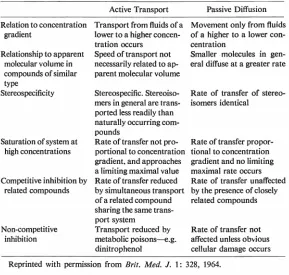
In concluding, it is of some
use to compare the criteria for
active and passive transport
pre-sented in Table 8. Although
some-what more sophisticated subdivi-sions of this table can be made,
I think it is useful to review the
data in this way. In active
trans-port, the substance is transported
from a fluid of a lower
concentra-tion to that of a higher; passive
30
AMINO-ACID ABSORPTION
3000
>.
:~
2000...
0
<
..
··
8 .
.
. ·
··"
·
··
.·
··
.
.
...
·
·
.. ··
····
····
·
·
·
·
·
·
...
A
o ... ... _ _ _ ...c? _ __ _ _ _ _----
-
-0 TO 20
JO
J/.O50
60
70
O'Omin.st?
Fig. 12-Specific activity (CPM/ml) in serum after oral application of 3H-L
-Leucine (23 µC/kg )---phenylketonuria patient 7 years, normal diet (A);
· · · · ·phenylketonuria, the same case, diet poor in phenylalanine
(B);:----control subject, 3 years ( C). (Translated from the German. Reprinted with
permission from Klin. Wochschr. 41:253, 1963.)
TABLE 7
Amino-Acid Transport by Jejuna! Slices from Rats Fed 30 Per Cent
D-Galactose for 2-3 Months
Distribution Ratios
Intestinal
Hydroxy-L-Slices L-alanine L-valine Glycine L-lysine pro line
Control 4.0 ± 0.6 4.2 ± 0.9 2.3 ± 0.5 J.6 ± 0.4 1.9 ± 0.3
Galactose-fed 2.8 ± 0.8 2.3 ± 0.3 l.5 ± 0.7 1.3 ± 0.3 1.3 ± 0.5
p <0.01 <0.01 <0.05 <0.05 <0.05
Adapted with permission from Nature 205: 700, 1965.
diffusion movement is only from a
higher concentration to a lower.
The rate of transport is not
neces-sarily related to molecular volume,
whereas in passive diffusion, by and
large, smaller molecules diffuse at
a greater rate. In active transport
there is stereospecificity, that is,
stereoisomers are generally trans-ported less efficiently than the
naturally occurring compounds. It
suggests that there is a specific kind
of receptor site to which the
trans-ported molecule must be attached.
In active transport the rate of
transport is not proportional to the
concentration gradient. It shows
the saturation phenomenon at high
substrate concentrations. This
does not occur in passive diffusion.
In active transport one can show competitive inhibition by related compounds; the rate of transfer is unaffected by the presence of closely related compounds in
pas-sive diffusion. Transport rate is
TABLE 8
Comparison of Active Transport and Passive Diffusion
Active Transport Passive Diffusion
Relation to concentration Transport from fluids of a Movement only from fluids
gradient lower to a higher concen- of a higher to a lower
con-tration occurs centration
Relationship to apparent Speed of transport not Smaller molecules in
gen-molecular volume in necessarily related to ap- eral diffuse at a greater rate
compounds of similar parent molecular volume
type
Stereospecificity Stereospecific. Stereoiso- Rate of transfer of
stereo-mers in general are trans- isomers identical ported less readily than
naturally occurring com-pounds
Saturation of system at Rate of transfer not pro- Rate of transfer
propor-high concentrations portional to concentration tional to concentration
gradient. and approaches gradient and no limiting
a limiting maximal value maximal rate occurs
Competitive inhibition by Rate of transfer reduced Rate of transfer unaffected
related compounds by simultaneous transport by the presence of closely
of a related compound related compounds
sharing the same trans-port system
Non-competitive Transport reduced by Rate of transfer not
inhibition metabolic poisons--e.g. affected unless obvious
dinitrophenol cellular damage occurs
Reprinted with permission from Brit. Med. J. 1: 328, 1964.
unless cellular damage has
oc-curred.
Thus, there are many character-istics by which one can attempt to
assess whether or not materials are
transported passively or require a specific energy-dependent system for their movement. There is
rea-son to believe on the basis of the
evidence presented here, on the
basis of in vitro studies with
everted gut sacs and other tissue preparations, on the basis of
phys-iologic perfusion studies, and,
per-haps most significantly, on the
basis of studies in various disease
states with specific amino acid
transport defects, that amino acid
transport by the gut is an active
process which may be affected by a
variety of inhibitors in
pathophysi-ologic states. We are just beginning
to understand the importance of
these defects in human metabolism.
References
Boom, C. C. Absorption from the
small intestine. In The Scientific Basis of Medicine Annual Reviews.
London: The Athlone Press, 1963,
pp. 171-196.
FLESHLER, B., J. H. Burr, AND J. D.
WISMAR. Absorption of glycine
and L-alanine by the human
jeju-num. J. Clin. Inv. 45: 1433-1441,
1966.
LINNEWEH, F., M. EHRLICH, E. H.
GRAUL, AND H. HUNDESHAGEN.
Ober den aminosliuren-transport bei
phenylketonurischer oligophrenie.
Klin. Wocl•schr. 41: 253-255. 1963.
Berlin -Gottingen -Heidelberg :
Springer. Miinchen: Bergmann.
MILNE, M. D. Disorders of amino
acid transport. Brit. Med. J. 1:327-336, 1964.
MILNE, M. D., M. A. CRAWFORD,
C. B. GIRAO, AND L. W.
LOUGH-RIDGE. The metabolic disorder in
Hartnup disease. Quart. J. Med.
29: 407-421, 1960.
SAUNDERS, S. J. AND K. J. ISSELBACHER.
Inhibition of intestinal amino-acid
transport by sugars. Nature 205:
700-701, 1965.
THIER,
s.,
M. Fox,s.
SEGAL, ANDL. E. ROSENBERG. Cystinuria: in
vitro demonstration of an intestinal
transport defect. Science 143: 482
-484, 1964.
WILSON, T. H. Intestinal Absorption.
Philadelphia: W. B. Saunders, 1962,
pp. 33, 123.
WISEMAN, G. Active transport of
amino acids by sacs of everted
small intestine of the golden hamster (Mesocricetus auratas). J.