O R I G I N A L A R T I C L E
Open Access
Face and content validity of the virtual
reality simulator
‘
ScanTrainer®
’
Amal Alsalamah
1*, Rudi Campo
2, Vasilios Tanos
3, Gregoris Grimbizis
4, Yves Van Belle
2, Kerenza Hood
5,
Neil Pugh
6and Nazar Amso
1Abstract
Background:Ultrasonography is a first-line imaging in the investigation of women’s irregular bleeding and other gynaecological pathologies, e.g. ovarian cysts and early pregnancy problems. However, teaching ultrasound, especially transvaginal scanning, remains a challenge for health professionals. New technology such as simulation may potentially facilitate and expedite the process of learning ultrasound. Simulation may prove to be realistic, very close to real patient scanning experience for the sonographer and objectively able to assist the development of basic skills such as image manipulation, hand-eye coordination and examination technique.
Objective:The aim of this study was to determine the face and content validity of a virtual reality simulator (ScanTrainer®, MedaPhor plc, Cardiff, Wales, UK) as reflective of real transvaginal ultrasound (TVUS) scanning.
Method:A questionnaire with 14 simulator-related statements was distributed to a number of participants with differing levels of sonography experience in order to determine the level of agreement between the use of the simulator in training and real practice.
Results:There were 36 participants: novices (n= 25) and experts (n= 11) who rated the simulator. Median scores of face validity statements between experts and non-experts using a 10-point visual analogue scale (VAS) ratings ranged between 7.5 and 9.0 (p> 0.05) indicated a high level of agreement. Experts’median scores of content validity statements ranged from 8.4 to 9.0.
Conclusions:The findings confirm that the simulator has the feel and look of real-time scanning with high face validity. Similarly, its tutorial structures and learning steps confirm the content validity.
Keywords:Ultrasound, Validation, Virtual reality simulation, Medical education, Transvaginal ultrasonography, ScanTrainer
Background
Simulation tools are either simplistic models or complex applications, and regardless of the technology used, a simulator must demonstrate validity to be an effective education tool [1]. This entails gathering evidence from multiple sources to show that the interpretation of image, examination or assessment is sound and sensible [1, 2]. At the outset, validation will usually attempt to confirm the fundamental reasons that these tools need to exist for learning [3–6]. From an educational perspective, a simu-lated performance should appear realistic when creating a cognitive-sensory mechanism known as‘sense of presence’
because it allows the trainee/operator to interact with the remote environment as if s/he were present within the environment [7]. With regard to the role of simulation in developing ultrasound knowledge and skills, the validity and reliability of a simulator system for educational goals must be proven, through structured face, content and construct validity studies [1, 8–10].
Face validity is defined as the extent of a simulator’s realism and appropriateness when compared to the actual task [11–13], whereas content validity is defined as the ex-tent to which a simulator’s content is representative of the knowledge or skills that have to be learnt in the real envir-onment. This is based on detailed examination of the learning resources, tutorials and tasks [3, 14–16]. Hence, in the context of ultrasound, face validity addresses the question of how realistic is the simulator, for example, in
* Correspondence:[email protected]
1School of Medicine, College of Biomedical and Life Sciences, Cardiff
University, Office 220, 45 Salisbury road, Cathays, Cardiff CF24 4AB, UK Full list of author information is available at the end of the article
examining the female pelvis and how realistic is the simu-lated feel (haptic sensation) experienced during the exam-ination. Similarly, content validity addresses the question of how useful is the ultrasound simulator in learning rele-vant skills such as measuring endometrial thickness and foetal biometry [13, 17, 18].
According to McDougall and colleagues [4], Kenney and colleagues [19] and Xiao and colleagues [16], face val-idity is expressed as the assessment of virtual realism by novices, while content validity refers to experts’ assess-ment of the suitability of a simulator as a teaching tool. However, reports in the literature are diverse and some authors undertake face validity of a simulator by seeking the opinion of any user including expert and non-expert subjects [12, 13, 15, 20–22]. Others have argued that sub-jects’experience is required for face validity of any educa-tional instrument [18, 23–26]. With regard to content validity, it widely refers to experts’judgement towards the learning content and tasks of a simulator [14, 17, 27–29]. Nevertheless, many published studies rely on subjects with different levels of experience in evaluating content validity of a simulator [12, 13, 22, 30–32].
The ultrasound simulator [33] enables the student to acquire transabdominal (TAS) or transvaginal ultra-sound scanning (TVUS) skills through a series of simu-lation tutorials, each with one or more assignments that include specified tasks reflecting real ultrasound prac-tice. Upon completion of the tasks, the simulator pro-vides computer-generated individualised student/trainee feedback. The hypotheses were that the simulator was (1) realistic for the purpose of developing ultrasound skills and reflects real-life scanning and (2) the content of its structured learning approach represents the know-ledge and psychomotor skills that must be learnt when scanning patients.
The aim of this study was to determine face and con-tent validity of TVUS ScanTrainer. The objectives were (1) to recruit practitioners with varying levels of ultra-sound experience from attendees of an international conference and (2) instruct study volunteers to under-take relevant simulator tutorials and complete a struc-tured questionnaire including statements on face and content validity.
Methods
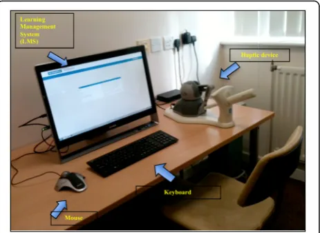
Subjects were voluntarily recruited from delegates visit-ing the ‘ESGE Simulation Island’during the 23rd Euro-pean Congress of Obstetrics and Gynaecology (2014) in Glasgow, Scotland, UK. Each delegate was given a brief, general introduction on the purpose of the study and in-structions on how to use the simulator and the relevant tutorials. They gave verbal consent to participate and proceeded to explore specific tasks in three tutorials with the TVUS ScanTrainer (Fig. 1). These were (1) core
skills gynaecology which has assignments on assessing the uterus, ovaries and adnexa and measuring the endo-metrial thickness, (2) core skills early pregnancy which has assignments on assessing the gestational sac, yolk sac as well as evaluating foetal viability and measure-ments and (3) advanced skills that consisted of several case studies, e.g. ovarian cyst, ectopic pregnancy and twin pregnancy. At the conclusion of the session, sub-jects completed a short questionnaire. Participants took between 10 and 15 min to complete the three tutorials.
The structured questionnaire (Additional file 1) con-sisted of two sections: one detailed subjects’ demo-graphic information, previous ultrasound experience and any previous experience with VR simulation or ultra-sound mannequins. The other section included simulation-related statements. An expert was defined as a subject who had ultrasonography experience of nearly 2 years or more, conducted daily scanning sessions and considered her/himself as an independent practitioner. Some experts with many years of independent ultra-sound experience had less than daily or weekly sessions due to other commitments. A non-expert was defined as having limited experience with ultrasound, had less than 2 years TVUS experience, with occasional or very lim-ited scanning sessions, e.g. once/month, or considered her/himself as a trainee under supervision, newly quali-fied or not yet competent in TVUS scanning.
Fourteen simulation-related statements/parameters were subjectively scored along a 10-cm visual analogue scale (VAS) line by marking the point that subjects felt most appropriate, with (0) at one end (very bad) and (10) at the other (very good). Statements 1 to 6 assessed face validity, 7 to 12 evaluated the simulator’s learning content and 13 and 14 were general statements on the
value of the simulator as training tool (for practical skill acquisition purpose) and testing tool (for assessment pur-pose). Ratings on the scale were defined in ‘mm’as 0–9 (very strongly disagree), 10–19 (strongly disagree), 20–29 (disagree), 30–39 (moderately disagree), 40–49 (mildly disagree), 50 (undecided), 51–59 (mildly agree), 60–69 (moderately agree), 70–79 (agree), 80–89 (strongly agree), 90–100 (very strongly agree). Millimetres were considered for accurate readings of subjects’ marking on scale and later converted to centimetres for final analysis.
The study was conducted in accordance with the general terms and conditions of the South East Wales Re-search Ethics Committee SEWREC (NHS REC Reference 10/WSE02/75) approval and approval of the study proto-col by the congress organising committee.
Statistical data analysis
IBM SPSS Statistics software version 20.0 was used for statistical analysis. Median values were chosen in prefer-ence to mean values as the data were not normally dis-tributed. Median scores and box plots were constructed for each statement as rated by non-experts and experts. Box plots and whiskers represented the median, first and third quartiles, minimum, maximum and outliers of scores obtained by expert and non-expert ratings of the 13 statements. Face validity and general statement items were stratified by expert and non-expert status, while content validity data were reported for experts only. Dif-ferences between experts and non-expert ratings were analysed using the Mann-WhitneyUtest using apvalue
≤0.05 to indicate significance.
Results
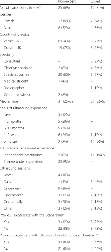
Demographic: Thirty-six subjects, 24 females (67%) and 12 males (33%), participated in this pilot study. Nine were UK-based and 27 were based in other European countries. Eleven subjects (31% expert group) rated themselves as skilled with more than 2 years of experience and practiced independently (n= 10) or with 1 to 2 years of experience and had daily ultrasound sessions (n = 1). Twenty-five subjects (69% non-expert group) were trainees under supervision and included two subjects with more than 2 years TAS experience and limited TVUS scanning. Me-dian age for the expert group was 51 years (range 32–67) and 31 years (range 25–39) for the non-expert group. The median ultrasound experience for experts was more than 2 years and for non-experts was 6 to 11 months. Further breakdown of demographics and years of ultrasound ex-perience is detailed in Table 1.
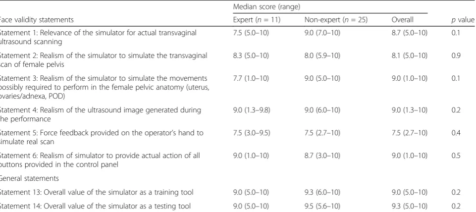
Face validity: Median scores of face validity statements are detailed in Table 2. In summary, experts’ and non-experts’ ratings ranged between 7.5 and 9.0 and were slightly higher than those by experts in two statements (2 and 6) relating to‘realism of the simulator to simulate
the TVUS scan of female pelvis and realism of the simu-lator to provide actual action of all buttons provided in the control panel’. Two statements (1 and 3) were rated lower by experts and related to‘relevance of the simula-tor for actual TVUS scanning and the realism of the simulator to simulate the movements possibly required
Table 1Participants’demographics and ultrasonography
experience
Non-expert Expert No. of participants (n= 36) 25 (69%) 11 (31%) Gender
Female 17 (68%) 7 (64%) Male 8 (32%) 4 (36%) Country of practice
Within UK 6 (24%) 3 (27%) Outside UK 19 (75%) 8 (73%) Speciality
Consultant – 3 (27%) Obs/Gyn specialist 2 (8%) 4 (36%) Specialist trainee 20 (80%) 3 (27%) Medical student 1 (4%) – Radiographer – 1 (10%) Other (midwives) 2 (8%) – Median age 31 (25–39) 51 (32–67) Years of ultrasound experience
Never 3 (12%) – < 6 months 5 (20%) – 6–11 months 9 (36%) – 1–2 years 6 (24%) 1 (10%) > 2 years 2 (8%) 10 (90%) Transvaginal ultrasound experience
Independent practitioner 2 (8%) 11 (100%) Trainee under supervision 23 (92%) – Ultrasound sessions
Never 4 (16%) – Daily 1 (4%) 5 (46%) Once/week 9 (36%) – Once/month 3 (12%) 2 (18%) Occasionally 5 (20%) 2 (18%) Other 3 (12%) 2 (18%) Previous experience with the ScanTrainer®
Yes 3 (12%) 3 (27%) No 22 (88%) 8 (73%) Previous experience with ultrasound model, i.e. blue Phantom™
to perform in the female pelvic anatomy (uterus, ovar-ies/adnexa, Pouch of Douglas POD)’. The remaining two statements (4 and 5) referring to ‘realism of the ultra-sound image generated during the performance and force feedback provided on the operator’s hand to simu-late real scan’ were equally rated. Two general state-ments (13 and 14) were also rated lower by experts. However, there were no statistically significant differ-ences between the two groups’ ratings in all statements (Table 1). Median values and box plots of the eight state-ments in the two groups are shown in Figs. 2 and 3.
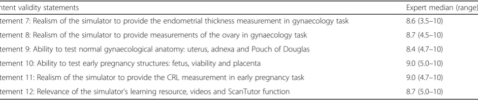
Content validity: Experts’ median scores of content validity statements ranged from 8.4 to 9.0 and are de-tailed in Table 3. Median values and box plots of the six statements are shown in Fig. 4.
Discussion
In this study, the ScanTrainer® simulator demonstrated high face and content validity and its overall value as a
training and testing tool received high ratings as well. To accurately measure participants’ level of agreement with relevant statements, VAS method was used in the question-naire [34]. Higher ratings were given by non-experts than experts with regard to‘relevance of the simulator to actual TVUS’and‘its realism to simulate the movements required to perform in the examination of the female pelvis’ (state-ments 1 and 3) highlighting the fact that such realism is crucial for non-experts for several reasons. This may be be-cause experts need to develop greater understanding of the strengths and limitations of the simulator compared to trainees [35]. Alternatively, beginners in the early stages of learning ultrasound skills are able to address their learning needs through simulated learning compared to the experts who expect variety and advanced or more complex per-formance rather than basic tutorials [12].
There are no comparable ‘face and content’ validity studies addressing virtual reality simulators for TVUS in obstetrics and gynaecology have been published in the
Table 2Face validity‘median scores’ratings by experts and non-experts (n= 36)
Median score (range)
Face validity statements Expert (n= 11) Non-expert (n= 25) Overall pvalue Statement 1: Relevance of the simulator for actual transvaginal
ultrasound scanning
7.5 (5.0–10) 9.0 (7.0–10) 8.7 (5.0–10) 0.1 Statement 2: Realism of the simulator to simulate the transvaginal
scan of female pelvis
8.3 (5.0–10) 8.0 (5.9–10) 8.1 (5.0–10) 0.9 Statement 3: Realism of the simulator to simulate the movements
possibly required to perform in the female pelvic anatomy (uterus, ovaries/adnexa, POD)
7.7 (1.0–10) 9.0 (5.0–10) 9.0 (1.0–10) 0.1
Statement 4: Realism of the ultrasound image generated during the performance
9.0 (1.3–9.8) 9.0 (6.0–10) 9.0 (1.3–10) 0.2 Statement 5: Force feedback provided on the operator’s hand to
simulate real scan
7.5 (3.0–9.5) 7.5 (2.7–10) 7.5 (2.7–10) 0.4 Statement 6: Realism of simulator to provide actual action of all
buttons provided in the control panel
9.0 (1.0–10) 8.7 (3.0–10) 9.0 (1.0–10) 0.5 General statements
Statement 13: Overall value of the simulator as a training tool 9.0 (5.0–10) 9.3 (6.0–10) 9.0 (5.0–10) 0.2 Statement 14: Overall value of the simulator as a testing tool 9.0 (5.0–10) 9.5 (5.6–10) 9.3 (5.0–10) 0.2
literature. In a face validity study of the dVT robotic sur-gery simulator, experts rated the simulator as less useful for training experts than for students/juniors and pointed out to the experts’ need for more critical and advanced procedures in gynaecological surgery and that simulators specifically designed for learning basic skills are less preferable to experts [32]. Creating simulated scenarios to correspond to real ones is always a chal-lenge [3, 29, 36, 37].
Experts’ ratings were higher for two statements relat-ing to the realism of the simulator to simulate the TVUS scan of a female pelvis and in providing actual action of all buttons in the control panel (statements 2 and 6) This may stem from non-experts’limited knowledge and experience, or they might not be familiar with the meas-urement possibilities of virtual simulators [20, 23]. Simi-larly, Weidenbach and colleagues [1] argued that experts gave a better grading for the realism of the EchoCom echocardiography simulator because they were not dis-tracted to drawbacks such as mannequin size and its surface properties, which were harder and more slippery than the human skin, and that experts scanned more
instinctively. The author noted that this mental flexibil-ity seemed to be as yet underdeveloped in beginners.
Non-experts’ and experts’ ratings were similar when evaluating the realism of the ultrasound image generated during the performance and the force feedback provided onto the operator’s hand (statements 4 and 5). Force feedback (haptics) scored 7.5 out of 10, the lowest score in this study. Similar to this study, Chalasani and col-leagues [38] reported low face validity ratings for the haptic force-feedback device of a transrectal ultrasound TRUS-guided prostatic biopsy virtual reality simulator (experts’ lifelike rating 64% and novices’ 67%) even though the author pointed out that haptics, often very difficult to replicate in a simulator environment, were realistic. Haptics will not replace the real-patient scan experience but should enhance the learning approach and improve self-confidence. A further factor is that the ScanTrainer’s haptic device can be tailored to three force feedback levels: normal resistance (most realistic), re-duced and minimal (lowest) designed to avoid overheat-ing duroverheat-ing heavy use, and it is likely that a lower force
Fig. 3Box plots represented the median, first and third quartiles, minimum, maximum and outliers of scores obtained by expert and non-expert ratings of the two general validity statements on the simulator as training and testing tool. Dots (outliers) represented those experts who scored lower than others and the number referred to participant’s code number in data analysis and that did not relate to score value
Table 3Content validity‘median scores’ratings by experts (n= 11)
Content validity statements Expert median (range) Statement 7: Realism of the simulator to provide the endometrial thickness measurement in gynaecology task 8.6 (3.5–10)
Statement 8: Realism of the simulator to provide measurements of the ovary in gynaecology task 8.7 (4.5–10) Statement 9: Ability to test normal gynaecological anatomy: uterus, adnexa and Pouch of Douglas 8.4 (4.7–10) Statement 10: Ability to test early pregnancy structures: fetus, viability and placenta 9.0 (5.0–10) Statement 11: Realism of the simulator to provide the CRL measurement in early pregnancy task 9.0 (4.7–10) Statement 12: Relevance of the simulator’s learning resource, videos and ScanTutor function 8.7 (5.0–10)
feedback setting might have contributed to the lower scores.
The role of force feedback in laparoscopic surgery is not clear [20]. Improving the realism of the simulator and its anatomical structures increases costs consider-ably due to increased demands for more complex hard-ware and softhard-ware. In contrast, Lin and colleagues [39] encouraged learning of bone-sawing skills with simula-tors that provide force feedback rather than not, con-firming the importance of force feedback when seeking to enhance hand-eye coordination. With regard to Scan-Trainer, virtual ultrasound and haptics are used instead of a mannequin allowing measurement of the force ap-plied to the probe and provide a somewhat realistic force-feedback during scanning. However, it still has the limitation of allowing a lower range of movements to the probe while lacking a simulated environment exem-plified by the absence of a physical mannequin [40].
There are numerous simulator systems in usage par-ticularly in the fields of laparoscopy and endoscopy, and several authors emphasised the importance of evaluating their content, including reviewing each learning task and assessing its overall value to determine whether it is ap-propriate for the test and whether the test contains sev-eral steps and skills for practice [12, 17, 31, 38]. In this study, experts’data were used to assess content validity. They had adequate time to review the simulator’s learn-ing resources, help functionality ‘ScanTutor’, read the task-specific instructions and undertake specified tasks before going on to the next step in the same tutorial. In addition, participants had the opportunity to review feedback on their performance in the respective tasks. The results of this study demonstrated that the simula-tor’s content and metrics were appropriate and relevant for ultrasound practice.
There are a number of published content validity stud-ies in ultrasound simulation, such as the educational curriculum for ultrasonic propulsion to treat urinary tract calculi [41], web-based assessment of the extended focused assessment sonography in trauma (EFAST) [2] and validation of the objective structured assessment of technical skills for duplex assessment of arterial stenosis (DUOSATS) [42] which is not based on virtual reality simulator devices. Shumard and colleagues [43] reported on face and content validity of a novel second trimester uterine evacuation task trainer designed to train doctors to perform simulated dilatation and evacuation under ultrasound guidance. Although all respondents were res-idents with limited ultrasound experience, they rated the task trainer as excellent.
Other studies evaluated the effectiveness of simulation-based training in obstetrics and gynaecology ultrasound, whether to investigate the construct validity of a simulator system [9, 40, 44, 45] or to compare simulation training to
conventional methods such as theoretical lectures and hands-on training on patients [10, 46].
Feedback that is automatically generated immediately after a practical simulator session should enhance trainees’ knowledge and ability to reflect critically on their performance and improve their skills [47]. How-ever, the big challenge is to determine how accurate, realistic and trusted the feedback is and, thus, should also be validated appropriately.
Validation studies at national scientific meetings have been reported previously [25, 48]. They offer researchers a rich environment where subjects from different back-grounds and levels of experience are present in one place at the same time. A potential limitation of the study is that it did not determine in advance the sample size required to obtain a reliable result for face and con-tent validation. There is no agreement on the adequacy of sample size in such studies [12, 13]. The number of subjects in this study was higher, and the findings are consistent with others [18, 22, 31, 49]. In addition, many face and content validity studies of simulators were based on smaller sample size compared to the current study [13, 19, 30, 36, 50, 51]. A larger number of partici-pants in this study might have improved the confidence in the results [2]. Participants in this study were from different UK and European institutions unlike others who were from single academic institution [41]; thus, it may be more widely generalizable.
Conclusions
In summary, this study confirms that ScanTrainer simula-tor has the feel and look (face validity) and tusimula-torial struc-ture (content validity) to be realistic and relevant for actual TVUS scanning. This study also concurs with the notion that advancing computer technologies have been able to incorporate virtual reality into training to facilitate the practice of basic skills as well as complex procedures that leave little room for error or mistake [3, 10, 24, 20]. Equally, such simulators should be part of the skill train-ing labs in teachtrain-ing hospitals as it is recommended for endoscopic surgery [52, 53]. It should be subject to an on-going validation to address trainees’learning needs, pro-vide a structured training path and propro-vide validated test procedures with the global and final aim to improve pa-tient care and safety [30, 31, 36, 52].
Additional file
Additional file 1:Face Validity Questionnaire: the ScanTrainer Ultrasound Simulator. (DOCX 207 kb)
Acknowledgements
European Society for Gynaecological Endoscopy for their support and for providing the facilities at simulation island during the 23rd European Congress of Obstetrics and Gynaecology (2014) in Glasgow, Scotland, UK. We are thankful to all the participants for sharing their experience and valuable feedback.
Authors’contributions
AA and NA are the principal investigators and conceived the study. NA and NP were the co-supervisors of the PhD thesis. AA, NA, RC, VT, GG, and YvB designed the study and questionnaire. AA undertook the study and carried out the statistical analysis under the supervision of KH. All authors critically reviewed the manuscript and approved it before submission.
Competing interests
Amal Alsalamah was a PhD student funded by the Government of Saudi Arabia. Nazar Amso is a founder of, owns stocks in and is a board member of MedaPhor, a spin-off company of Cardiff University. He is a co-inventor of a patent for ultrasound simulation training system.
Publisher’s Note
Springer Nature remains neutral with regard to jurisdictional claims in published maps and institutional affiliations.
Author details
1School of Medicine, College of Biomedical and Life Sciences, Cardiff
University, Office 220, 45 Salisbury road, Cathays, Cardiff CF24 4AB, UK.
2European Academy of Gynaecological Surgery, Leuven, Belgium.3Aretaeion
Medical Center, Nicosia, Cyprus.4First Department Obstetrics/Gynecology, Aristotle University of Thessaloniki, Thessaloniki, Greece.5Centre for Trials
Research, College of Biomedical & Life Sciences, Cardiff University, Cardiff, UK.
6Department of Medical Physics and Radiology, University Hospital of Wales,
Cardiff and Vale University Health Board, Cardiff, UK.
Received: 26 June 2017 Accepted: 31 August 2017
References
1. Weidenbach M, Rázek V, Wild F, Khambadkone S, Berlage T, Janousek J, Marek J (2009) Simulation of congenital heart defects: a novel way of training in echocardiography. Heart 95(8):636–641
2. Markowitz JE, Hwang JQ, Moore CL (2011) Development and validation of a web-based assessment tool for the extended focused assessment with sonography in trauma examination. J Ultrasound Med 30(3):371–375 3. Carter FJ, Schijven MP, Aggarwal R, Grantcharov T, Francis NK, Hanna GB,
Jakimowicz JJ (2005) Consensus guidelines for validation of virtual reality surgical simulators. Surg Endosc 19:1523–1532
4. McDougall M, Corica FA, Boker JR, Sala LG, Stoliar G, Borin JF, Chu FT, Clayman RV (2006) Construct validity testing of a laparoscopic surgical simulator. J Am Coll Surg 202(5):779–787
5. Gilliam AD, Acton ST (2007) Echocardiographic simulation for validation of automated segmentation methods. Image Processing, ICIP 2007. IEEE Int Conf 5:529–532
6. Wilfong DN, Falsetti DJ, McKinnon JL, Daniel LH, Wan QC (2011) The effects of virtual intravenous and patient simulator training compared to the traditional approach of teaching nurses: a research project on peripheral i.v. catheter insertion. J Infus Nurs 34(1):55–62
7. Aiello P, D’Elia F, Di Tore S, Sibilio M (2012) A constructivist approach to virtual reality for experiential learning. E-Lear Digital Media 9(3):317–324 8. Wright MC, Segall N, Hobbs G, Phillips-Bute B, Maynard L, Taekman JM
(2013) Standardized assessment for evaluation of team skills: validity and feasibility. Soc Simul Healthc 8:292–303
9. Madsen ME, Konge L, Norgaard LN, Tabor A, Ringsted C, Klemmensen A, Ottesen B, Tolsgaard M (2014) Assessment of performance and learning curves on a virtual reality ultrasound simulator. Ultrasound Obstet Gynecol 44(6):693–699
10. Tolsgaard M, Ringsted C, Dreisler E, Nørgaard LN, Petersen JH, Madsen ME, Freiesleben NL, Sørensen JL, Tabor A (2015) Sustained effect of simulation-based ultrasound training on clinical performance: a randomized trial. Ultrasound Obstet Gynecol 46(3):312–318
11. Byrne A, Greaves J (2001) Assessment instruments used during anaesthetic simulation: review of published studies. Br J Anaesth 86(3):445–450
12. Hung AJ, Zehnder P, Patil MB, Cai J, Ng CK, Aron M, Gill IS, Desai MM (2011) Face, content and construct validity of a novel robotic surgery simulator. J Urol 186(3):1019–1024
13. Alzahrani T, Haddad R, Alkhayal A, Delisle J, Drudi L, Gotlieb W, Fraser S, Bergman S, Bladou F, Andonian S, Anidjar M (2013) Validation of the da Vinci surgical skill simulator across three surgical disciplines. Can Urol Assoc J 7(7–8):520–529
14. Nicholson W, Patel A, Niazi K, Palmer S, Helmy T, Gallagher A (2006) Face and content validation of virtual reality simulation for carotid angiography. Simul Healthc 1(3):147–150
15. Schreuder HW, van Dongen KW, Roeleveld SJ, Schijven MP, Broeders IA (2009) Face and construct validity of virtual reality simulation of laparoscopic gynecologic surgery. Am J Obstet Gynecol 200(5):540 e541– 540 e548
16. Xiao D, Jakimowicz JJ, Albayrak A, Buzink SN, Botden SM, Goossens RH (2014) Face, content, and construct validity of a novel portable ergonomic simulator for basic laparoscopic skills. J Surg Educ 71(1):65–72
17. Seixas-Mikelus S, Stegemann AP, Kesavadas T, Srimathveeravalli G, Sathyaseelan G, Chandrasekhar R, Wilding GE, Peabody JO, Guru KA (2011) Content validation of a novel robotic surgical simulator. BJU Int 107(7): 1130–1135
18. Dulan G, Rege RV, Hogg DC, Gilberg-Fisher KK, Tesfay ST, Scott DJ (2012) Content and face validity of a comprehensive robotic skills training program for general surgery, urology, and gynecology. Am J Surg 203(4):535–539 19. Kenney PA, Wszolek MF, Gould JJ, Lobertino JA, Moinzadeh A (2009) Face,
content, and construct validity of dV-trainer, a novel virtual reality simulator for robotic surgery. Urology 73(6):1288–1292
20. Verdaasdonk EG, Stassen LP, Monteny LJ, Dankelman J (2006) Validation of a new basic virtual reality simulator for training of basic endoscopic skills: the SIMENDO. Surg Endosc 20(3):511–518
21. Seixas-Mikelus S, Kesavadas T, Srimathveeravalli G, Chandrasekhar R, Wilding GE, Guru KA (2010) Face validation of a novel robotic surgical simulator. Urology 76(2):357–360
22. Kelly D, Margules AC, Kundavaram CR, Narins H, Gomella LG, Trabulsi EJ, Lallas CD (2012) Face, content, and construct validation of the da Vinci skills simulator. Urology 79(5):1068–1072
23. Schijven M, Jakimowicz J (2002) Face-, expert, and referent validity of the Xitact LS500 laparoscopy simulator. Surg Endosc 16(12):1764–1770 24. Sweet R, Kowalewski T, Oppenheimer P, Weghorst S, Satava R (2004) Face,
content and construct validity of the University of Washington virtual reality transurethral prostate resection trainer. J Urol 172(5):1953–1957
25. Maithel S, Sierra R, Korndorffer J, Neumann P, Dawson S, Callery M, Jones D, Scott D (2006) Construct and face validity of MIST-VR, Endotower, and CELTS, are we ready for skills assessment using simulators? Surg Endosc 20:104–112 26. Aydin A, Ahmed K, Brewin J, Khan MS, Dasgupta P, Aho T (2014) Face and
content validation of the prostatic hyperplasia model and holmium laser surgery simulator. J Surg Educ 71(3):339–344
27. Fisher J, Binenbaum G, Tapino P, Volpe NJ (2006) Development and face and content validity of an eye surgical skills assessment test for ophthalmology residents. Ophthalmology 113(12):2364–2370
28. Scott DJ, Cendan JC, Pugh CM, Minter RM, Dunnington GL, Kozar RA (2008) The changing face of surgical education: simulation as the new paradigm. J Surg Res 147(2):189–193
29. Gould D (2010) Using simulation for interventional radiology training. Br J Radiol 83:546–553
30. Vick LR, Vick KD, Borman KR, Salameh JR (2007) Face, content, and construct validities of inanimate intestinal anastomoses simulation. J Surg Educ 64(6):365–368
31. Gavazzi A, Bahsoun A, Haute W, Ahmed K, Elhage O, Jaye P, Khan M, Dasgupta P (2011) Face, content and construct validity of a virtual reality simulator for robotic surgery (SEP Robot). Ann R Coll Surg Engl 93(2):152–156 32. Schreuder HR, Persson JE, Wolswijk RG, Ihse I, Schijven MP, Verheijen RH
(2014) Validation of a novel virtual reality simulator for robotic surgery. Sci World J 2014:30
33. Medaphor® Plc, The ScanTrainer (2016) [online] Available at http://www. medaphor.com/scantrainer/ [Accessed 30 June 2016]
34. Jensen MP, Chen C, Brugger AM (2003) Interpretation of visual analog scale ratings and change scores: a reanalysis of two clinical trials of postoperative pain. J Pain 4(7):407–414
colectomy: what metrics have construct validity? Dis Colon rectum 57(2): 210–214
36. O'Leary SJ, Hutchins MA, Stevenson DR, Gunn C, Krumpholz A, Kennedy G, Tykocinski M, Dahm M, Pyman B (2008) Validation of a networked virtual reality simulation of temporal bone surgery. Laryngoscope 118(6):1040–1046 37. de Vries AH, van Genugten HG, Hendrikx AJ, Koldewijn EL, Schout BM, Tjiam IM, van Merriënboer JJ, Muijtjens AM, Wagner C (2016) The Simbla TURBT simulator in urological residency training: from needs analysis to validation. J Endourol 30(5):580–587
38. Chalasani V, Cool DW, Sherebrin S, Fenster A, Chin J, Izawa JI (2011) Development and validation of a virtual reality transrectal ultrasound guided prostatic biopsy simulator. Can Urol Assoc J 5(1):19–26 39. Lin Y, Wang X, Wu F, Chen X, Wang C, Shen G (2014) Development and
validation of a surgical training simulator with haptic feedback for learning bone-sawing skill. J Biomed Inform 48:122–129
40. Chalouhi GE, Bernardi V, Gueneuc A, Houssin I, Stirnemann JJ, Ville Y (2015) Evaluation of trainees’ability to perform obstetrical ultrasound using simulation: challenges and opportunities. Am J Obstet Gynecol 214(4):525.e1–525.e8 41. Hsi RS, Dunmire B, Cunitz BW, He X, Sorensen MD, Harper JD, Bailey MR,
Lendvay TS (2014) Content and face validation of a curriculum for ultrasonic propulsion of calculi in a human renal model. J Endourol 28(4):459–463 42. Jaffer U, Singh P, Pandey VA, Aslam M, Standfield NJ (2014) Validation of a
novel duplex ultrasound objective structured assessment of technical skills (DUOSATS) for arterial stenosis detection. Heart Lung Vessel 6(2):92–104 43. Shumard KM, Akoma UN, Street LM, Brost BC, Nitsche JF (2015)
Development of a novel task trainer for second trimester ultrasound-guided uterine evacuation. Simul Healthc 10(1):49–53
44. Maul H, Scharf A, Baier P, Wüstemann M, Günter HH, Gebauer G, Sohn C (2004) Ultrasound simulators: experience with the SonoTrainer and comparative review of other training systems. Ultrasound Obstet Gynecol 24(5):581–585
45. Merz E (2006) Ultrasound simulator—an ideal supplemental tool for mastering the diagnostics of fetal malformations or an illusion? Ultraschall in Med 27(4):321–323
46. Williams CJ, Edie JC, Mulloy B, Flinton DM, Harrison G (2013) Transvaginal ultrasound simulation and its effect on trainee confidence levels: a replacement for initial clinical training? Ultrasound 21(2):50–56 47. Cline BC, Badejo AO, Rivest II, Scanlon JR, Taylor WC, Gerling GJ (2008)
Human performance metrics for a virtual reality simulator to train chest tube insertion. IEEE Systems Inf Eng Des Symp:168–173 doi:10.1109/SIEDS. 2008.4559705
48. Stefanidis D, Korndorffer JR, Markley S, Sierra R, Heniford BT, Scott DJ (2007) Closing the gap in operative performance between novices and experts: does harder mean better for laparoscopic simulator training? J Am Coll Surg 205(2):307–313
49. White MA, Dehaan AP, Stephens DD, Maes AA, Maatman TJ (2010) Validation of a high fidelity adult ureteroscopy and renoscopy simulator. J Urol 183(2):673–677
50. Bright E, Vine S, Wilson MR, Masters RS, McGrath JS (2012) Face validity, construct validity and training benefits of a virtual reality turp simulator. Int J Surg 10(3):163–166
51. Shetty S, Panait L, Baranoski J, Dudrick SJ, Bell RL, Roberts KE, Duffy AJ (2012) Construct and face validity of a virtual reality-based camera navigation curriculum. J Surg Res 177(2):191–195
52. Campo R, Puga M, Meier Furst R, Wattiez A, De Wilde RL (2014) Excellence needs training“Certified programme in endoscopic surgery”. Facts Views Vis Obgyn 6(4):240–244