RADIATION
PROTECTION
European guidelines
on radiation protection in dental radiology
The safe use of radiographs
in dental practice
EUROPEAN COMMISSION
Radiation Protection 136
European guidelines on radiation protection in dental radiology
The safe use of radiographs in dental practice
Directorate-General for Energy and Transport Directorate H — Nuclear Safety and Safeguards
Unit H.4 — Radiation Protection
This report was produced by the Victoria University of Manchester (United Kingdom) for the European Commission and represents that organisation’s views on the subject matter. These views have not been adopted or in any way approved by the Commission and should not be relied upon as a statement of the Commission’s views.
The European Commission does not guarantee the accuracy of the data included in this report, nor does it accept responsibility for any use made thereof.
Europe Direct is a service to help you find answers to your questions about the European Union
New freephone number:
00 800 6 7 8 9 10 11
A great deal of additional information on the European Union is available on the Internet. It can be accessed through the Europa server (http://europa.eu.int).
Luxembourg: Office for Official Publications of the European Communities, 2004 ISBN 92-894-5958-1
© European Communities, 2004
Reproduction is authorised provided the source is acknowledged.
Printed in Belgium
PRINTED ON WHITE CHLORINE-FREE PAPER Text completed on 1 December 2003 Contract ID: B4-3040/2001/326435/MAR/C4
PREFACE
The aim of this study is to provide a practical guide to radiation protection for professional groups of dentists and their assistants, based upon the two relevant Council Directives of the European Union:
· Directive 96/29/Euratom, of 13 May 1996, laying down the basic safety
standards for the protection of the health of workers and the general public against the dangers arising from ionising radiation
· Directive 97/43/Euratom of 30 June 1997, on health protection of individuals against the dangers of ionising radiation in relation to medical exposure (Medical Exposures Directive).
The 1996 Basic Safety Standards Directive mentioned above ensures the protection of workers exposed to ionising radiation, including dentists and their assistants, and of members of the public.
Directive 97/43/Euratom provides for a high level of health protection to ionising radiation in medical exposure. All the measures adopted in the Directive are concerned not only with avoiding unnecessary or excessive exposure to radiation but also with improving the quality and effectiveness of medical uses of radiation.
No exposure to X-rays can be considered completely free of risk, so the use of radiation by dentists and their assistants implies a responsibility to ensure appropriate protection.
In order to help Member States to implement the Directives, the Commission decided to update and extend the technical guidelines in Radiation Protection 81 ( Radiation protection and quality assurance in dental radiology: "The safe use of radiographs in dental practice” (1995)). A contract was awarded to the University of Manchester, UK, to carry out the study “European Guidelines on Radiation Protection in Dental Radiology”.
The project was designed to give clear and comprehensive information on dental radiological practices, taking into account relevant knowledge and available technology, and give guidance on the application of radiation protection principles in dental radiology to all individuals, including the patient and the personnel.
This document provides general guidelines on the safe use of radiographs in dental practice. Guidelines are not a rigid constraint on clinical practice. Local variations may be required according to healthcare practice and provision.
I am confident that the results of the study will be of help to professional groups of dentists and their assistants, and will contribute to optimising the use of ionising radiation in dentistry.
A. JANSSENS Acting Head of Unit
Contents
Chapter/
Section
Topic
Page
Preface
1Contents
3-6Panel
7Acknowledgements
7Foreword
8Chapter 1
Introduction 9-101.1. Why radiographs in dentistry? 9
1.2. Guideline development 9
1.3. References 10
Chapter 2 Radiation dose and risk
11-172.1. X-rays 11
2.2. Radiation damage 11
2.3. Radiation dose 11
2.4. The risks 12
2.5. Doses and risks in context 15
2.6. References 16-17
Chapter 3 Justification: referral criteria
18-403.1. Dental caries diagnosis 19
3.1.1. Children 20
3.1.2. Adults 23
3.1.3. Alternative methods to radiography for caries
diagnosis 23
3.2. Radiographs in the management of the developing dentition
24
3.2.1. Orthodontic radiographs 24
3.2.2. Other views 25
3.3. Radiography in periodontal assessment 28
3.4. Radiography in endodontics 28
3.4.1. Pre-operative 28
3.4.2. Working length estimation 29
3.4.3. Pre-condensation 29
3.4.4. Post-operative 29
3.4.5. Review 29
3.5. New adult patients 30
3.6. The edentulous patient 32
3.7. Radiography in implantology 32
3.7.1. Pre-operative planning 32
3.7.2. Choice of radiographic techniques 34
3.7.3. During surgery 34
Chapter/
Section
Topic
Page
3.8. Radiography prior to oral surgery and tooth
extraction 36
3.9. Radiography of pregnant patients 36
3.10. Consent in dental radiology 37
3.11. Previous radiographs and reports 37
3.12. Information for patients 38
3.13. References 39-40
Chapter 4 Equipment factors in the reduction of
radiation doses to patients
41-52
4.1. X-ray generation and kilovoltage 41
4.2. Filtration 42
4.3. Collimation, field-size trimming 43
4.3.1. Intraoral radiography 43
4.3.2. Panoramic radiography 44
4.3.3. Cephalometric radiography 45
4.4. Choice of image receptor 45
4.4.1. Intraoral radiography 45
4.4.2. Extraoral radiography 46
4.4.3. Digital receptors 46
4.5. Lead protection of patients 49
4.5.1. Leaded aprons 49
4.5.2. Thyroid collar 49
4.7. References 50-52
Chapter 5 Quality standards and quality assurance
53-835.1. Quality assurance programme 53
5.2. Image quality assessment 53
5.2.1. Targets for radiographic quality 56
5.3. Practical radiographic technique 61
5.3.1. Intraoral radiography 61
5.3.2. The paralleling technique 61
5.3.3. Panoramic radiography 62
5.3.4. Cephalometric radiography 62
5.4. Patient dose and X-ray equipment - Diagnostic
Reference Levels 63 5.4.1. Intraoral 63 5.4.2. Digital equipment 64 5.4.3. Panoramic radiography 68 5.4.4. Cephalometry (teleradiography) 68 5.4.5. Using DRLs 69
5.5. Dental X-ray equipment 69
5.5.1. Maintenance and testing 70
5.5.2. Critical examination 70
5.5.3. Acceptance test 71
Chapter/
Section
Topic
Page
5.5.5. Assessment of representative patient doses 73 5.6. Darkroom, film, cassettes and processing 73 5.6.1. Darkroom and desktop processing units 73
5.6.2. Film 73
5.6.3. Cassettes 73
5.6.4. Processing 73
5.6.5. Digital systems and quality control 74 5.6.6. Viewing and reporting the radiograph 75
5.7. Training 75
5.7.1. Procedures 76
5.8. Quality assurance audit 76
5.9. Common problems in radiography 77
5.9.1. All radiographs 77
5.9.2. Intraoral radiographs 78
5.9.3. Panoramic radiographs 80
5.10. References 81-83
Chapter 6 Staff protection
84 - 926.1. Overall responsibility of dental practice 84
6.1.1. Own country legislation 84
6.1.2. Reporting use of X-ray equipment to competent authorities
84
6.1.3. Assessing risk 84
6.1.4. Seeking advice of qualified expert 85
6.2. Staff dose levels 85
6.2.1. Typical dose level 85
6.2.2. Dose limits 85
6.2.3. Applying ALARA 86
6.2.4. Need for personnel monitoring 86
6.2.5. Pregnant staff 86
6.3. Principles of protection 86
6.3.1. Primary and scattered radiation 86
6.3.2. Use of distance 87
6.3.3. Use of protective screens etc 88
6.3.4. Classification of areas 88
6.3.5. Holding patients 88
6.3.6. Written procedures and supervision 89
6.4. Design of the facility 90
6.4.1. Protection for adjacent areas 90
6.4.2. Room layout 90
6.4.3. Signs and warning lights 91
6.5. Training staff 91
6.6. Dealing with incidents 92
6.7. References 92
Appendix 1 Methodology 93-96
Appendix 2 Summary of recommendations/statements 97-107
List of Tables and Figures
Table/Figure
Topic
Page
Table 1.1 Criteria used to grade recommendations 10 Table 2.1 Nominal lifetime probability coefficients for
stochastic effects
12
Table 2.2 Risk in relation to age 13
Table 2.3 Effective doses and risks of stochastic effects – tabular summary of literature review.
14 Table 2.4 Estimated annual numbers of radiographs in
European Union countries for which data are available
15
Table 3.1 Caries risk factors 21
Table 3.2 Various radiographic views and their function in orthodontic practice.
26 Table 3.3 A simplified flow chart to determine whether a
pre-treatment cephalogram is needed. 27 Table 3.4 Flow chart of radiographic management of
dentate patients.
31 Table 3.5 Summary of radiographic techniques for
implantology
33 Table 3.6 Appropriate imaging techniques for pre-operative
planning 35
Table 4.1 The effect upon dose of equipment modification when compared with a baseline of a 70 kV AC dental X-ray set with a 60 mm cylindrical beam used with E-speed film.
50
Table 5.1 Film fault frequency for intraoral radiography carried out within general dental practice.
54 Table 5.2 Film fault frequency within panoramic
radiographs taken in general dental practice.
55 Table 5.3 Quality standards for bitewing radiography 57 Table 5.4 Quality standards for periapical radiography 58 Table 5.5 Quality standards for panoramic radiography 59 Table 5.6 Quality standards for cephalometric radiography 60 Table 5.7 Summary of surveys of intraoral dose quantities
and DRLs. 65-6
Table 5.8 Summary of surveys of panoramic radiography
dose quantities and DRLs. 67
Table 5.9 Summary of surveys of cephalometric dose quantities and DRLs.
67 Figure 6.1 Radiation scattered from the primary beam. 87 Figure 6.2 Example of a controlled area designation around
an intraoral radiography unit.
About the Panel Members
NAME CURRENT JOB
TITLE AFFILIATION Professor Keith Horner BChD MSc PhD FDSRCPSGlasg FRCR DDRRCR
Professor of Oral and
Maxillofacial Imaging University of Manchester,UK
Dr. Vivian Rushton PhD MDS BDS DDRRCR MFGDP Senior Lecturer in Dental and Maxillofacial Radiology University of Manchester, UK Mrs. Anne Walker
BSc MSc MIPE MSRP Consultant MedicalPhysicist North Western MedicalPhysics, Christie Hospital, Manchester, UK
Dr. Kostas Tsiklakis DDS MSc PhD
Associate Professor Athens University, School of Dentistry, Greece Mr. Peter N Hirschmann MSc FDS RCS FRCR DDR RCR Consultant Dental Radiologist
Leeds Dental Institute, Leeds Teaching Hospitals NHS Trust
Leeds, UK Professor Paul F van
der Stelt DDS PhD
Professor of Oral and Maxillofacial Radiology
Dept. of Oral and Maxillofacial Radiology Academic Centre for Dentistry Amsterdam (ACTA),The Netherlands Ms. Anne-Marie Glenny BSc(Hons) MMedSci Lecturer in Evidence Based Oral Health Care
Cochrane Oral Health Centre, University Dental Hospital of Manchester, UK Dr. Xandra L Velders DDS PhD Dentist, Radiation Protection (level 3) Algemeen Stralingsdeskundige (RPA)
Academic Medical Centre (AMC) Amsterdam, The Netherlands Dr. Sue Pavitt BSc(Hons) PhD EU Project Co-ordinator University of Manchester, UK
Acknowledgements
In particular, the panel members wish to thank the Article 31 Group for their many hours of work in initiating the project and their comments and suggestions. In particular, we thank Nico Harpes, Division de la Radioprotection, Direction de la Santé, Luxembourg. To reviewers for their helpful comments Professor
Luciano González García, Professor Eliseo Vaño, Dr. L.K.Harding. The panel members would also like to acknowledge the help of several people: We are
Foreword
The radiation protection activities of the Commission of the European Union in the medical field are based on two Council Directives:
· Directive 96/29/Euratom, of 13 May 1996, laying down the basic safety standards for the protection of the health of workers and the general public against the dangers arising from ionising radiation (European Basic Safety Standards); and
· Directive 97/43/Euratom of 30 June 1997, on health protection of individuals against the dangers of ionising radiation in relation to medical exposure (Medical Exposures Directive).
Although doses incurred during dental examinations are in general relatively low, dental radiology accounts for nearly one third (1)of the total number of radiological examinations in the European Union and therefore merits specific attention with regard to radiation protection.
Article 7 of the ‘Medical Exposures Directive’ stipulates that dental
practitioners must have adequate theoretical and practical training for the purpose of radiological practices as well as relevant competence in radiation protection. Article 7 also requires continuing education and training after qualification.
To facilitate the implementation of this article by Member States, the Commission decided to develop the present document, updating and
extending the technical guide Radiation Protection 81 (2) in order that it takes into account the technological developments and the new requirements of the two Council Directives. It is designed to give clear and comprehensive
information on dental radiological practices, taking into account relevant knowledge and technology available, and give guidance on the application of the radiation protection principles in dental radiology for all individuals,
including both the patient and the personnel.
It is our hope that this handbook will be of help to professional groups of dentists and their assistants, and that it will contribute to optimise the use of ionising radiation in dentistry.
Professor Keith Horner Professor of Oral and Maxillofacial Imaging, University of Manchester, UK. References
1. 2001. United Nations Scientific Committee on the Effects of Atomic Radiation UNSCEAR Report to the General Assembly with Scientific Annex.
2. van der Stelt, P. F. 1995. Radiation protection and quality assurance in dental radiography. The safe use of radiographs in dental practice. Office for Official Pulications of the European Communities, Luxemborg.
1. Introduction
The aim of this book is to provide a practical guide to radiation protection for dentists working in a primary care setting, based upon the two relevant Council Directives of the European Union (EU).
· Directive 96/29/Euratom of 13 May 1996 laying down basic safety standards for the protection of the health of workers and the general public against the dangers arising from ionising radiation.
· Directive 97/43/Euratom of 30 June 1997 on health
protection of individuals against the dangers of ionising radiation in relation to medical exposure.
Laws derived from these Directives exist within individual EU States that impose specific enforceable requirements upon dentists. This document sets general guidelines on good practice in the use of X-rays by dentists.
Guidelines are systematically developed statements to assist practitioner and patient in decisions about appropriate health care for certain specific clinical circumstances (1). As this
implies, guidelines are not a rigid constraint on clinical practice, but a concept of good practice against which the needs of the individual patient can be considered(2).
1.1. Why radiographs in dentistry?
Radiographs are essential to dentists for:
· Diagnosis
· Treatment planning
· Monitoring treatment or lesion development
However, an integral part of radiography is exposure of patients and,
potentially, clinical staff to X-rays. No exposure to X-rays can be considered completely free of risk, so the use of radiation by dentists is accompanied by a responsibility to ensure appropriate protection.
1.2. Guideline development
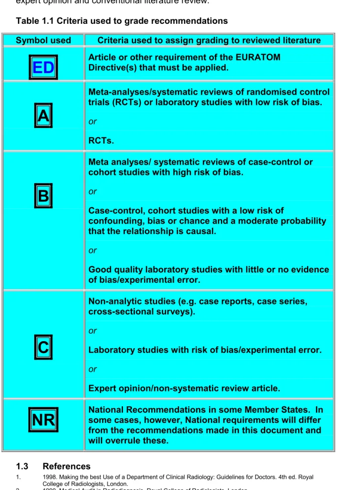
There is now widespread acceptance in medicine and dentistry that clinical practice should be as ‘evidence-based’ as possible. This document was developed using such an approach. The project team collected and analysed relevant published literature, guidelines that have proved effective in the past to arrive at recommendations that will contribute to optimisation of the use of ionising radiation in dentistry. Details of the methodology used in identifying relevant literature and the appraisal process are given in Table 1.1 and Appendix 1.
It should be clearly understood that the approach adopted for different sections within this document has not been uniform. This is because the volume of evidence available for review varies. Some sections have involved more comprehensive sifting of the evidence, while others rely heavily on expert opinion and conventional literature review.
Table 1.1 Criteria used to grade recommendations
Symbol used Criteria used to assign grading to reviewed literature
ED
Article or other requirement of the EURATOMDirective(s) that must be applied.A
Meta-analyses/systematic reviews of randomised control trials (RCTs) or laboratory studies with low risk of bias.
or
RCTs.
B
Meta analyses/ systematic reviews of case-control or cohort studies with high risk of bias.
or
Case-control, cohort studies with a low risk of
confounding, bias or chance and a moderate probability that the relationship is causal.
or
Good quality laboratory studies with little or no evidence of bias/experimental error.
C
Non-analytic studies (e.g. case reports, case series, cross-sectional surveys).
or
Laboratory studies with risk of bias/experimental error.
or
Expert opinion/non-systematic review article.
NR
National Recommendations in some Member States. Insome cases, however, National requirements will differ from the recommendations made in this document and will overrule these.1.3 References
2 Radiation dose and risk
2.1 X-rays
X-rays are a type of electromagnetic (EM) radiation. EM radiation also
includes visible light, radio waves, microwaves, cosmic radiation, and several other varieties of ‘rays’. All can be considered as ‘packets’ of energy, called
photons, which have wave properties, most importantly a wavelength and frequency. X-rays are short wavelength, high frequency EM radiation. The importance of this is that high frequency means high energy. When X-rays hit atoms this energy can be transferred, producing ionisation of atoms.
2.2 Radiation damage
When patients undergo X-ray examinations, millions of photons pass through their bodies. These can damage any molecule by ionisation, but damage to the DNA in the chromosomes is of particular importance. Most DNA damage is repaired immediately, but rarely a portion of a chromosome may be
permanently altered (a mutation). This may lead ultimately to the formation of a tumour. The latent period between exposure to X-rays and the clinical diagnosis of a tumour may be many years. The risk of a tumour being
produced by a particular X-ray dose can be estimated; therefore, knowledge of the doses received by radiological techniques is important. While doses and risks for dental radiology are small, a number of epidemiological studies
have provided evidence of an increased risk of brain (19, 22), salivary gland
(16, 22) and thyroid(15, 27) tumours for dental radiography.
The effects described above are believed to have no threshold radiation dose below which they will not occur(2). They can be considered as ‘chance’ (stochastic) effects, where the magnitude of the risk is proportional to the radiation dose. There are other known damaging effects of radiation, such as cataract formation, skin erythema and effects on fertility, that definitely have threshold doses below which they will not occur. These threshold doses vary in size, but all are of a magnitude far greater than those given in dental
radiography. Thus, except in extraordinary circumstances, these deterministic
effects are given no further consideration.
2.3 Radiation dose
The terms ‘dose’ and ‘exposure’ are widely used but often misunderstood. ‘Doses’ may be measured for particular tissues or organs (e.g. skin, eye, bone marrow) or for the whole body, while ‘exposure’ usually refers to equipment settings (time, mA, kV). A commonly used measure of dose in surveys is ‘entrance dose’, measured in milligrays (mGy).This has an advantage of being
The aim of this section is to describe the: · The nature of X-rays
· The nature of radiation damage · Radiation dose
· Radiation risk
· Dental radiography doses and risks in a life context
reference levels (DRLs), based upon entrance dose surveys, may be set as standards against which X-ray equipment can be assessed as part of quality assurance (see Chapter 5 Section 5.4 for a discussion of DRLs in dental radiography).
In this chapter, however, radiation dose is expressed as effective dose (refer
to Glossary for definition), measured in units of energy absorption per unit
mass (Joules / kg) called the Sievert (more usually the microSievert, µSv,
representing one millionth of a Sievert). In practice, effective dose is
calculated for any X-ray technique by measuring the energy absorption in a number of ‘key’ organs in the body, so that the final figure is a representation of ‘whole body’ detriment. While effective dose is an impossible quantity to
measure in vivo, it is possible to determine it from laboratory studies or
computer modelling. This can then be used to estimate radiation risk. Many studies have measured doses of radiation for dental radiography, but only a few have estimated effective dose. There are still a number of
radiographic techniques for which no published data are available and some for which very different results have been reported. In many cases this reflects controversy about whether salivary glands should be given special weighting in calculation of dose. Furthermore, variation in the technical parameters of the X-ray sets and image receptors used in studies means that care should be taken when comparing dose estimations from different studies.
2.4 The risks
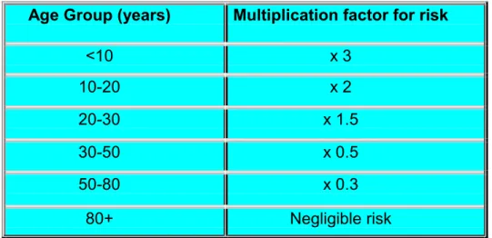
Radiation detriment can be considered as the total harm experienced by an irradiated individual. In terms of stochastic effects, this includes the lifetime risk of fatal cancer, non-fatal cancer and hereditary effects. The probability of radiation-induced stochastic effects for the whole population is 7.3 x 10⁻²Sv-1. Table 2.1 (derived from (3) ) gives the breakdown of this summed figure into its constituent elements. Hereditary effects are believed to be negligible in dental radiography (26).
Table 2.1 Nominal lifetime probability coefficients for stochastic effects Detriment (10-2 Sv-1)
Fatal cancer 5.0
Non-fatal cancer* 1.0
Severe hereditary effects 1.3
Total 7.3
*The lifetime probability co-efficient for non-fatal cancer represents detriment rather then true incidence, which would be significantly greater.
Risk is age-dependent, being highest for the young and least for the elderly. Here, risks are given for the adult patient at 30 years of age. These should be modified using the multiplication factors given in Table 2.2 (derived from (3)). These represent averages for the two sexes; at all ages risks for females are slightly higher and those for males slightly lower.
Table 2.2 Risk in relation to age
These data are derived from (3) and represent relative attributable lifetime risk based upon a relative risk of 1 at age 30 (population average risk). It assumes the multiplicative risk projection model, averaged for the two sexes. In fact,
risk for females is always relatively higher than for males.
Age Group (years) Multiplication factor for risk
<10 x 3 10-20 x 2 20-30 x 1.5 30-50 x 0.5 50-80 x 0.3 80+ Negligible risk
Beyond 80 years of age, the risk becomes negligible because the latent period between X-ray exposure and the clinical presentation of a tumour will probably exceed the life span of a patient. In contrast, the tissues of younger people are more radiosensitive and their prospective life span is likely to exceed the latent period.
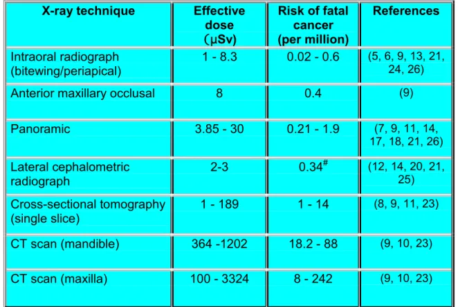
Table 2.3 gives doses and risks for the dental radiographic techniques likely to be used in dentistry. However, care should be taken to adjust the risk
estimates according to the age of patients using Table 2.2.
As mentioned above, a particular problem arises from the inclusion or
exclusion of the salivary glands in the calculation of dose. The salivary glands are not specifically included as an organ in effective dose calculations as described by the International Commission on Radiation Protection (3), leading to an underestimation of risk. However, in view of the apparent relationship between dental radiography and increased risk of salivary gland tumours, many researchers have applied a special weighting factor so that salivary gland doses, that would otherwise be excluded, are incorporated into dose calculation. By following this practice, effective doses and risks are increased.
Table 2.3 Effective doses and risks of stochastic effects – tabular summary of literature review.
The paper by White (26) represented a recalculation of largely pre-ICRP 60 publications. Only papers subsequent to 1990 are specifically referenced, in addition to White. The use of E-speed film and rare-earth intensifying screens has been assumed for intraoral and panoramic radiography, respectively. Round (60 mm diameter) collimation is assumed for intraoral radiography.
X-ray technique Effective
dose (µSv) Risk of fatal cancer (per million) References Intraoral radiograph (bitewing/periapical) 1 - 8.3 0.02 - 0.6 (5, 6, 9, 13, 21, 24, 26)
Anterior maxillary occlusal 8 0.4 (9)
Panoramic 3.85 - 30 0.21 - 1.9 (7, 9, 11, 14, 17, 18, 21, 26) Lateral cephalometric radiograph 2-3 0.34 # (12, 14, 20, 21, 25) Cross-sectional tomography (single slice) 1 - 189 1 - 14 (8, 9, 11, 23) CT scan (mandible) 364 -1202 18.2 - 88 (9, 10, 23) CT scan (maxilla) 100 - 3324 8 - 242 (9, 10, 23)
(5): Data derived for single intraoral film by halving figures to allow for E-speed film and by dividing original data for full mouth survey by 19. No adjustment made for high kV (90) used in this study.
(6): Data derived for single intraoral film by halving figures to allow for E-speed film and by dividing original data for full mouth survey by 19. No adjustment made for high kV (90) used in this study.
(26): White excluded salivary glands from consideration in dose and risk estimations, accounting for lower figures. His data for intraoral radiography are derived by halving figures to allow for E-speed film and by dividing original data for full mouth survey by 20.
#Based upon risks to brain, salivary glands and thyroid gland only.
Despite their principal use in hospital practice, doses for cross-sectional tomography and CT are given because of their increasing use for implant treatment planning by dentists.
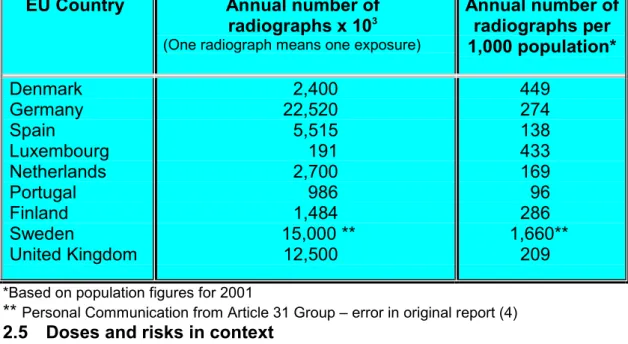
From the figures presented above, it can be seen that conventional dental radiography is associated with low doses and risks for the individual patient. However, while dental radiography is generally ‘low dose’, it is a high volume procedure, with many millions of radiographs taken annually in the European Union (Table 2.4).
Table 2.4 Estimated annual numbers of dental radiographs in EU
countries for which data are available(4).
EU Country Annual number of
radiographs x 103
(One radiograph means one exposure)
Annual number of radiographs per 1,000 population* Denmark Germany Spain Luxembourg Netherlands Portugal Finland Sweden United Kingdom 2,400 22,520 5,515 191 2,700 986 1,484 15,000 ** 12,500 449 274 138 433 169 96 286 1,660** 209
*Based on population figures for 2001
** Personal Communication from Article 31 Group – error in original report (4)
2.5 Doses and risks in context
Life is a risky business. Among the many risks to which we are prone, we are all constantly exposed to normal background radiation, which averages about
2400 µSv (2)each year (average world figures). Medical exposures (including
dental) add substantially to this figure, with wide variation from country to country. With this in mind, a panoramic radiograph may be associated with an effective dose the same as 1-5 days’ additional background radiation, while two bitewing radiographs would be equivalent to about one day. For
comparative purposes, a chest X-ray (20 µSv) would be equivalent to around
three days of additional background radiation. Comparisons can be made between radiation doses in dental radiography associated with increased exposure to cosmic rays (another high energy form of EM radiation). For example, a long haul flight from Brussels to Singapore is estimated to lead to
an additional effective dose of 30 µSv, while a short flight from Brussels to
Athens incurs an estimated dose of about 10 µSv (1).
Dental radiography doses and risks are minimal, comparable in most cases (with the exception of CT and multiple cross-sectional tomography) with exposure to a few days of natural background radiation.
Statement 2.A
Individual doses in basic dental radiography (intra-oral, panoramic and cephalometric) are low, being equivalent to those associated with a few days of background radiation. Individual doses from more complex imaging (CT scans and multiple slice cross-sectional tomography) can be substantially higher.
Statement 2.B
2.6 References
1. 1997. European Commission. Radiation Protection 88. Recommendations for the implementation of Title VII of the European Basic Safety Standards (BSS) Directive concerning significant increase in exposure due to natural radiation sources. Office for Official Publications of the EC.
2. 2001. European Commission. Radiation Protection 125: Low dose ionizing radiation and cancer risk. Office for Official Publications of the EC,Luxembourg. http://europa.eu.int/comm/environment/radprot/publications. 3. 1990. ICRP Publication 60. Recommendations of the International Commission on Radiatiological
Protection. Annal of the ICRP 21.
4. 2001. United Nations Scientific Committee on the Effects of Atomic Radiation UNSCEAR Report to the General Assembly with Scientific Annex.
5. Avendanio, B., N. L. Frederiksen, B. W. Benson, and T. W. Sokolowski. 1996. Effective dose and risk
assessment from detailed narrow beam radiography. Oral Surg Oral Med Oral Pathol Oral Radiol Endod 82:713-9.
6. Cederberg, R. A., N. L. Frederiksen, B. W. Benson, and T. W. Sokolowski. 1997. Effect of the geometry
of the intraoral position-indicating device on effective dose. Oral Surg Oral Med Oral Pathol Oral Radiol Endod 84:101-9.
7. Danforth, R. A., and D. E. Clark. 2000. Effective dose from radiation absorbed during a panoramic
examination with a new generation machine. Oral Surg Oral Med Oral Pathol Oral Radiol Endod 89:236-243.
8. Dula, K., R. Mini, J. T. Lambrecht, P. F. van der Stelt, P. Schneeberger, G. Clemens, H. Sanderink,
and D. Buser. 1997. Hypothetical mortality risk associated with spiral tomography of the maxilla and mandible prior to endosseous implant treatment. Eur J Oral Sciences 105:123-9.
9. Dula, K., R. Mini, P. F. van der Stelt, and D. Buser. 2001. The radiographic assessment of implant
patients: decision-making criteria. Int J Oral Maxillofac Implants 16:80-9.
10. Frederiksen, N. L., B. W. Benson, and T. W. Sokolowski. 1995. Effective dose and risk assessment from
computed tomography of the maxillofacial complex. Dentomaxillofac Radiol 24:55-8.
11. Frederiksen, N. L., B. W. Benson, and T. W. Sokolowski. 1994. Effective dose and risk assessment from
film tomography used for dental implant diagnostics. Dentomaxillofac Radiol 23:123-7.
12. Gijbels, F., C. Bou Serhal, G. Willems, H. Bosmans, G. Sanderink, M. Persoons, and R. Jacobs.
2001. Diagnostic yield of conventional and digital cephalometric images: a human cadaver study. Dentomaxillofac Radiol 30:101-5.
13. Gijbels, F., R. Jacobs, G. Sanderink, E. De Smet, B. Nowak, J. Van Dam, and D. Van Steenberghe.
2002. A comparison of the effective dose from scanography with periapical radiography. Dentomaxillofac Radiol 31:159-63.
14. Gori, C., F. Rossi, A. Stecco, N. Villari, and G. Zatelli. 2000. Dose evaluation and quality criteria in
dental radiology. Radiat Prot Dosimetry 90:225-227.
15. Hallquist, A., L. Hardell, A. Degerman, G. Wingren, and L. Boquist. 1994. Medical diagnostic and
therapeutic ionizing radiation and the risk for thyroid cancer: a case-control study. Eur J Cancer Prevention 3:259-67.
16. Horn-Ross, P. L., B. M. Ljung, and M. Morrow. 1997. Environmental factors and the risk of salivary gland
cancer. Epidemiology 8:414-9.
17. Lecomber, A. R., S. L. Downes, M. Mokhtari, and K. Faulkner. 2000. Optimisation of patient doses in
programmable dental panoramic radiography. Dentomaxillofac Radiol 29:107-12.
18. Lecomber, A. R., and K. Faulkner. 1998. Conference Proceeding: Dose and risk in Dental Radiography,
Luxembourg 1997. Reference Doses and Quality in Medical Imaging: What the referring practitioner and directing medical staff should know. Radiat Prot Dosimetry 80:23-25.
19. Longstreth, W. T., Jr., L. K. Dennis, V. M. McGuire, M. T. Drangsholt, and T. D. Koepsell. 1993.
Epidemiology of intracranial meningioma. Cancer 72:639-48.
20. Maillie, H. D., and J. E. Gilda. 1993. Radiation-induced cancer risk in radiographic cephalometry. Oral
Surg Oral Med Oral Pathol 75:631-7.
21. Pasler, F. A., and H. Visser. 1999. Zahnmedizinische Radiologies, Vol. 5. Georg Thieme, Auflage.
22. Preston-Martin, S., and S. C. White. 1990. Brain and salivary gland tumors related to prior dental
radiography: implications for current practice. J Am Dent Assoc 120:151-8.
23. Scaf, G., A. G. Lurie, K. M. Mosier, M. L. Kantor, G. R. Ramsby, and M. L. Freedman. 1997. Dosimetry
and cost of imaging osseointegrated implants with film-based and computed tomography. Oral Surg Oral Med Oral Pathol Oral Radiol Endod 83:41-8.
Individual risks in dental radiography are small but are greater in the younger age groups (below 30 years) in which (in many Member States) dental radiography is most frequently performed.
24. Velders, X. L., J. van Aken, and P. F. van der Stelt. 1991. Risk assessment from bitewing radiography. Dentomaxillofac Radiol 20:209-13.
25. Visser, H., T. Rodig, and K. P. Hermann. 2001. Dose reduction by direct-digital cephalometric
radiography. Angle Orthodontist 71:159-63.
26. White, S. C. 1992. Assessment of radiation risk from dental radiography. Dentomaxillofac Radiol
21:118-26.
27. Wingren, G., A. Hallquist, and L. Hardell. 1997. Diagnostic X-ray exposure and female papillary thyroid
3 Justification:
referral criteria
Any X-ray exposure entails a risk to the patient. Under normal circumstances the risk from dental radiography is very low. Nonetheless, it is essential that any X-ray examination should show a net benefit to the patient, weighing the total potential diagnostic benefits it produces against the individual detriment that the exposure might cause. The efficacy, benefits and risk of available alternative techniques having the same objective but involving no or less exposure to X-rays should be taken into account.
Recommendation 3 A
In order that the justification process can be carried out, it is essential that selection of appropriate radiography is based on the individual patient’s
history and a clinical examination. The ‘routine’ use of radiography on patients based on a generalised approach rather than individual prescription is
unacceptable. A ‘routine’ or ‘screening’ examination is defined as one in which a radiograph is taken regardless of the presence or absence of clinical signs and symptoms.
Recommendation 3 B
*
The statement/recommendation although not specifically stated in the European Directive is intrinsic to the process of justification as defined by the Directive. There are no randomised controlled trials to support the recommendation; such a study design would neither be possible nor ethical to perform.All X-ray examinations must be justified on an individual patient basis by demonstrating that the benefits to the patient outweigh the potential detriment.
The anticipated benefits are that the X-ray examination would add new information to aid the patient’s management.
ED
No radiographs should be selected unless a history and clinical examination have been performed.
‘Routine’ radiography is unacceptable practice.
ED*
The aim of this section is to:· Explain the concept of radiographic justification
· To provide specific guidelines for a range of clinical conditions commonly encountered in general dental practice
Choosing the appropriate radiographic examination should also be based upon consideration of the prevalence of diseases, their rates of progression and the diagnostic accuracy of the imaging techniques in question.
Consulting guidelines facilitates the process of selecting radiographs. Such guidelines, called ‘referral criteria’ or ‘selection criteria’ exist for both medical and dental radiography. Radiographic Referral Criteria have been defined as “descriptions of clinical conditions derived from patient signs, symptoms and history that identify patients who are likely to benefit from a particular
radiographic technique". As with any guideline, these are not intended to be rigid constraints on clinical practice, but a concept of good practice against which the needs of the individual patient can be considered.
The term ‘referral criteria’ is appropriate for medical practitioners, where radiography is usually arranged by referral to a specialist in radiology. However, some dentists may refer patients for radiography to hospitals or dental colleagues where they do not have the necessary equipment in their own practices. When acting as a referrer, the dentist should ensure that
adequate clinical information about the patient is provided to the person taking responsibility for the exposure.
Recommendation 3 C
Evidence-based guidelines (9) have been devised for selection of dental radiography. The following parts of this section are a representation of selected guidelines from that document. In isolated cases guidelines have been adjusted to take into account evidence in a European context.
3.1
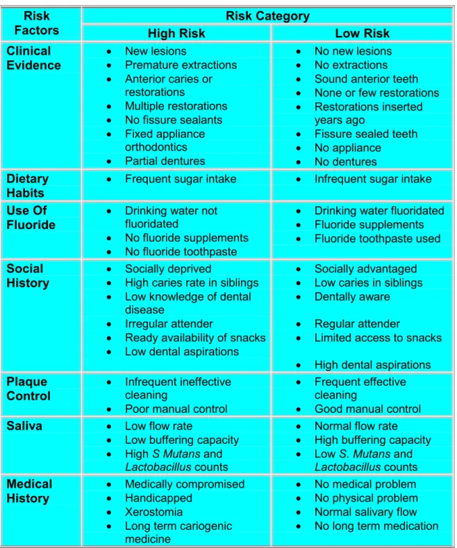
Dental caries diagnosisCaries risk must be assessed for all new patients and then subsequently at recall appointment as risk factors may change in the intervening period. By identifying patients who are at the greatest risk of dental decay, clinicians can effectively implement prevention techniques to maintain low caries risk status. Caries is a multifactorial disease requiring a wide-ranging assessment of categories of risk. The important categories identified during the systematic review (9) were:
When referring a patient for a radiographic examination, the dentist should supply sufficient clinical information (based upon a history and clinical examination) to allow the practitioner taking clinical responsibility for the X-ray exposure to perform the justification process.
· Clinical evidence of previous disease · Dietary habits · Social history · Use of fluoride · Plaque control · Saliva · Medical history
When combined with the clinical judgement of the dentist, the use of these factors have been found to be an extremely efficient predictor of caries risk(6, 7). Table 3.1 expands on each of the categories by sub-dividing them into high and low risk. Obviously, the moderate risk patient will lie in between the two levels.
3.1.1. Children
The early enamel lesion progresses at a relatively slow rate taking at least two years to progress into dentine, although progression is not inevitable(6). Early diagnosis of these enamel lesions is important, as with intervention lesion progression can be slowed or reversed (6).
Posterior bitewing radiographs are an essential adjunct to clinical
examination(9). The initial clinical examination must include an assessment of caries risk (as high, medium or low). As outlined previously, the
assessment of risk is relevant in determining when to take radiographs and therefore must be carried out at each subsequent recall examination ensuring that the time interval for radiography becomes patient-specific. It is feasible that adoption of the following recommendation may lead to more radiographs being taken. However, this is justified as it will result in better patient care. Recommendation 3 D
Prescription of bitewing radiographs for caries diagnosis should be based upon caries risk assessment.
Intervals between subsequent bitewing radiographic examinations must be reassessed for each new period, as individuals can move in and out of caries risk categories with time.
Table 3.1: Caries Risk Factors (9)
Risk Category Risk
Factors High Risk Low Risk
Clinical Evidence · New lesions · Premature extractions · Anterior caries or restorations · Multiple restorations · No fissure sealants · Fixed appliance orthodontics · Partial dentures · No new lesions · No extractions
· Sound anterior teeth
· None or few restorations
· Restorations inserted years ago
· Fissure sealed teeth
· No appliance
· No dentures
Dietary
Habits ·
Frequent sugar intake · Infrequent sugar intake
Use Of
Fluoride ·
Drinking water not fluoridated
· No fluoride supplements
· No fluoride toothpaste
· Drinking water fluoridated
· Fluoride supplements
· Fluoride toothpaste used
Social
History ·
Socially deprived
· High caries rate in siblings
· Low knowledge of dental disease
· Irregular attender
· Ready availability of snacks
· Low dental aspirations
· Socially advantaged
· Low caries in siblings
· Dentally aware
· Regular attender
· Limited access to snacks
· High dental aspirations
Plaque
Control ·
Infrequent ineffective cleaning
· Poor manual control
· Frequent effective cleaning
· Good manual control
Saliva · Low flow rate
· Low buffering capacity
· High S Mutans and
Lactobacillus counts
· Normal flow rate
· High buffering capacity
· Low S. Mutans and
Lactobacillus counts Medical History · Medically compromised · Handicapped · Xerostomia
· Long term cariogenic medicine
· No medical problem
· No physical problem
· Normal salivary flow
· No long term medication
In high caries risk children there is good evidence to support taking posterior bitewing radiographs at the initial examination, even in the absence of
clinically detectable decay. The benefit is reported as being between 167% and 800% of the diagnostic yield from clinical diagnosis with or without fibre optic transillumination assistance. Where a child is classified as high caries risk the subsequent bitewing examination should be after 6 months. Bitewing radiographs should not be taken more frequently than this and it is imperative to reassess caries risk in order to justify using this interval again. Evidence of no new or active lesions would be an indicator that the child had entered the moderate or low risk category.
Recommendation 3 E
In moderate caries risk children the evidence also supports the diagnostic use of bitewing radiographs. Many authors report significant addition to the
diagnostic yield from the use of bitewing radiographs, varying from 150% to 270% of the yield from clinical examination alone. Where a child is classified as moderate caries risk the subsequent bitewing examination should be after 12 months. Evidence of no new or active lesions would be an indicator that the child had entered the low risk category.
Recommendation 3 F
In low caries risk children there is less good evidence to support the taking of posterior bitewing radiographs: diagnostic yield is lower than that with higher risk groups. Nevertheless, radiographs reveal 2-3 times more caries lesions than clinical examination alone. In low caries prevalence populations, it is suggested that selective radiography should be conducted of surfaces suspected clinically as being carious. Where caries population prevalence is not low, but a child is classified as low caries risk, the subsequent bitewing examination should be after 12-18 months in the deciduous dentition and 24 months in the permanent
dentition. More extended recall intervals may be employed if there is explicit evidence of continuing low caries risk. Selective radiography of suspect surfaces may be appropriate as an alternative to bitewing radiography where caries
prevalence is low. Recommendation 3 G
3.1.2. Adults
It is recommended that when children are designated as high caries risk they should have six-monthly posterior bitewing radiographs taken. This should continue until no new or active lesions are apparent and the individual has entered a lower risk category.
B
It is recommended that when children are designated as moderate caries risk they should have annual posterior bitewing radiographs. This should continue until no new or active lesions are apparent and the individual has entered a lower risk category.
B
Radiography for caries diagnosis in low caries risk children should take into account population prevalence of caries.
Intervals of 12-18 months (deciduous dentition) or 24 months (permanent dentition) may be used, although longer intervals may be appropriate where there is continuing low caries risk.
There is comparatively little evidence evaluating the diagnostic yield of radiographs for caries in adults. Therefore, in the absence of research data guidelines have been devised by extrapolation of studies in children and young adults.
Recommendation 3 H
Recommendation 3 I
Recommendation 3 J
3.1.3. Alternative methods to radiography for caries diagnosis
Clinicians have recommended flossing teeth and the temporary separation of teeth, using orthodontic separators or wooden wedges, to assist in caries diagnosis during the clinical examination.
Alternative methods to ionising radiation with which to diagnose caries have also been developed. These include established techniques such as fibreoptic transillumination (FOTI) and electrical conductance measurements (ECM). Other newer emerging technologies include Quantitative Light-induced Fluorescence (QLF), Infrared Laser Fluorescence (DIAGNOdent) and Digital Imaging Fiber Optic Transillumination (DIFOTI).
Some of these techniques have limitations that affect their diagnostic or commercial availability and in some cases, their practicality within the dental
It is recommended that adults designated as moderate caries risk have annual posterior bitewing radiographs taken until no new or active lesions are apparent and the individual has entered another risk category.
C
It is recommended that adults designated as low caries risk have posterior bitewing radiographs taken at approximately 24-month intervals. More extended intervals may be used where there is continuing low caries risk.
C
It is recommended that adults designated as high caries risk have six-monthly posterior bitewing radiographs taken until no new or active lesions are apparent and the individual has entered another risk category.
surgery. Others require further in vivo research and validation. However, several of these techniques have shown promise and may well become an accepted part of the routine diagnostic armamentarium of the practicing clinician in the future(12, 27, 44).
Recommendation 3 K
3.2
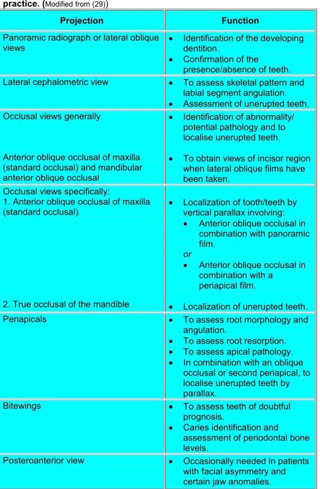
Radiographs in the management of the developing dentition Many children seek orthodontic treatment. When such treatment is clinically required, most children are appropriately treated at around 12-13 years of age and will require radiographs to confirm the presence and condition of all teeth. Occasionally, there will be a need for a radiographic examination at an earlier age where there is a serious departure from normal dental development or when a child attends in pain or after trauma.Children are subject to higher risks from X-ray exposure than are adults. Consequently the importance of justification for radiography is underlined. Basic information on radiography for orthodontics is available on the following pages and Table 3.2. For further details, refer to the literature (29).
Usually the radiographic examination will consist of a panoramic radiograph (or right and left oblique lateral radiographs). Upper anterior occlusal
radiographs are invariably required to supplement oblique lateral radiographs, but this is not the case for panoramic radiographs. Such films only provide additional information to the panoramic film in a minority of cases (25, 29). Therefore they should be prescribed only after being justified by examining the panoramic radiograph.
3.2.1. Orthodontic radiographs
Radiography is needed following clinical examination in a proportion of orthodontic patients. In addition, a patient in the mixed dentition stage may well require radiography to determine if interceptive treatment is appropriate. When previous radiographs are available, these may already contain all the information that the clinician needs for further management.
A clinical examination is necessary to ensure that the radiographs requested will be appropriate for the patient's specific orthodontic problem. Similarly, the need for radiography to monitor treatment progress is dependent upon a careful clinical assessment. Table 3.2 gives a broad overview of the function of the various radiographic projections used in orthodontic practice.
Alternative methods to using ionising radiation in caries diagnosis should be considered once their diagnostic validity has been clearly established.
Various studies have confirmed that a clinical examination supplemented by study models is often sufficient for treatment planning (26). Furthermore research using algorithms (14) and clinical indicators (28), has shown that a marked reduction in the numbers of orthodontic films is possible without compromising patient treatment. From these studies, the effect of radiographs on changing orthodontic diagnosis and treatment plans is limited ranging from 16% to 37% and 4% to 20% respectively (13-15, 20).
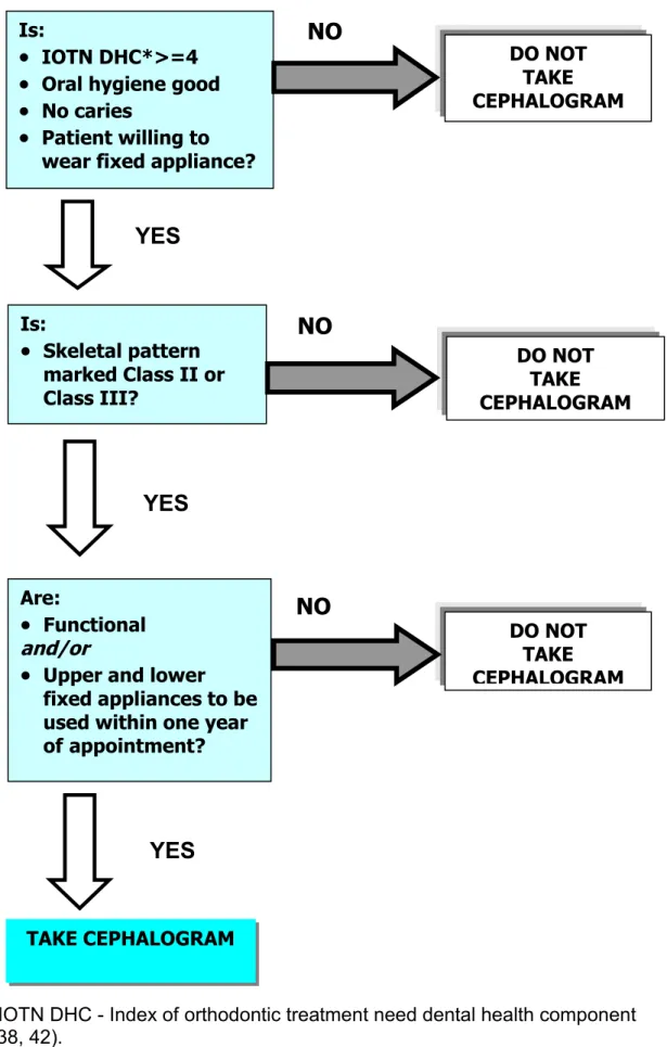
Cephalometric radiography is often requested for selected patients
undergoing orthodontic treatment. The flow chart in Table 3.3 gives a very simplified overview of those cases that require lateral cephalometry. In addition, a cephalogram should be taken at:
·
The end of functional appliance treatment to see the position to which the lowers anterior teeth have been proclined.·
The end of presurgical treatment for orthognathic cases.·
Just prior to the end of active fixed appliance treatment to assess the position of the lower incisors.When assessing the position of the lower incisors, the lateral cephalogram is recommended only if the information is going to change the orthodontist's decision on their finishing mechanics or retention regime.
3.2.2 Other views
The posteroanterior (PA) view of the face/headhas been advocated in cases of patients who present with facial asymmetry. The value of hand or wrist radiography in clinical orthodontics has been questioned, as these views lack the reliability to predict growth spurts. Similarly, radiography for
temporomandibular joint dysfunction cannot be justified (22, 29)and films taken for this reason have been shown to have no impact on treatment planning (34). A more detailed description of the frequency and use of all
types of orthodontic films can be obtained from published guidelines (29). Recommendation 3 L
Specialist guidelines on orthodontic radiography should be consulted as an aid to justification in the management of the developing dentition in
children.
Table 3.2: Various radiographic views and their function in orthodontic practice. (Modified from (29))
Projection Function
Panoramic radiograph or lateral oblique
views · Identification of the developingdentition. · Confirmation of the
presence/absence of teeth. Lateral cephalometric view · To assess skeletal pattern and
labial segment angulation. · Assessment of unerupted teeth. Occlusal views generally
Anterior oblique occlusal of maxilla (standard occlusal) and mandibular anterior oblique occlusal
· Identification of abnormality/ potential pathology and to localise unerupted teeth.
· To obtain views of incisor region when lateral oblique films have been taken.
Occlusal views specifically:
1. Anterior oblique occlusal of maxilla (standard occlusal)
2. True occlusal of the mandible
· Localization of tooth/teeth by vertical parallax involving: · Anterior oblique occlusal in
combination with panoramic film.
or
· Anterior oblique occlusal in combination with a
periapical film.
· Localization of unerupted teeth. Periapicals · To assess root morphology and
angulation.
· To assess root resorption. · To assess apical pathology. · In combination with an oblique
occlusal or second periapical, to localise unerupted teeth by parallax.
Bitewings · To assess teeth of doubtful prognosis.
· Caries identification and
assessment of periodontal bone levels.
Posteroanterior view · Occasionally needed in patients with facial asymmetry and certain jaw anomalies.
Table 3.3: A simplified flow chart to determine whether a pre-treatment cephalogram is needed.
*IOTN DHC - Index of orthodontic treatment need dental health component (38, 42). DO NOT TAKE CEPHALOGRAM
YES
NO
Are:·
Functional and/or·
Upper and lowerfixed appliances to be used within one year
of appointment?
YES
TAKE CEPHALOGRAMYES
Is:·
IOTN DHC*>=4·
Oral hygiene good·
No caries·
Patient willing towear fixed appliance?
DO NOT TAKE CEPHALOGRAM
NO
Is:·
Skeletal pattern marked Class II or Class III? DO NOT TAKE CEPHALOGRAMNO
3.3
Radiography in periodontal assessmentThe diagnosis of periodontal diseases depends on a clinical examination. This maybe supplemented by radiographs if they provide additional information, which could potentially change patient management and prognosis. However, there is no clear evidence to support any robust recommendations on selection of radiographs (45).
The posterior bitewing projection offers both optimal geometry and the fine detail of intraoral radiography for patients with small amounts of uniform bone loss (36). Bitewings have the additional advantage in that they may have already been indicated for caries assessment, providing information about bone levels without the need for an additional radiation dose. More complex or extensive bone loss would require different imaging. Vertical bitewing, periapical and panoramic radiographs all have uses, either alone or in
combination. Where periapical radiographs are used, the paralleling technique is indicated as this gives a better geometrical perspective on the periodontal bone than the bisecting angle technique.
Recommendation 3 M
Recommendation 3 N
3.4
Radiography in endodonticsRadiographs are essential for many aspects of endodontic treatment. It is appropriate to consider their role at the different stages of treatment(1).
3.4.1. Pre-operative
A periapical radiograph provides essential information about pulp and root canal anatomy that cannot be obtained in any other way (30). In addition it provides information about periradicular anatomy that may contribute to treatment planning or be essential if surgical endodontic treatment is being considered.
Radiographs should be used in the management of periodontal disease if they are likely to provide additional information that could potentially change patient management and prognosis.
C
There is insufficient evidence to propose robust guidelines on choice of radiography for periodontal diagnosis and treatment, but existing
radiographs e.g. bitewing radiographs taken for caries diagnosis should be used in the first instance.
3.4.2. Working length estimation
Some types of electronic apex locators are reliable at identifying the apical constriction and are useful for locating perforations. However, using these devices in certain clinical situations can result in a degree of inaccuracy. In view of this, periapical radiography is often still required during working length estimation. It may be necessary to take two (or more) radiographs in order to determine the length of all the root canals in multi-rooted teeth (23).
3.4.3. Pre-condensation
If there is doubt about the integrity of the apical constriction, a check radiograph should be taken of the master gutta-percha cone before final condensation/obturation.
3.4.4. Post-operative
A periapical radiograph should be taken immediately following obturation as this gives a basic assessment of the quality of the root filling and a reference image of the periapical condition for subsequent review.
3.4.5. Review
The peak incidence of healing and the peak incidence of emerging chronic apical periodontitis are seen at 1 year after treatment, with a high proportion (89%) of endodontically treated teeth demonstrating signs of healing at one year (35). This suggests that a one-year follow-up radiography may be sufficient for small asymptomatic apical lesions. Teeth that remain
symptomatic and those with large periapical lesions may require additional radiographic review to assess the treatment options.
Recommendation 3 O
It is recommended that radiographic examinations are carried out at the following stages of endodontic treatment:
1. Pre-operative assessment
B
2. Working length estimation*
B
3. Post-operative
B
4. At 1-year review or if symptomatic
C
* For those practitioners without access to electronic apex locators, a working length estimation will be required.
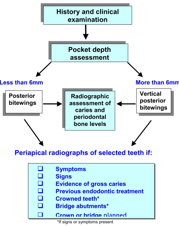
3.5 New adult patients
Many dentists follow a routine practice of examining new adult patients using panoramic or full-mouth intraoral radiography. As discussed above, such ‘routine’ practices are not acceptable (39-41).
Most evidence shows that conventional panoramic radiography has lower diagnostic accuracy for the common dental radiographic diagnostic tasks (caries diagnosis, periapical diagnosis) than intraoral (bitewing and periapical) radiography. Over and above these common tasks, routine panoramic
radiography in search of asymptomatic bony lesions without clinical signs is not justified because of the low prevalence of such abnormalities. There is no justification for review panoramic radiography at arbitrary time intervals.
Full-mouth periapical radiography can be criticised in the same way as routine panoramic radiography. ‘Routine’ radiography will inevitably lead to
unnecessary X-ray exposure. Selected periapical radiography of new adult patients will improve the relative risk/benefit for patients (17, 37). Taking periapical radiographs of teeth with clinical symptoms, and of those with a history of endodontic therapy and deep caries as shown on bitewing
radiographs, revealed 90% of periapical lesions in one research study (11). Others have also reported (19) the effectiveness of selection criteria for identification of periapical pathosis. Table 3.4 shows a flow chart (43) for the selection of radiography for new adult patients.
Recommendation 3 P
Recommendation 3 Q
For a new adult dentate patient, the choice of radiography should be based upon history, clinical examination and an individualised prescription as illustrated in Table 3.4.
C
For a new adult dentate patient, panoramic radiography may be indicated in a limited number of dental treatments, notably orthodontic assessment and certain oral surgical procedures (i.e. lower third molars).
Table 3.4 Flow chart of radiographic management of dentate patients. (Modified from (43)).
Less than 6mm
More than 6mm
Periapical radiographs of selected teeth if:
*If signs or symptoms present
This flow chart will not apply in every case. For example, the patient who has few remaining teeth due to advanced periodontal disease may not need a radiographic examination to plan treatment. The history and examination is therefore crucial and prescription of radiographs should be planned on this basis.
History and clinical
examination
Pocket depth
assessment
Vertical
posterior
bitewings
q
Symptoms
q
Signs
q
Evidence of gross caries
q
Previous endodontic treatment
q
Crowned teeth*
q
Bridge abutments*
q
Crown or bridge
planned
Posterior
bitewings
assessment of
Radiographic
caries and
periodontal
bone levels
3.6. The edentulous patient
In the absence of any clinical signs or symptoms, there is no justification for any radiographic examination (18, 31, 32). The obvious exception is if implant treatment is planned, although if treatment is extensive other more advanced imaging (cross-sectional imaging) may well be appropriate. Where clinical examination identifies the possible presence of an abnormality, such as a possible retained root, then an intraoral radiograph of the site is the appropriate radiographic examination.
Recommendation 3 R
3.7. Radiography in implantology
Imaging is essential in implantology. In treatment planning, radiographs provide information on the quantity and quality of bone in the proposed site of implant placement. Following treatment, imaging is used to assess implant osteointegration, bone healing and to periodically review the fixture.
The review of the literature displays a paucity of evidence-based guidelines on radiography for implantology. Evidence has, in the main, been derived from expert opinion and review papers (24, 33). An assessment of these papers revealed inconsistencies and little reliable information on the frequency of follow-up radiography.
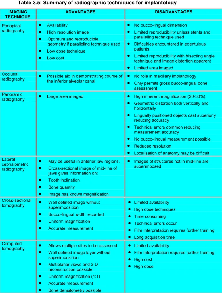
The imaging modality chosen is often a function of the treatment phase and a reflection of the number of proposed implants and their position in the oral cavity. Table 3.5 gives a broad overview of the advantages and
disadvantages of the various radiographic projections used in implantology.
3.7.1. Pre-operative planning
In evaluating a pre-operative site, the clinician requires information on:
·
The quality and quantity of bone·
The bucco-lingual width and height of available bone·
The inclination of bony contours·
The presence of osseous undercuts·
Evidence of atypical anatomy such as enlarged marrow spaces·
Presence of pathology·
Exact location of certain anatomic structures (i.e. the maxillary antrum, inferior alveolar canal, the mental foramen etc)There is no justification for radiography of edentulous patients without a specific indication such as implant treatment or clinical signs or symptoms.
Table 3.5: Summary of radiographic techniques for implantology
IMAGING
TECHNIQUE ADVANTAGES DISADVANTAGES
Periapical
radiography
·
Availability
·
High resolution image·
Optimum and reproduciblegeometry if paralleling technique used
·
Low dose technique·
Low cost·
No bucco-lingual dimension·
Limited reproducibility unless stents andparalleling technique used
·
Difficulties encountered in edentulouspatients
·
Limited reproducibility with bisecting angletechnique and image distortion apparent
·
Limited area imagedOcclusal
radiography
·
Possible aid in demonstrating course ofthe inferior alveolar canal·
No role in maxillary implantology·
Only permits gross bucco-lingual boneassessment Panoramic
radiography
·
Large area imaged·
High inherent magnification (20-30%)·
Geometric distortion both vertically andhorizontally
·
Lingually positioned objects cast superiorlyreducing accuracy
·
Technical errors common reducingmeasurement accuracy
·
No bucco-lingual measurement possible·
Reduced resolution·
Localisation of anatomy may be difficultLateral cephalometric radiography
·
May be useful in anterior jaw regions.·
Cross-sectional image of mid-line ofjaws gives information on:
·
Tooth inclination·
Bone quantity·
Image has known magnification·
Images of structures not in mid-line aresuperimposed
Cross-sectional
tomography
·
Well defined image withoutsuperimposition·
Bucco-lingual width recorded·
Uniform magnification·
Accurate measurement·
Limited availability·
High dose techniques·
Time consuming·
Technical errors occur·
Film interpretation requires further training·
Long acquisition timeComputed
tomography
·
Allows multiple sites to be assessed·
Well defined image layer withoutsuperimposition
·
Multiplanar views and 3-Dreconstruction possible.
·
Uniform magnification (1:1)·
Accurate measurement·
Bone densitometry possible·
Limited availability·
Film interpretation requires further training·
High costWith the exception of reformatted computed tomography (CT), all
radiographic projections are magnified. The magnification factor must be derived and any assessments of available bone height must be calculated having taken this factor into consideration. Magnification factors can be derived by use of a reference object in the same plane as the alveolus. Periapical radiographs taken for single tooth replacement require the use of film holders and the paralleling technique for optimum geometry. Optimum geometry is often difficult to achieve in the edentulous jaw. The magnification factor in panoramic radiography is particularly variable and the consensus of one expert committee (47)was to recommend that the panoramic film should be augmented by tomography, either conventional or computed, in order to provide the information necessary for optimum implant placement.
When imaging using either conventional or computed tomography to generate cross-sectional images, proposed implants sites and/or tomographic
landmarks should be identified using surgical stents consisting of metal rods, balls or radiopaque markers.
Conventional tomography is obtained either from dedicated software incorporated into panoramic equipment or from specifically designed X-ray machines for implantology. The latter comprises multimodal systems using narrow beam radiography and spiral tomography. In the past CT scanning has been restricted to general hospital facilities, however smaller dedicated head and neck CT imaging equipment is becoming more commonplace. Spiral CT techniques benefit from shorter scanning times and improved accuracy.
3.7.2. Choice of radiographic techniques
The number of implants and their proposed position in the oral cavity are often the main factors dictating the choice of imaging technique. A proportion of patients need advanced imaging especially in cases involving bone grafts and in those in which there are multiple potential implant sites. In these cases CT has been recommended (47). Table 3.6 details the range of imaging methods for pre-operative planning in various parts of the oral cavity.
3.7.3. During Surgery
If any radiography is needed then periapical radiographs are readily available and use of digital imaging should be considered which offers the benefits of 'real-time’ imaging.
3.7.4. Postoperative assessment
Radiography has been recommended to evaluate the implant
post-operatively. The frequency and timing of review radiographs appears to be purely subjective. During the healing phase, radiography would obviously needed if the patient has clinical symptoms. If not, the next radiographic review should occur at 12 months and is considered essential to assess marginal bone levels. Subsequent review intervals range from annual reviews to once every three years. More frequent radiography is obviously needed if the patient is symptomatic.
Table 3.6: Appropriate imaging techniques for pre-operative planning.
Cross-sectional imaging refers to either CT or specialised tomography equipment.
Implant number
Location Technique Complicating factors Supplementary techniques
·
Extensive bone resorption·
Enlarged incisive foramen Combinations of late