SCIENCEDOMAIN international
www.sciencedomain.org
Tibiotalar Joint Stabilization by Steinman Pins
in Oestern-tscherne Type III Open Fracture
Dislocation of the Ankle
P. K. Karampinas
1*, E. Kavroudakis
1, J. Vlamis
1,
D. Anagnostopoulos
1, V. Polyzois
1and Sp. Pneumaticos
11
University of Athens Medical School, III Department of Orthopaedics, KAT Hospital, 2 Nikis Str., 14561, Kifissia, Athens, Greece.
Authors’ contributions
This work was carried out in collaboration between all authors. Authors PK and JV designed the study, the protocol and the first draft of the manuscript. Author EK performed the statistical analysis of the study. Author DA managed the literature searches. Authors VP and SP revise the manuscript. All authors read and approved the final manuscript.
Received 2nd November 2013 Accepted 9th January 2014 Published 25th January 2014
ABSTRACT
Aim: Open fracture-dislocation of the ankle is a high-energy limp-threading injury, almost always associated with vary grades of soft tissue damage. Stabilization of the tibiotalar joint by Steinman pins is retrospectively evaluated and seems to favor the management of the soft tissue damage and probably minimize the rate of complications in diabetic patients.
Study Design: Retrospective case series study.
Place and Duration of Study: From 2003 to 2011, Department of Orthopedics and Traumatology, University of Athens Medical School, KAT Hospital Athens, Greece. Materials and Methods: 17 patients were admitted with a fracture dislocation of the ankle. Twelve were featured as Oestern-Tscherne type III and 5 as type V. There have been used Steinmann pins to provide a rigid stabilization of the ankle in anatomic position, and available enough space to observe, follow and manage the soft tissue
Results: At their last follow up visit, 14 patients were evaluated and the mean AOFAS score was 86, 5. The mean follow up period was 18, 1 months. Five patients underwent secondary arthrodesis of tibiofibular joint and in 4 patiens were observed non union of the fibula. Intra operatively in 10 patients were observed osteochondral post-traumatic lesions. In 3 patients the talus demonstrated signs of AVN at 7 months after trauma. There was no statistical difference between the two groups studied.
Conclusions: Immediate débridement, irrigation, antibiotic therapy and use of Steinmann pins to stabilize the tibiotalar joint are indicated in a way to reduce the complication rates in diabetic patients. This technique seems to be effective and low cost, evidenced by the unnecessary use of further wound and soft tissue treatment operations.
Keywords: Ankle; fracture; Oestern-Tscherne type III; treatment; diabetic foot.
1. INTRODUCTION
Open fracture-dislocation of the ankle is a high-energy trauma injury, usually resulting from motor-vehicle or contact sport accidents. Because of the ankles’ high stability established by the ankle mortise, the ligaments and the tendons, commonly there is associated to ankles’ injury a malleoli fracture [1,2]. Also these complex injuries of the ankle almost always are associated with vary grades of soft tissue damage [3]. Generally is regarded one of the most limp-threatening injuries. The complications vary from infection and soft tissue necrosis to amputation [4]. Depending on the soft tissue damage and their management strategy, the optimal surgical technique decided for the fracture treatment is crucial and sometimes could be very stressing to the treating surgeon and difficult to decide [5,6].
After meticulous surgical debridement, treatment options have been limited to minimal internal fixation, external fixation and combination of these two [3,7]. The use of Kirschner wires and Steinmann pins is also reported in this injures and recommended for diabetic patients [8]. Today, the most common treatment for an open ankle fracture-dislocation is considered to be the external fixation after repeated surgical debridement [3,4]. The internal fixation of open ankle fractures in patients with and co-morbidities (Diabetes mellitus), the complication rate and limp-threading is high [6,9]. Considering the soft tissue damage of the injured area, a specific management protocol must be always followed in a way to minimize the rate of complications and reoperations [1,10]. Negative pressure wound therapy (NPWT) facilitates the wound healing and prevent or facilitates secondary soft tissue defect coverage [11,12]. Free flaps are the gold standard for the treatment of soft tissue defects of the foot and ankle area [13].
2. MATERIALS AND METHODS
In the past nine years (from 2003 to 2011), 17 patients (12 males and 5 females), ranging in age from twenty-seven to forty-two years (mean age 32,7 years) were admitted to our level four trauma center with a fracture dislocation of the ankle and grave soft tissue damage. There were 9 right and 8 left ankles. The mechanism of injury was in all cases a motor vehicle accident. Two deferent groups of patients were encountered according to their co-morbidity of diabetes mellitus. In six patients referred to our institution there was medical history of diabetes mellitus, controlled by nutritional diet and per os medication at the time of surgery at the time of surgery and at the rest eleven patients without such a clinical history. All 17 cases involved an anterolateral dislocation with a concomitant fracture of the malleoli but with varying degrees of soft tissue damage. All the injuries are classified as Gustilo 3B. Twelve as Oestern-Tscherne type III and 5 as type V. During the radiological evaluation there were medial malleolar fractures in 2 patients, lateral malleolar fractures type Weber C in 13 and bimalleolar fractures in 2 patients. There was a need of Negative pressure wound therapy (NPWT) use for the soft tissue management in 7 patents and a free flap in 3 cases.
The associations between categorical variables were evaluated using the Fisher’s exact test. Student’s t-test was used for comparisons between the groups. We performed all statistical tests using SPSS® 20 statistical software (SPSS Inc, Chicago, IL, USA). P-values less than 0.05 were considered significant.
Fig. 2. Photographs preoperatively (a) and postoperatively (b) with the two Steinman stabilization of the limb
The Steinman needles were removed at six weeks and were replaced by a cast. A removable cast was applied in 10th week allowed range of motion exercises of the tibiocalcaneal and subtalar joints and partial weight bearing was allowed (Fig. 3). All patients were treated on an outpatient basis after hospital discharge. The demographic details of our patients were documented together with the time elapsed between injury and definitive surgical treatment and the complications encountered.
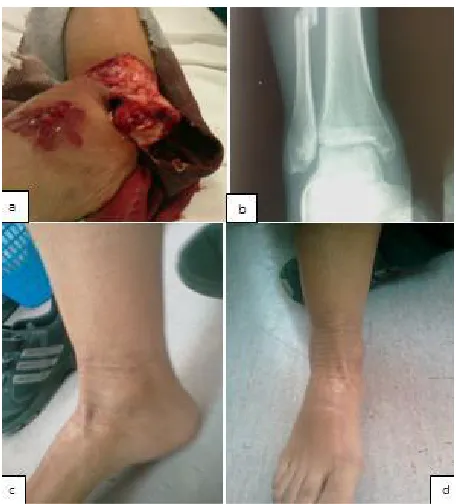
Fig. 3. Radiographs and pictures of an open fracture dislocation of the ankle(a), preoperatively(b), ten weeks(c) and 21 weeks postoperatively(d) presenting excellent
3. RESULTS AND DISCUSSION
The same surgical technique was followed in all patients. The mean time elapsed between injury and final operative treatment was 7, 4 hours (range from 5 to 19 h). The mean follow up period was 18, 1 month (range from 15 to 21). At their last follow up visit 3 patients were missed and 14 patients were evaluated both radio graphically and functionally with the AOFAS Ankle-Hind foot Scale score.
The mean AOFAS Ankle-Hindfoot Scale score at the time of the last follow up visit was 82,1 (range from 64 to 96) (Table 1). Intra operativelly were observed osteochondral post-traumatic lesions in 7 out of 17 patient’s (Picture 3). In 3 patients the talus demonstrated signs of AVN at 7 months after trauma. Five patients underwent secondary procedures and arthrodesis of tibiofibular joint due to instability and pain of the ankle. Non union of the fibula was observed in 4 patients who necessitated second operation whereby internal fixation of the fibula and tibiofibular arthrodesis was done. In 8 patients, radiographic signs of subtalar and tibiotalar arthritis were evident at 21 months after injury. There were 4 patients following the treatment protocol that had a post operative infection controlled with repeated débridement and intravenous antibiotic therapy according to antibiogram (Table 1). There was no statistical difference between the two groups (co-morbidity of diabetes mellitus and free of co-morbidities) with open fracture dislocation treated with this soft tissue management protocol and surgical technique.
Pure talo-tibial dislocation without a combined malleoli injury is rare [1,2]. High energy injuries interesting the ankle usually are associated with extensive soft tissue damage [3,4]. Even in a level four trauma center it is encountered approximately twice per year. Limp salvage instead of amputation must be the primary goal for the treating surgeon. The complications after these injuries varying from infection, avascular necrosis of the talus, tibio-tala or/and subtibio-talar post-traumatic arthritis to amputation [4,7]. Infection is a limp-threatening situation and especially in patients with co-morbidities (diabetes mellitus). Complications presented in diabetic patients sustained an open ankle dislocation-fracture are also the impaired wound healing, malunion, non union, Charcot arthropathy. Most of the time complications arrive by the injured soft tissue [7,8,14]. Treatment options and decision is challenging with high rates of infection recurrence and amputation [6,8].
Table1. Clinical and trauma characteristics of single patient, final outcome and AOFA Score
Sl no.
COMORBIDITY OESTERN-TSCHERNE
FRACTUTR TYPE
SOFT TISSUE TREATMENT
ASSOCIATED LESSONS
AVN OSTEOARTHRITIS ARTHRODESIS AOFAS
1 DM III WEBER C FF OCL AVN OA AD 64
2 - III WEBER C NPWT - - - - 96
3 - V WEBER C NPWT OCL - OA AD 71
4 DM III M.M.FRC - - - 90
5 - V WEBER C NPWT - - - - 82
6 - III WEBER C - OCL - OA - 87
7 - III WEBER C - - - 96
8 DM V BIM. FRC NPWT - AVN OA AD 64
9 - V WEBER C FF OCL - - AD 87
10 DM III BIM.FRC NPWT - - OA - 77
11 - III WEBER C - OCL AVN - AD 71
12 DM III WEBER C - - - 90
13 - III M.M.FRC - - - OA - 87
14 - III WEBER C NPWT OCL - OA - 77
15 - III WEBER C - OCL - - - 96
16 - III WEBER C FF - - OA - 71
17 DM V WEBER C NPWT - - - - 90
DM=Diabetes mellitous, FCR=Fracture, M.M FRC=Medial maleolar fracture, BIM=Bimaleolar Fracture, FF=Free Flap, NPWT= Negative Pressure Wound Therapy, OCL= Osteochondral Post-traumatic Lesions, AVN=Avascular Necrosis, AD=Arthrodesis, AOFAS=American Orthopaedic Foot
Controversy exists regarding the fracture-dislocations’ treatment of these high energy injuries. Minimal invasive stabilization, open reduction and internal fixation, external fixation or combinations of these rigid osteosynthesis are the surgical options for dislocation-fractures of the ankle [14,15]. The minimal invasive includes the external fixation, the Kirschner wires or the Steinman pins [8,16].Complications from the osteosynthesis or the rigid minimal invasive stabilization of the fracture-dislocation are staged in: perioperative (malreduction, inadequate fixation, hardware penetration), early postoperative (wound healing problems, tissue necrosis, compartment syndrome) and late (stiffness, tibiofibular synostosis, osteoarthritis) [9]. Grade I and clean Grade II open injuries are indicated for reduction and minimal internal fixation [7]. External fixation with or without internal fixation is an option in open grade III floating ankle injuries [3]. Also external fixation has been used with great success in high level trauma centers for limp salvage in ankle fracture-dislocation, especially associated with extensive soft tissue damage or in diabetic patients [4,14]. Kirschner wires or the Steinman pins were used in patients with high energy and soft tissue damage injuries in diabetic patients [8,14,16]. Many authors conclude that management of the soft tissue injuries is the key for the successful treatment. This case series adds a new concept of minimal rigid stabilization of ankles’ fracture-dislocation in a way to achieve optimal management and control of the soft tissue damage in patients with or without co-morbidities as diabetes mellitus.
Infection and arthritis should be expected establishing the necessity of a second operation. The posttraumatic osteoarthritis is not a rare complication of this injury. It is reasonable to expect at least some degree of subtalar or tibiotalar arthritis due to the use of Steinman needles. Although only in almost half of cases in our series was identified subtalar arthritis, we believe that our follow up time is relatively short and thus we are unable to safely assess the incidence of arthritis. The prognosis for avascular necrosis of the talus is better with no infections in closed injuries, whereas the risk of avascular necrosis was 20% in close and 18.2% in open injuries. Magnetic resonance imaging may be helpful in early diagnosis of talus necrosis [17]. In our case series all our patients had open injuries with minimal amount of soft tissues remaining attached to the talus. Nevertheless, only 3 out of 17 patients developed AVN. The Steinman pins provided additional stability to the talus and maybe this enhanced revascularization. Moreover, this technique shares some similarities with subchondral bone decompression. Our small group of patients does not allow us to draw safe conclusions about the risk factors that predispose to either complication.
4. CONCLUSION
Our results dictate that it is of great importance the immediate meticulous soft tissue management and a careful open trauma protocol application, reduction of the open ankle dislocation and rigid stabilization. Immediate débridement, irrigation, antibiotic therapy and use of Steinman pins to stabilize the tibiotalar joint are indicated in a way to reduce the complication rates in diabetic patients. This technique seems to be effective, low cost and easy, facilitating the soft tissue treatment, evidenced by the use of further wound and soft tissue treatment options.
CONSENT
ETHICAL APPROVAL
All authors hereby declare that "Principles of laboratory animal care" (NIH publication No. 85-23, revised 1985) were followed, as well as specific national laws where applicable. All experiments have been examined and approved by the appropriate ethics committee.
All authors hereby declare that all experiments have been examined and approved by the appropriate ethics committee and have therefore been performed in accordance with the ethical standards laid down in the 1964 Declaration of Helsinki.
COMPETING INTERESTS
The authors PK, EK, JV, DA, VP and SP declare that they have no competing interests and any conflicts of interest financial or otherwise, with individuals or organizations that could influence the author’s work inappropriately. The authors disclose any actual or potential conflict of interest and all patients taking part of this study consent to use all the necessary data from their medical files for the study.
REFERENCES
1. Tarantino U, Cannata G, Gasbarra E, et al. Open medial dislocation of the ankle without fracture. J Bone Joint Surg Br. 2008;90:1382-1384.
2. Karampinas PK, Stathopoulos IP, Vlamis J, Polyzois VD, Pneumatikos SG. Conservative treatment of an anterior-lateral ankle dislocation without an associated fracture in a diabetic patient: a case report. Diabet Foot Ankle. 2012;3.
3. Debnath UK, Maripuri SN, Guha AR, et al. Open grade III "floating ankle" injuries: a report of eight cases with review of literature. Arch Orthop Trauma Surg. 2007;127:625-631.
4. Ross AJ, Little J, Seligson D. Limb salvage of ankle fracture dislocation with LIMA external fixator: a case report. Foot Ankle Spec. 2013;6:50-53.
5. Calvert JW, Kohanzadeh S, Tynan M, Evans GR. Free flap reconstruction for infection of ankle fracture hardware: case report and review of the literature. Surg Infect (Larchmt). 2006;7:315-322.
6. Zalavras CG, Christensen T, Rigopoulos N, Holtom P, Patzakis MJ. Infection following operative treatment of ankle fractures. Clin Orthop Relat Res. 2009;467:1715-1720 7. Johnson EE, Davlin LB. Open ankle fractures. The indications for immediate open
reduction and internal fixation. Clin Orthop Relat Res. 1993:118-127.
8. Chaudhary SB, Liporace FA, Gandhi A, et al. Complications of ankle fracture in patients with diabetes. J Am Acad Orthop Surg. 2008;16:159-170.
9. Leyes M, Torres R, Guillen P. Complications of open reduction and internal fixation of ankle fractures. Foot Ankle Clin. 2003;8:1. 131-147, ix
10. Sirkin M, Sanders R, DiPasquale T, Herscovici D, Jr. A staged protocol for soft tissue management in the treatment of complex pilon fractures. J Orthop Trauma. 2004;18:S32-38.
11. Lee HJ, Kim JW, Oh CW, et al. Negative pressure wound therapy for soft tissue injuries around the foot and ankle. J Orthop Surg Res. 2009;4:14.
12. Baechler MF, Groth AT, Nesti LJ, Martin BD. Soft tissue management of war wounds to the foot and ankle. Foot Ankle Clin. 2009;15:113-138.
14. Facaros Z, Ramanujam CL, Stapleton JJ. Combined circular external fixation and open reduction internal fixation with pro-syndesmotic screws for repair of a diabetic ankle fracture. Diabet Foot Ankle. 2010;1.
15. Herscovici D. Jr., Scaduto JM. Management of high-energy foot and ankle injuries in the geriatric population. Geriatr Orthop Surg Rehabil. 2012;3(1):33-44.
16. Jani MM, Ricci WM, Borrelli J. Jr., Barrett SE, Johnson JE. A protocol for treatment of unstable ankle fractures using transarticular fixation in patients with diabetes mellitus and loss of protective sensibility. Foot Ankle Int. 2003;24:838-844.
17. Vallier HA, Nork SE, Barei DP, Benirschke SK, Sangeorzan BJ. Talar neck fractures: results and outcomes. J Bone Joint Surg Am. 2004;86-A:1616-1624.
© 2014 Karampina et al.; This is an Open Access article distributed under the terms of the Creative Commons Attribution License (http://creativecommons.org/licenses/by/3.0), which permits unrestricted use, distribution and reproduction in any medium, provided the original work is properly cited.
Peer-review history: