Copyright 0 1996 by the Genetics Society of America
Fine-Structure Mapping of Meiosis-Specific Double-Strand
DNA Breaks at a
Recombination Hotspot Associated With
an
Insertion of Telomeric
Sequences Upstream of the HIS4 Locus in Yeast
Fei
X u
and
Thomas D.
PetesDepartment of Biology and Curriculum in Genetics and Molecular Biology, University of North Carolina, Chapel Hill, North Carolina 27599-3280
Manuscript receivedJanuary 2, 1996 Accepted for publication March 25, 1996
ABSTRACT
Meiotic recombination in Saccharomyces cerevisiae is initiated by double-strand DNA breaks (DSBs). Using two approaches, we mapped the position of DSBs associated with a recombination hotspot created
by insertion of telomeric sequences into the region upstream of HZS4. We found that the breaks have no obvious sequence specificity and localize to a region of -50 bp adjacent to the telomeric insertion. By mapping the breaks and by studies of the exonuclease I11 sensitivity of the broken ends, we conclude that most of the broken DNA molecules have blunt ends with 3’-hydroxyl groups.
C
ERTAIN regions of the Saccharomyces cerevisiae ge- nome have unusually high levels of meiotic re- combination (reviewed by PETES et al. 1991; LICHTEN and GOLDMAN 1995). Such hotspots are associated with elevated levels of meiosis-specific double-strand breaks (DSBs). DSBs, initially described at the ARG4 locus (SUN et al. 1989), have been associated with many other recombination hotspots in yeast (CAO et al. 1990; ZEN-FIRTH et al. 1992; DE MASSY and NICOM et al. 1993;
GAME 1993), including HIS4 (NAG and PETES 1993; FAN
et al. 1995). Several lines of evidence indicate that the DSBs are likely to be the lesion responsible for the initiation of meiotic recombination. First, the level of DSB is proportional to the recombination frequency at the ARG4 (DE MAssYand NICOM 1993) and HIS4 (FAN
et al. 1995) loci. Second, DSBs occur at a time in meiosis consistent with a role as an initiator of recombination
(PADMORE et al. 1991). Third, the position of the DSB corresponds to the region of highest recombination activity, the peak of the polarity gradient of gene con- version (SUN et al. 1989; NAG and PETES 1993).
Hotspots of recombination in yeast are usually lo- cated between rather than within genes (PETES et al.
1991). The rules governing the position and intensity of the DSBs have not yet been established. Two types of experiments indicate that chromatin structure is likely to be important. First, hotspot activity and DSB formation at the HIS4 locus requires binding of tran- scription factors (WHITE et al. 1991, 1993; FAN et al.
1995), although a high rate of transcription is not re- quired for hotspot activity (WHITE et al. 1992). Second, most DSBs occur in chromosomal regions that are sensi- tive to DNase I or micrococcal nuclease (OHTA et al.
1994; WU and LICHTEN 1994; FAN and PETES 1996).
Corresponding author: Thomas D. Petes, Department of Biology, Uni- versity of North Carolina, Chapel Hill, NC 27599-3280.
E-mail: [email protected]
Genetics 1 4 3 1115-1125 (July, 1996)
In wild-type yeast cells, the ends resulting from DSBs are degraded 5‘ to 3’, resulting in 3‘ single-stranded “tails” (SUN et al. 1991). In strains containing the rad50Smutation, this degradation does not occur, facili- tating the mapping of DSBs (CAO et al. 1990). Using
rad5OS strains, three groups have done fine-structure mapping of DSBs associated with the following recombi- nation hotspots: ARG4 and YCR74c-YCR48w (LIU et al.
1995), HIS4/LEU2 (XU and KLECKNER 1995), and CYS3 (DE MASSY et al. 1995). These mapping experiments
showed that the DSBs were spread over regions of 70- 200 bp and that break sites had no obvious sequence specificity. A covalent attachment of the 5‘ strand of the broken DNA end to a protein was also observed (DE MASSY et al. 1995; KEENEY and KLECKNER 1995; LIU
et al. 1995). The nature of the broken end in these studies is controversial. Whereas LIU et al. (1995) found that the termini had a two base 5’ overhang, DE MASSY
et al. (1995) concluded that the ends were blunt. The results of XU and KLECKNER (1995) are most consistent with termini with a 4-6bp 5’ overhang.
WHITE et al. (1993) showed that an insertion of te- lomeric DNA upstream of the HIS4 gene created a very strong recombination hotspot. This hotspot was associ- ated with a high level of DSB near the site of the te- lomeric insertion (FAN et al. 1995). Below, we describe the fine-structure mapping of the DSBs associated with this hotspot. We conclude the following: (1) DSBs are formed with blunt ends, (2) the DSBs occur in a 50- bp region adjacent to (but not within) the telomeric sequences, and (3) at least some of the 3‘ termini associ- ated with the broken ends have a hydroxyl group.
MATElUALS AND METHODS
Strains and plasmids: The diploid yeast strain FX4 used for
1116 F. Xu and T. D. Petes
1995) : a / a his4-202/his4-202 his4-lopc/HZS4 radSOS/radSOS trpI/TRPl arg4/ARG4 tyr7/TYR7 LEU2/leu2 adetj/adetj. The his4-lopc allele is a mutation caused by insertion of a palin- dromic oligonucleotide (NAG et al. 1989). The homozygous his4-202 allele is a 50-bp telomeric sequence inserted u p stream of HIS4 that creates a strong recombination hotspot (WHITE et al. 1993). The plasmid pMW50 (WHITE et al. 1993) has a Sau3Al fragment (containing the HIS4 upstream region with the his4-202 subsitution, Figure 1) inserted into the BamHI site of B142 (a YIp5 derivative lacking the PuuII site). Plasmids pDN42 [XhoI-BgRI HIS4 fragment inserted in XhoI/ BamHI-treated BluescriptII SK( -) (NAG and PETES 1993)] and pFX2b [SauSA fragment containing normal BZKI-HIS4 in- tergenic region inserted in the B a d 1 site of the vector (Stra- tagene Cloning Systems) BluescriptII SK(-)I were used as hybridization probes (Figure 1).
Yeast DNA isolation and Southern analysis of DNA frag- ments separated by agarose gel electrophoresis: Yeast geno- mic DNA was prepared as described previously (NAG and PETES 1993). Meiotic DNA was isolated from cells collected after 24 hr incubation in sporulation medium; this time point was chosen based on previous studies of the kinetics of DSB formation in FX4 (FAN et al. 1995). DNA derived from meiotic cells was treated with EcoRI, and DNA derived from mitotic cells was treated with EcoRI and h I I or EcoRI and BbsI. The resulting fragments were separated by agarose gel electropho- resis, transferred to a Hybond N+ membrane (Amersham), and hybridized to an EcoRI-XhoI fragment derived from pDN42 (Figure 1).
Two-dimensional agarose gel electrophoresis was done as described by LIU et al. (1995). Meiotic DNA samples (treated with BglII) were run in the first dimension using standard conditions. Gels were then soaked twice for 40 min in 50 mM NaOH, 1 mM EDTA, rotated go", and run in alkaline buffer (50 mM NaOH, 1 mM EDTA). The separated DNA fragments were transferred to Hybond-N+ and hybridized to labeled pDN42 DNA.
Oligonucleotides used for PCR, primer extension and se- quencing: In addition to an identifymg number and the se- quence of the oligonucleotides, we indicate (in parentheses) whether the sequences are derived from vector or the BZKl or HIS4 region. The number in parentheses represents the position of the 5' base of the oligonucleotide within the BgRI fragment containing the BZKl and HIS4 gene, with
+
1 repre- senting the first base in the BgEI site in BZKl; the GenBank accession number for the sequences between the BZKl Sac1 and the HIS4 BgRI restriction sites (Figure 1) isV01310. Oligo- nucleotides 10083, 10193, 10918 and 10929 were used in the primer extension experiments and had 5' ends located at (10083, 10193) or one base pair displaced (10929, 10918) from BstUI or Hue111 sites, as indicated.1233, AGCGGATAACAATTTCACACAGG (Bluescript I1
7278, GGGAATTCCCCCCCCCCCCCCCCCCCC
7492, CGCGGATCCCACTGGTGCCAACGAAGATGTCG
7680, CGCGGATCCATCCAAAAGTACCTGACCAACAAG
7981, CAGCTGGAACAATTACGCAACATAG (BZKI, 634) 7982, CGTATTCCTTCTTACTATTCCATGAG (HIS4, 1256) 8172, ATCGCAATGCTCACACCACTG (BZKI, 960)
9905, TATGAACAGTAGTATACTGTGT (HIS4, 1049) 10083, CGAACAATTGAAAATGCATAACGATTCG (BZKI,
101 93, CCAGATCATCAATTAACGGTAGAATCGG (HZS4,
10918, GAACAATTGAAAATGCATAACGATTCG (BZKI, SK(-))
(BZKI, 595)
(HZS4, 1287)
BstUI, 702)
HaeIII, 1229)
BstUI
+
1, 703)10929, CAGATCATCAATTAACGGTAGAATCGG (HZS4, HaeIII- 1, 1228)
Mapping DSB sites by genomic sequencing, primer exten- sion and Southern analysis: The protocol that we used is based on that of LIU et al. (1995). The 3' termini of the DSBs were mapped directly by Southern analysis of restriction fragments of meiotic DNA separated by polyacrylamide gels; the mobility of bands was detected by hybridization to strand- specific probes. Ten micrograms of yeast DNA (meiotic or mitotic) were cut with BstUI (to map the termini on the BZKl side of the DSB, called DSB(L)) or Hue111 (to map the termini on the HIS4 side of the DSB, called DSB(R) )
.
For mapping 5' termini of DSBs, eight rounds of primer extension were performed using Tug DNA polymerase. Two micrograms of meiotic yeast DNA (untreated with restriction enzymes) were used as templates for primer extension. The
20 p1 of reactions contained 2 pl of 1.25 mM dNTP, 2 p1 of 1OX Taq DNA polymerase buffer [ 100 mM Tris (pH 8.3), 500 mM KCl, 0.01% glycerol, 20 mM MgC12], 0.5 pl of 40 p M primer (primer 10083 for DSB(L), 10193 for DSB(R)), 0.5 p1 of Taq DNA polymerase (5 units/pl) and 15 p1 of a DNA solution; in some experiments, we used primers that were one base displaced from the BstUI or Hue111 sites, 1091 8 (DSB (L) ) and 10929 (DSB(R)). Samples were heated to 94" for 1.5 min, and then eight cycles of primer extension (94" for 1.5 min, 58" for 2 min, and 72" for 2 min) were performed. After the last cycle, the samples were incubated at 72" for 7 min.
The DNA samples used for mapping the 3' and 5' termini were extracted with phenol, ethanol precipitated, and dis- solved in 10 pl of H 2 0 (65" incubation for 1 hr). To control for the lane-to-lane variation in electrophoretic mobility, DNA samples for mapping were mixed with a size standard. For DSB(L) mapping, the size standard was a PUuII-BstUI frag- ment (BZKl region) derived from pMW50; for DSB(R) map- ping, the size standard was a PCR product (HIS4 region) obtained by amplifymg pMW50 DNA with primers 9905 and 10193 using Pfu DNA polymerase (Stratagene). Pfu DNA poly- merase is
-
12-fold more accurate than Tuq DNA polymerase(AUSUBEL et al. 1993).
To map DSBs at single-base pair resolution, it is necessary to use DNA sequence ladders in adjacent lanes that are de- rived from the same region of the genome. These ladders were prepared using 0.6 pg of plasmid pMW50 and primers 10083 (DSB(L)) or 10193 (DSB(R)) and the thermal cycle DNA sequencing kit (fmol DNA sequencing system, Promega). The DNA was divided among four individual sequencing reac- tions, each containing a volume of 6 pl. The conditions rec- ommended by the manufacturer were used in the reactions. About 0.3 p1 of each sequencing reaction was used for each gel lane.
Before DNA samples were loaded for gel electrophoresis, the DNA was denatured by boiling for 10 min. The samples were run on 6% polyacrylamide gels (0.4 mm thick) con- taining 7 M urea at 1500 V for 4 hr. DNAwas then transferred to Hybond N f membranes with a Hoeffer Gene Sweep trans- fer apparatus. The DNA was fixed to the membrane by incuba- tion at 80" for 30 min followed by crosslinking of the DNA by using ultraviolet irradiation (300 nm) for 4 min (dose of
-
1700 J/m2).Mapping Meiosis-Specific DNA Breaks 1117
and dTTP, 0.5 p1 of Taq DNA polymerase (5 units/pl), 1 p1 of 1 pM primer (8172 for the DSB(L) probe, 9905 for the DSB(R) probe), 0.5 p1 (20 ng) of the double-stranded DNA template, and 19 pl of a-32PdATP(3000 Ci/mmol, Amer- sham). Twenty-five cycles of primer extension (94" for 1 min,
55" for 1 min and 72" for 2 min) were performed. The probe was purified with a Sephadex G50 column. We used the hy- bridization protocols recommended for Hybond-N+ by the manufacturer (Amersham).
As described above, the lanes containing the DSB samples also contained size standards. To map the DSB sites, we aligned the DNA fragments representing the size standards to the appropriate sequence on the DNA sequencing ladders. The fragments representing the DSB sites could then be aligned to the sequencing ladder at single base pair resolu- tion. Excellent agreement was found for alignments estabi- lished independently by two different people.
Addition of an untemplated nucleotide by Taq DNA poly- merase: Taq DNA polymerase adds an untemplated base during PCR reactions (HEMSLEY et al. 1989). To determine whether this reaction occurred under our experimental con- ditions, we did primer extensions with PuuII-treated pMW50 DNA (0.01 pg in a 20 p1 reaction mixture) performed under the same conditions described above; the primer 10083 was used and eight cycles of primer extension were done. The primer 10083 begins at the BstUI cleavage site. Thus, if no untemplated terminal base is added, the primer extension product should be identical in size to the PuuII-BstUI frag- ment derived from pMW50. By analyzing the product in a DNA sequencing gel, however, we found that the primer ex- tension product was one base longer than the PuuII-BstUI fragment (data not shown), indicating the addition of an untemplated base.
Analysis of DNA termini using exonuclease 111: As shown in Table 1, DNA termini with blunt ends, 5' overhangs or 3'
overhangs less than four bases are degraded by exonuclease I11 by a 3' to 5' activity (BRUTLAG and KORNBERG 1972; HENI-
KOFF 1984). If 5' overhangs are "filled in" by DNA polymerase using a-phosphorothioate dNTPs (aSdNTPs) , then the modi- fied ends are resistant to exonuclease I11 (PUTNEY et al. 1981).
Both modified and unmodified termini were examined in our experiments.
Yeast meiotic DNA (84 pg) were cut with Ha11 and Sad for DSB detection; both H d I and Sac1 generate four base 3'
overhangs ( SAMBROOK et al. 1989). Four micrograms of pBR322
were cut with A d , BamHI and RruI as controls for 5' overhang termini ( A d and BamHI fragments) and blunt termini ( R s u I fragment) (SAMBROOK et al. 1989). These treated DNAs were mixed, extracted with phenol, precipitated with ethanol and dissolved into 80 pl of TE (10 mM Tris, 1 mM EDTA pH
8.0). Forty microliters of the DNA solution was removed for treatment with DNA polymerase and aSdNTPs. The re- maining 40 pl of DNA were analyzed without modification. To mod+ the termini, we added to the 40 pl of DNA, 10 p1 of 1OX Klenow buffer (NEB), 10 pl of aSdNTPs (0.4 mM, Promega), 1 pl of dithiothreitol (0.1 M ) , 1 p1 of Klenow/exo- DNA polymerase (5 units/pl, NEB), and 38 p1 of H 2 0 . The solution was incubated at 37" for 10 min, and DNA was then extracted with phenol and precipitated with ethanol. The mod- ified DNA was dissolved in 40 pl of TE.
From the 40-pl aliquots of the modified and unmodified DNA samples, we used 32 p1 of DNA for exonuclease I11 treatment and 8 pl as an untreated control. For the exo- nuclease I11 reaction, we mixed the 32-p1 DNA solutions with
5 p1 of 1OX exonuclease I11 buffer (NEB), 4 pl of exonuclease 111 (100 units/pl, NEB) and 9 p1 of H20. The solutions were incubated at 30". Seventeen-microliter samples were removed after 30 sec, 1 min and 3 min and placed on dry ice. Exo- nuclease I11 was inactivated by incubating the samples at 70"
DSB(L) DSB(R)
BS Bb Pv Ps 70\0
Su Ha
972 9y91932
-1086 -44
;"
466 555 700-"
1228 1288 1910A B C
FIGURE 1.-Restriction map of the chromosomal region flanking the HIS4 hotspot associated with a telomeric inser- tion. The directions of transcription are shown by arrows, and the labeled rectangles represent the BIKl and HIS4 coding sequences. DSB(L) and DSB(R) indicate the regions to the left and right, respectively, of the hotspot-associated DSBs. Numbers under the restriction sites represent the distances from the BglII site in BIKl. H, the telomeric insertion. The stippled rectangle marked A shows the location of the single- stranded hybridization probe (595-960) used to analyze DSB(L) and the stippled rectangle marked B indicates the location of the single-stranded probe (1049-1223) used to analyze DSB(R); the cross-hatched rectangle (marked C) r e p resents the sequences derived from pDN42 used as a probe in the Southern analyses shown in Figure 2. Not all restriction sites for the indicated enzymes are shown. Ec, EcoRI; Xh, XhoI; Bg, BgnI; Sc, Sad; Su, Sau3AI; Bs, BstUI; Bb, BbsI; P v , PuuII; Ps, PstI; Ha, HaeIII; He, HaeII.
for 20 min. For the 8-p1 DNA samples that were not treated with exonuclease 111, we added 1.7 p1 of 1OX exonuclease I11 buffer and 7 p1 of water to generate a 17-pI sample size.
The end products of exonuclease I11 treatment of appro- priate substrates are single-stranded DNA molecules. To com- plete degradation of the DNA, we digested the exonuclease 111-treated samples with mung bean nuclease, an enzyme that degrades single-stranded DNA (SAMBROOK et al. 1989). Each 30-pl reaction contained 17 p1 of DNA, 3 p1 of 10 mM ZnSO4,
1.3 p1 of lox exonuclease I11 buffer, 2 pl of mung bean nuclease (10 units/pl, NEB) and 6.7 pl of H 2 0 . The samples were incubated at 30" for 30 min and then examined by South- ern analysis. The HIS4specific hybridization probe was an EcoRI-XhoI fragment derived from pDN42.
Mapping DSB sites by anchor-PCR For each experiment,
3 pg of yeast DNA were treated with XhoI, extracted with phenol/chloroform, and precipitated with ethanol. The DNA was resuspended in 42 p1 of H20 and denatured at 100" for 10 min. Twenty-one microliters of the denatured DNA was used for the terminal deoxynucleotide transferase (TdT) "tailing" reaction. We then added 6 p1 of 5X TdT buffer (USB) , 2 p1 of 1 mM dGTP, and 1 pl of TdT (USB, 17 units/ pl). The reaction was incubated at 37" for 30 min then diluted with H 2 0 to 100 pl. These conditions add a single-stranded poly dG tail of -20 bases to the 3' ends of the substrate DNA, and this modified DNA was used as a substrate for PCR
(BOLLUM 1974).
The primers used to examine DSB(L) were 7492 and the poly dC primer 7278. DSB(R) was studied using 7680 and
7278. Each 50-pl reaction had 5 p1 of the substrate DNA (described above), 8 pl of dNTP mix (1.25 mM each of dCTP, dGTP, dATP and dTTP), 1.2 pl of primer 7492 or 7680 (40
pM concentration of oligonucleotide), 1.2 p1 of the 7278
1118 F. Xu and T. D. Petes
1.6+
e
I1.4+
'
,,
ii'
0.9+
*
0.8
+0.6
+i
FIGURE Z.-Southern analysis of the hotspot-associated DSB at HIS4. A mixture of yeast DNA derived from pDN42 (position of probe shown in Figure 1) and lambda DNA was used as a hybridization probe. Lane 1, HindIII/EcoRI-treated lambda DNA, lane 2, DNA isolated from meiotic cells of FX4 and treated with EcoRI. The top band represents the intact EcoRI fragment and the bottom band represents the DSB generated fragment. Lane 3, DNA isolated from mitotic cells treated with EcoRI and BbsI; lane 4, DNA isolated from mitotic cells treated with EcoRI and h I I .
temperatures and times for each cycle: 94" for 1 min, 65' for 2 min, 72" for 3 min. After the last cycle, the sample was incubated at 72" for 7 min.
The PCR products were checked using a 1.2% low-melting- temperature agarose gel. The PCR products representing the DSBs were cut out and purified using the protocols provided with the Promega Wizard PCR. To further confirm that the PCR products were from the HIS4 DSBs rather then a nonspe- cific product, this PCR product was checked by Southern anal- ysis using pFX2b as a probe or by a nested PCR reaction using primer 7981 (DSB(L)) or 7982 (DSB(R)) and primer 7278. The PCR product was cloned with the TA cloning kit (In- vitrogen). The resulting plasmids were sequenced by standard methods.
RESULTS
In previous studies (WHITE et al. 1993; FAN et al. 1995), we found that an insertion of telomeric se- quences greatly stimulated the frequency of meiotic re- combination and DSBs at the HIS4 locus. Below, we describe the mapping of these breaks and an analysis of some features of the broken ends.
Mapping the position of DSBs
by
agarose gel electro- phoresis: The diploid strain FX4 is homozygous for the rad50S mutation and for an insertion of telomeric se- quences upstream of HZS4. In meiosis, a DSB is ob-served in this strain near the site of the telomere inser- tion (FAN et al. 1995). To refine the mapping of the position of the DSB, we used restriction enzymes that
RE
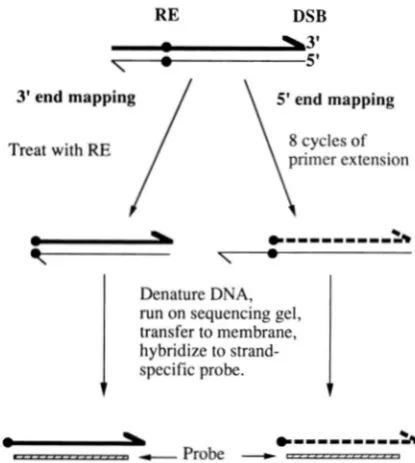
DSB3' end mapping
/ \
5' end mapping-
-
7
*"""""A
Denature DNA,
run on sequencing gel, transfer to membrane, hybridize to strand-
1
specific probe.1
-
,...,... -Probe-
p . . - - ..,... -.., *""""SAFIGURE 3.-Mapping 3' and 5' DSB-generated termini. The mapping of sequences on only one side of the break is de- picted. The 3' ends (thick lines) were mapped by treating the DNA with a restriction enzyme (RE) that cuts at a site close (within 200-300 bp) to the DSB site (BstUI for DSB(L) and HaeIII for DSB(R)). The resulting DNA was denatured, run on DNA sequencing gels, transferred to a membrane, and hybridized to a strand-specific probe. The 5' ends (thin lines) were mapped by denaturing unrestricted DNA, and per- forming eight cycles of primer extensions using a primer l o -
cated at the same restriction site used in the analysis of 3'
ends. This primer hybridized to the thin strand and was ex- tended to the 5' end; the resulting product is shown with a thick dashed line. The DNA molecules resulting from these manipultations were denatured and examined as described for the 3' termini.
generated restriction fragments shorter than those used in the previous study. As shown in Figure 1, the te- lomeric insertion is bracketed by PVuII and PstI sites, and there is a BbsI site in BZKl -50 bp from the PVUZZ site. When DNA was isolated from meiotic cells, treated with EcoA, and examined by Southern analysis (using a probe derived from the HZS4, Figure l ) , a DSB frag- ment of -1 kb was observed. This fragment comigrated with an EcoA-BbsI fragment derived from mitotic cells but was distinctly larger than an EcoA-huII fragment
(Figure 2). Thus, the DSB cleavages occur on the BZKl
side of the telomere insertion. In addition, since the band representing the DSB is only slightly wider than the bands representing restriction fragments, the DSB sites are restricted to a small chromosomal interval.
Mapping DSB sites by primer extension and Southern analysis: The method that we used to map DSB sites to single-base pair resolution (Figure 3) is based on methods developed for genomic sequencing (CHURCH and GILBERT 1984; HUIBRECSTE and ENCELKE 1991), as modified by LIU et al. (1995). A DSB within the HZS4 hotspot produces two DNA fragments with two DSB- generated termini. The terminus of the fragment with
Mapping Meiosis-Specific DNA BI -e aks 1119
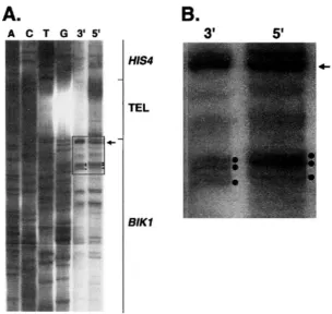
A.
A C T G 3 ' 5 '
HIS4
E L
BIKl
of the fragment with HIS4 sequences will be called DSB(R). Both ends were mapped by similar procedures, and we describe the mapping of DSB(L) first.
DNA was isolated from strain FX4 after the cells had been incubated in sporulation medium for 24 hr. To measure the position of breaks at the 3' end of DSB(L), we treated the meiotic DNA with BstUI, separated the fragments on a polvacrylamide DNA sequencing gel, and hybridized the separated fragments with a strand- specific probe. We included a DNA sequencing ladder from this region of the genome on the same gel (Figure
4A). Since this sequencing ladder was obtained using a primer that started at the
BstUI
site, it is aligned with the DSB ends. It is clear from Figure4A
that there are multiple 3' ends. Although there are faint bands at many positions in the gel lane, the strongest bands are mostly distributed within a 50-bp region. There are no strong breaks in the telomeric sequences. N o DSBs were detectable in DNA derived from mitotic cells by stan- dard Southern procedures (FAN et al. 1995) or by South- ern analysis using DNA sequencing gels (data not shown).The 5' ends of DSB(L) were mapped by primer ex- tension. We used primer extension, rather than directly examining the position of the DSBs using a different strand-specific probe, for two reasons. First, the primer extension analysis allowed us to compare DNA strands that had the same sequence and, therefore, the same molecular weight. The position of breaks in the two
strands could be established using the same DNA se- quencing ladder and the same size standard. Second, other researchers observed a protein attached to the 5' end of the DSB in yeast (DE MASSY et al. 1995; KEENEY
FIGURE 4.-Mapping the 3' and 5' termini
of DSB(L). (A) As outlined in Figure 3, the 3' ends were examined by direct Southern analysis and the 5' ends mapped by primer extension. For the 3' analysis, meiotic DNA was treated with BslUI. The oligonucleotide (10083) used for the primer extensions began at the BslUI site. The DNA sequencing ladder was also pre- pared using the 10083 primer. The arrow indi- cates the position of a PuuII-BstUI fragment (derived from pMW50) added to the 5' and 3' samples as a size standard. A strand-specific probe derived from the BIKl region (Figure 1 ) was used to detect the positions of the breaks. The outlined region of the gel indicates the area shown at higher magnification in Figure 4B. Dots indicate three prominent DSB bands. (B) Magnified region of gel lanes shown in Fig- ure 4A. The bands representing the control PuuII-BstUI fragment are in direct alignment, whereas the bands representing the 3' DSBs are one base pair shorter than those representing the 5' DSBs.
and KLECKNER 1995;
LIU
et al. 1995). This attached pro- tein would be expected to affect the migration of the 5' strand. The primer extension method allows one to map the position of the 5' end without this complica- tion. Using an oligonucleotide primer with sequences that begin at the BstUI site, we performed eight rounds of primer extensions (unidirectional PCR, details in MATERIALS AND METHODS). The resulting products were analyzed as described above for the 3' ends and showed a similar pattern of bands (Figure4A).
To compare relative mobilities of DNA representing the 3' and 5' ends, we included the same size standard in both lanes, the BstUI-hII restriction fragment shown in Figure 1. The alignment of the 5' and 3' ends of DSB(L) indi- cated that the 5' ends were displaced by one base pair (Figure 4B). SinceTug
DNA polymerase adds an extra terminal base during PCR amplification (HEMSLEY et al.1989), the simplest explanation of this result is that the 5' and 3' cleavages occur at the same position in the DNA molecule, indicating that ends generating by DSBs are blunt. We confirmed this conclusion by reanalyzing the
5'
ends using a DNA primer that was recessed one base from BstUI site (Figure 5). When the DNA samples representing the 3' ends and the primer extension product representing the 5' ends were mixed, it was clear that the5'
and 3' bands comigrated. When primer extension experiments were done on DNA isolated from mitotic cells, a few faint bands were seen, but the number and intensity of these bands varied in different primer extension reactions (data not shown).1120 F. Xu and T. D. Petes
3'
3'
5'
5'
FIGURE 5.-Mapping of DSB(L) using a different primer for primer extension. The 3' termini were mapped as de- scribed in Figure 4. The 5' termini were analyzed using a primer (10918) one base displaced from the BstUI site. Thus, since primer extension with T q DNA polymerase results in
the addition of an untemplated base with this protocol (HEMS
LEY et al. 1989; see MATERIAIS AND METHODS), the 5' and 3'
strands should comigrate if the DSB results in a blunt end. Since the lane containing a mixture of the 5' and 3' strands has the same pattern of bands as the lanes with the individual strands, the DSB-generated termini are blunt.
5'
mapping that are not represented in the 3' mapping (Figure 4); such bands could represent a low level of sequence-specific nicking or could reflect premature termination during the primer extension reaction. To confirm the conclusion that the DSB(L) ends were blunt, we also mapped the DSB(R) ends by similar pro- cedures (Figure 7). The 3' ends were mapped by treat- ing meiotic DNA with HueIII, separating the cleavage products with DNA sequencing gels and hybridizing to a strand-specific probe. The 5' ends were mapped using a primer that was displaced by one base from the Hap111 site. Relative to the DNA sequencing standard, the 3' ends mapped at the positions expected for a blunt end cleavage. Although most of the 5' and 3' ends derived from DSB(R) were at the same positions, there were fewer bands observed for the5'
ends, and the relative band intensities were different for the 5' and 3' ends. Although some of these discrepancies may represent a low level of site-specific strand nicks, it is more likely that additional bands or differences in band intensities reflect artifacts of the primer extension procedure. In summary, most of the ends generated by DSBs at the HIS4 hotspot appear to be blunt, in agreement with the results of DE MASSY et al. (1995) at the CYS3 hotspot.Since the methods described above involve denatur- ing the DNA, some of the observed break positions
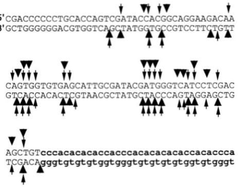
t Y
tv
t
S'CGACCCCCCTGCACCAGTCGATACCACGGCAGGAAGACAA S'GCTGGGGGGACGTGGTC
v
m
CAGTGGTGTGAGCATTGCGATACGATGGGTCATCCTCGAC ""
AGCTGTcccacacacaccacccacacacacaccacaccca TCG
c
99gtgtgtgtsgtssgtgtstststsststssst
sF3
4
caccacaccaacccacTCTGCAGTCGAGGTATGAACAGTA gtggtgtggttgggtgAGACGTCAGCTCCATACTTGTCAT
FIGURE 6.-Patterns of break sites determined by direct Southern analysis and primer extension. This figure summa-
rizes the mapping of DSB(L) and DSB(R) that was performed by analyzing gels such as those shown in Figures 4, 5 and 7;
more than five independent gels for both DSB(L) and DSB(R) were used to generate these data. The slender and
thick arrows represent mapping of 3' and 5' ends, respec- tively. Arrows above and below the sequence show cleavage sites on the upper and lower strands, respectively. Thus, the mapping of DSB(L) is represented by thin arrows on the top strand and thick arrows on the bottom strand, and the map ping of DSB(R) is shown by thick arrows on the top strand and thin arrows on the bottom strand. The telomeric repeats are shown in lowercase letters, and sequences to the left of the telomeric repeats are BIKl sequences.
could reflect nicks rather than DSBs. Consequently, we did two-dimensional agarose gel electrophoresis (neu- tral gel electrophoresis followed by alkaline gel electro- phoresis) to look for site-specific nicks. No specific nicks were detected in this analysis (data not shown), in agreement with similar studies done by the three other groups of workers mapping DSBs at recombination hot- spots (DE MASSY et al. 1995; LIU et al. 1995; XU and
KLECKNER 1995).
Although the methods used in our study were not designed to detect proteins bound to the
5'
end of DSB generate termini, several other groups (DE MASSY et al.Mapping Meiosis-Specific DNA Breaks 1121
3'
T
G
A
C
3'
5'
5'
&
L
bk
4IBIKl
TEL
HIS4
FIGLIRE 7.-Mapping the 3' and 5' termini of DSB(R). The 3' lane contains meiotic DNA treated with HwIII. The 5' lane
contains the products of a reaction in which meiotic DNA was used as a substrate for primer extension with the oligonu- cleotide 10929 (one base recessed from the Hap111site). The
DNA sequencing ladder was prepared using a primer (10193) at the Had11 site. The arrow shows the position of migration of a size standard mixed with the 3' and 3' DNA samples. As
described in MATERIALS AND METHODS, the 181-bp size stan- dard was generated by PCR amplification of pMW50, using primers 10193 (which align at the HneIII site) and 9905; this fragment has DNA sequences identical to those in the DNA
sequencing ladder. The rightmost lane contains a mixture
of 5' and 3' DNA samples. After electrophoresis, the DNA
fragments were transferred to Hybond-N+ and hybridized to
a strand-specific probe (described in MATERIALS AND METH- ODS) containing HIS4 sequences.
supports the conclusion that the DSFbgenerated ends are blunt. This conclusion is also reinforced by the exo- nuclease digestion experiments described below.
Analysis of DSB-produced DNA ends with exo- nuclease 111: Exonuclease 111 catalyzes the stepwise re- lease of mononucleotides from the 3' O H e n d s of the following DNA substrates (BRUTLAC and KORNBERC 1972; SAMRROOK et al. 1989): ends with 5' overhangs, blunt ends, ends with 1-3-base 3' overhangs, and nicked double-stranded DNA molecules. Exonuclease 111 is unable to degrade linear DNA with a 3' overhang that is greater than or equal to four bases (HENIKOFF 1984) o r a DNA molecule in which a 5' overhang has been filled in with a-phosphorothioate dNTPs
(as
dNTP) ( PUTNEY et al. 198 1 ).
To analyze the nature of the termini produced by
TABLE 1
Diagnosis of DNA termini by treatment with exonuclease III
Sensitivity to exonuclease 111 treatment"
Unmodified Modified Type of DNA ends DNA ends DNA ends"
3' overhang ( 2 4 bases) R R
5' overhang S R
Blunt or 3' overhang
(<4 bases) S S
" R indicates resistance and S indicates sensitivity to exo- nuclease 111 digestion.
'The modification is treatment of the DNA with the Klenow fragment of DNA polymerase and cuMNTPs. For DNA ter- mini with a 5' overhang, this treatment will result in blunt- ended termini that are resistant to exonuclease 111 digestion.
the meiosis-specific DSB, we examined the sensitivity of these ends to exonuclease 111, before and after treat- ment of the ends with DNA polymerase and aSdNTPs. We monitored the sensitivity of the ends to exonuclease
111 by Southern analysis. A summary of the results ex- pected for exonuclease digestion of various types of termini is given in Table 1. As indicated in this table, termini with a 3' overhang greater than or equal to four bases in length will be resistant to exonuclease 111, with or without treatment of the DNA with DNA polymerase a n d a M N T P s . Both Sac1 and Hue11 cleave DNA to generate termini with four base 3' overhangs (SAMBROOK rt al. 1989). The HIS4 hotspot region is included in a 2.3-kb S u d - H a d fragment (Figure 1), and the sites of DSBs are located -500 bp from the Sad site and 1.8 kb from the Had1 site. As shown in Figure 8A, the 2.3kb Sad-Ha11 fragment without the DSB was resistant to digestion with exonuclease 111, as expected. T h e l.&kb band representing the DSB was sensitive to exonuclease 111. Since this band should represent fragments with one exonuclease 111-resistant HaeII ter- minus, this result indicates that the end produced by the DSB does not have a 3' overhang that is four bases or longer (Table 1).
As a control, plasmid pBR322 DNA treated with the enzymes BamHI and AvaI (which result in termini with
4-bp
5' overhangs), and RsaI (which results in blunt- ended termini) was mixed with the yeast DNA samples. The four plasmid DNA fragments visualized by staining of the gel with ethidium bromide (indicated by arrows in Figure 8) represent (from largest to smallest), RtaI- RraI, BumHI-AvaI, AvaI-RsuI and RsuI-RsuI fragments. As expected, all fragments are sensitive to exonuclease 111 in DNA samples that have not been treated with DNA polymerase a n d a M N T P s .1122 F. Xu and T. D. Petes
A-
u
9.
T
"
0
0.5
1
2
0 0.5 1 2
M
FIGURE 8.-Analysis of DSB-generated termini using exo- nuclease 111. DNA samples isolated from meiotic cultures of FX4 were digested with Sad and Had1 (enzymes that result in termini with 3' overhangs of four bases) and were mixed with AvnI/RnmHI/RsnI-digested pBR322 DNA, AvaI and RamHI result in termini with four base 5' overhangs, whereas RsaI results in blunt termini. These samples were either treated with exonuclease 111 (time of treatment 0, 0.5, 1 or 2 min) immediately (A) or were modified by the addition of DNA polymerase and aSdNTPs before treatment with exo- nuclease 111 (B). As detailed in MATERIAIS AND METHODS, the DNA samples were then treated with mung bean nuclease and analyzed by gel electrophoresis. The top portions of the figure show the ethidium bromide staining pattern of the gel, and the bottom portions represent Southern analysis of the same gels (after an additional period of electrophoresis) using HIS4-specific fragments derived from pDN42 (Figure 1) as a
hybridization probe. The four bands marked by arrows on the top part of A and B represent (from largest to smallest) RsaI-RsaI, BnmHI-AvnI, Avnl-RsaI, and RsaI-RtaI fragments of pBR322; the numbers next to the arrows indicate the sizes of the fragments in kb. All fragments were exonuclease III- sensitive in the untreated ( U ) samples (A), but the BnmHI- AvaI fragment became resistant to exonuclease I11 in the treated (T) samples. As shown in the bottom part of the fig- ure, the 2.3kb SacI-Huc.II fragment (representing meiotic DNA without a DSB) was mostly resistant to exonuclease 111
treatment, whereas the 1.8-kb fragment (representing the DSB) was sensitive to exonuclease 111 with or without modifi- cation. The diffuse staining near the top of the gels represents SncI-Had1 yeast genomic DNA fragmena. The lane marked M contained HindIWEcoRI-treated lambda DNA.
sensitive to exonuclease 111 following this treatment. The control plasmid DNA BamHI-AvaI fragment was rendered resistant to digestion by exonuclease 111, indi- cating that the DNA polymerase utilized the aSdNTPs to generate exonuclease III-resistant termini. The sim- plest interpretation of this result is that the terminus caused by the DSB is blunt or has a 3' overhang of less than four bases. These results also argue that the 3' end of the DSB is not covalently attached to a protein since such an attachment would be expected to make the termini resistant to exonuclease 111.
Several other points should be discussed. First, we observed some exonuclease digestion of the 2.3kb Sad-
Had1 fragment (Figure 8). A fraction of the Sad or
Ha11
ends may be sensitive to exonuclease 111 as a con- sequence of loss of one or more base pairs of the 3' overhangs because of a small amount of a contaminat- ing exonuclease. In some preparations of DNA, the exonuclease-sensitive component is only a small frac- tion of the total (data not shown). Second, a very small fraction of the DSB termini appears resistant to exo- nuclease 111 after treatment with DNA polymerase and aSdNTPs (bottom of Figure 8B). These termini may represent a small fraction of ends that has 5' overhangs allowing modification to exonuclease III-resistance, or a small amount of DNA polymerase-mediated exchange of the terminal nucleotides. Third, although the plas- mid control BamHI-AvaI fragment was made resistant to exonuclease 111 digestion by treatment with the modi- fied nucleotides, it could be argued that a terminus with a smaller 5' overhang might be refractory to this modification. We found, however, that RstNIgenerated termini, which have only a single base 5' overhang, were efficiently modified to resistance to exonuclease I11 with aSdNTPs (data not shown). Fourth, we showed that treatment of the plasmid control RamHI-AvaI fragment with DNA polymerase and unmodified nucleotides did not result in the fragment becoming exonuclease 111resistant (data not shown). Thus, the resistance does not reflect the activity of a contaminating nuclease of the DNA polymerase that converts 5' overhangs to 3' overhangs. In summary, the exonuclease 111 studies are consistent with our conclusion that most DSB-gener- ated termini at the HIS4 hotspot are blunt.
Mapping meiotic DSBs by PCR The yeast S. cermisiae
has a very high rate of meiotic recombination per kb relative to most higher eukaryotes, and the rad5OSmuta- tion prevents the loss of DNA lesions by subsequent steps of meiotic recombinaton. In organisms without these advantages, the procedures for fine-structure mapping that we and others have used may be difficult to employ. Consequently, we modified the technique of anchor-PCR (FROHMAN et al. 1988) to investigate the placement of DSBs in yeast.
Mapping Meiosis-Specific DNA Breaks 1129
3+
2+
1.6+
1+
0.5+
0.4+
A.
Mitotic
B.
Meiotic
~'CGACCCCCCTGCACCAGTCGATACCACGGCAGGAAGACAAYf
1
*
- 1
1
2
3
1
2
3
f
*
A A
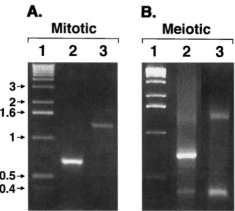
FIGURE 9.-Detection of DSBs by anchor-PCR. DNA was
isolated from either mitotic (A) or meiotic (R) cultures of
FX4. This DNA was treated with XhoI, terminal transferase and dGTP, and then PCR analysis was performed. To examine
DSB(L) (lanes 2 in A and B), we used a primer containing poly dC sequences (7278) and a primer from the BIKl side of the DSB (7492); to examine DSB(R) (lanes 3 in A and B), we employed primer 7278 and a primer from the HIS4 side
of the DSB (7680). The single bands in lanes 2 and 3 of A
represent the PCR products containing sequences between the primers and the XhoI sites. The additional bands in lanes
2 and 3 of B represent the PCR products containing sequences between the primers and the DSB-generated termini. Lane 1 contains marker DNA of I-kb ladder (GIBCO BRL).
cells, in addition to the 700-bp fragment, we observed a fragment of -380 bp (Figure SB, lane 2). From our previous mapping analysis, this fragment had the ex- pected size to represent the DSB. The detection of this fragment indicates that the DSBs result in 3' termini with a hydroxyl group, since terminal transferase re- quires such a substrate (SAMRROOK et al. 1989).
The second set utilized the same poly dC primer and a primer derived from the HIS4 gene (primer 7680, described in MATERIALS AND METHODS). In DNA isolated from mitotic cells, a single 1300-bp fragment (the dis- tance between the primer and the BIKl XhoI site) was observed (Figure SA, lane 3), whereas in DNA isolated from meiotic cells, an additional 360-bp fragment was present (Figure 9B, lane 3). This PCR product was the size expected to represent the DSB (Figure 1).
The PCR product.. representing the DSBs were puri- fied from the gel, cloned and sequenced. Since termi- nal transferase adds nucleotides to 3' termini, the se- quences obtained represent 3' break sites, with the first set of primers mapping DSB(L) and the second m a p ping DSB(R). The mapping of these breaks is summa- rized in Figure 10. Breaks representing DSB(R) were mapped in two independent experiments with upward- pointing arrows below the sequence representing the mapped sites. Since we cloned the DNA ends by addi- tion of poly dG, the position at the breakpoint is ambig-
3
f
CaccacaccaacccacTCTGC TCGAGGTATGAACAGTA gtggtgtggttgggtgA CGTCAGCTCCATACTTGTCAT
I'
A
FIGURE 10.-Mapping of DSB sites using anchor-PCR. The meiosis-specific PCR fragments shown in Figure 9B were cloned and sequenced. Each arrow represents a single clone, except for the arrows marked 22 and 16, which represent 22
and 16 clones with that breakpoint. Arrows with different shapes represent clones obtained in different experiments. Arrows above the line represent 3' breaks in the upper strand
(the 3' termini of DSB(L)), and arrows below the line reflect
3' breaks in the lower strand (the 3' termini of DSB(R)).
Since the ends were defined by adding a poly dG tail to the termini, breakpoints mapping at Gs in the sequence are am-
biguous. This ambiguity is indicated by horizontal lines above or below the sequence.
uous if it maps next to a G in the chromosomal se- quence; such ambiguities are indicated by horizontal lines adjacent to the arrows in Figure 10.
As expected
from the other mapping study, most of the breaks were located on the BIKl side of the telomere insertion with few breaks within the telomere insertion. Sixty of sixty- four sequenced clones for DSB(R) are shown in the figure; an additional four clones had breakpoints u p stream and downstream of the sequences in Figure 10.1124 F. Xu and T. D. Petes
changes in experimental conditions, since this site was not strongly represented in two of the experiments. A second alternative is that DNA contamination occurred in two of the experiments. Although this alternative is difficult to rule out, the two experiments in which the site were overrepresented were done with completely different sets of solutions.
In summary, mapping by the PCR procedure is in reasonably good agreement with the previous mapping protocol. Since we found that the PCR procedure a p peared to result in an artifactually strong site in two experiments, however, the direct blotting procedure is the method of choice in organisms with a high level of meiosis-specific DNA lesions. In organisms in which the level of DNA lesions is low, the PCR procedure is a reasonable alternative.
DISCUSSION
A telomere insertion upstream of HIS4 results in a very high level of meiotic recombination (WHITE et al. 1993) associated with a high level of DSB (FAN et al.
1995). The fine-structure mapping of the DSB indicates the following: (1) Most of the DSBs occur in a 50-bp region within the BIKl gene adjacent to the telomeric sequences; DSBs do not occur in the telomeric se- quences.
(2)
The DSB appears to involve a concerted cleavage of both strands to generate a blunt-ended DNA molecule, and (3) since the 3' ends are substrates for terminal transferase, these termini have a 3' hydroxyl group.In yeast (WHITE et al. 1993; FAN et al. 1995), Cuenorhub
&tis ehgans ( CANGIANO and LA VOLPE 1993), and mam-
malian cells (ALVAREZ et al. 1993), interstitial telomeric sequences stimulate recombination and/or DSB forma- tion. One interpretation of these effects is that these sequences are particularly susceptible to the endonucle- ase responsible for DSB formation. Our observation that DSBs are adjacent to rather than within the te- lomeric repeats rules out this interpretation. Our pre- ferred model is that the hotspot activity of the telomeric sequences is related to the binding of the transcription factor Raplp. The 50-bp telomeric insertion contains three near consensus Raplp binding sites, and dupli- cated Raplp binding sites upstream of HIS4 strongly stimulate meiotic recombination (WHITE et al. 1993). We previously suggested that this stimulation is a conse- quence of Raplp binding increasing the accessibilty of meiotic chromatin to the meiotic endonuclease that causes the DSB or a consequence of local targeting of the endonuclease to the hotspot region by Raplp (FAN et al. 1995). By either of these mechanisms, the breaks could occur within the telomeric sequences or adjacent to these sequences.
In studies using DNase I and micrococcal nuclease on meiotic chromatin of the HIS4 region, we failed to find dramatic alterations in chromatin structure associ- ated with different levels of hotspot activity (FAN and
PETES 1996). Consequently, we prefer the model that Raplp targets the meiotic nuclease to the chromatin, either directly or through bridging proteins. If this mechanism is correct, the targeting is sufficiently flexi- ble that multiple sites of cleavage are allowed. Since the DSBs do not occur within the telomeric insertion or on the HIS4 side of the insert, cleavage may also be pre- vented by DNA binding proteins. By this model, a strong DSB site may require two properties, a binding site for a transcription factor and an adjacent sequence that is not protected by a DNA-binding protein.
Although we favor a model in which the positions of the cleavages are controlled by interactions of the endonuclease with transcription factors, we cannot rule out the possibility that the DSB sites are determined by accessibility of chromatin affected indirectly by tran- scription factor binding. Previous studies (OHTA et al. 1994; WU and LICHTEN 1994) at several recombination hotspots in yeast showed a correlation between nuclease-sensitive sites and hotspots for DSB formation. The nature of the correlation is that all hotspots for DSB formation appear in nuclease-sensitive regions, but not all nuclease-sensitive sites are hotspots for DSB for- mation (LIU et al. 1995; FAN and PETES 1996).
DSBs have been mapped at recombination hotspots in three previous studies (DE MASSY et al. 1995; LIU et al. 1995; Xu et al. 1995). In these studies, as in our study, DSB sites are distributed over a relatively small region
(<200
bp). The studies disagree about the na- ture of the termini produced by the break. LIU et al.(1995) concluded the termini have a two base 5' exten- sion, and DE MASSY et al. (1995) found that the termini are blunt. This discrepancy could reflect different strain backgrounds, the different loci examined, or an experi- mental artifact. We have three independent arguments that the termini at the HIS4 locus do not have 5' over- hangs: (1) mixtures of 5' and 3' strands have no extra bands on sequencing gels, (2) DSB(L) and DSB(R) sites map to identical positions, and (3) the termini are sensitive to exonuclease I11 with or without treatment with aSdNTPs. If the different termini detected in these studies do not represent an experimental artifact, then there is apparently more than one type of enzy- matic activity that can produce DSBs in yeast. One inter- esting explanation of such a difference is that different transcription factors may target different endonucle- ases to hotspot regions.
We thank QINGQING FAN and DAVID KIFWATRICK for frequent dis- cussions concerning the research and for comments on the manu- script. We thank s. PORTER, D. NAG, and M. WHITE for strains o r plasmids and M. LICHTEN for advice on mapping protocols. The re- search was supported by the National Institutes of Health (GM- 241 10).
LITERATURE CITED
-I, E., R. PADMORE and N. KLECKNEK, 1990 Analysis of wild-type and rad50 mutants of yeast suggests an intimate relationship
Mapping Meiosis-Specific DNA Breaks 1125
ALVAREL, L., J. W. EV.~NS, R. WILKS, J. N. L u c s , J. M. BROWN et al., 1993 Chromosomal radio sensitivity at intrachromosomal
telomeric sites. Genes Chromosomes Cancer 8: 8-14.
AUSUBEI., F. M., R. BRENT, R. E. KINGSTON, D. D. MOORE, J. G. SEID-
MAN et al., 1993 Current Protocols in Molecular Biology. Vol. I: 3.17.4. John Wiley & Sons, New York.
BISHOP, D. R , D. PARK, L. Xu and N. KLECKNER, 1992 DMCI: a
meiosis-specific yeast homolog of E. coli recA required for meiotic
recombination, synaptonemal complex formation and cell cycle progression. Cell 69: 439-456.
BOLLUM, F. J., 1974 Terminal deoxynucleotidyl transferase, pp.
145-171 in TheEnzymps, Vol. 10, edited by P. D. BOYER. Academic Press, New York.
BRL~TIAG, D., and A. KORNRERG, 1972 Enzymatic synthesis of deoxy-
ribonucleic acid. .J. Biol. Chem. 247: 241-248.
CANMANO, G., and A. L a VOLPE, 1993 Repetitive DNA sequences
located in the terminal portion of the Canamhabditis ekgans
chromosomes. Nucleic Acids Res. 21: 1133-1 139.
C A o , L.., E. A I A N I and N. KLECKNER, 1990 A pathway for generation and processing of double-strand breaks during meiotic recombi- nation in S. cereuisiae. Cell 61: 1089-1101.
CHITRCEI, G . M., and W. GIL.BERT, 1984 Genomic sequencing. Proc.
Natl. Acad. Sci. USA 81: 1991-1995.
DE MASSY, B., and A. N r c o ~ . . ~ , 1993 The control in cis of the position
and amount of the ARC4 meiotic double-strand break of Saccharo-
myces rweuisiae. EMBO J. 12: 1459-1466.
DE MASSY, B., V. R o c c o and A. NICOLAS, 1995 The nucleotide m a p
ping of DNA double-strand breaks at the CYS3 initiation site of
meiotic recombination i n Sarrharomyces cereuisiae. EMBO J. 14:
4589-4598.
FAN, Q., and T. PETES, 1996 Relationship between nuclease hyper-
sensitive sites and meiotic recombination hotspot activity at the
HIS4 locus of Saccharomyces cereuisiae. Mol. Cell. Biol. (in press).
FAN, Q., F. Xu and T. PETES, 1995 Meiosis-specific double-strand
breaks at the HIS4 recombination hot spot in the yeast Saccharo- myces cermisinr: control in cis and trans. Mol. Cell. Biol. 15: 1679- 1688.
FROHMAN, M. A,, M. K. DUSH and G. R. MARTIN, 1988 Rapid produc- tion of full-length cDNA from rare transcripts: amplification us- ing a single gene-specific oligonucleotide primer. Proc. Natl.
Acad. Sci. USA 85: 8998-9002.
GAME, J. C., 1993 Pulsed-field gel analysis of pattern of DNA double-
strand breaks in the Saccharomyces meuisiaegenome during meio-
sis. Dev. Genet. 13: 485-497.
HEMSLEY, A,, N. ARNHEIM, M. D. TONEY, G. CORTOPASSI and D. J.
GALAS, 1989 A simple method for site-directed mutagenesis us-
ing the polymerase chain reaction. Nucleic Acids Res. 17: 6545-
6551.
HENIKOFF, S., 1984 Unidirectional digestion with exonuclease I11
creates targeted breakpoints for DNA sequencing. Gene 28:
35 1 - 359.
HUIBREGTSE, J. M., and D. R. ENGELKE, 1991 Direct sequence and
footprint analysis of yeast DNA by primer extension. Methods
Enzymol. 194 550-562.
KEENEY, S., and N. KLECKNER, 1995 Covalent protein-DNA com- plexes at the 5’ strand termini of meiosicspecific double-strand
breaks in yeast. Proc. Natl. Acad. Sci. USA 9 2 11274-11278.
LICHTEN, M., and A. S. H. GOLDMAN, 1995 Meiotic recombination
hotspots. Annu. Rev. Genet. 29: 423-444.
LIU, J., T.-C. WU and M. LICHTEN, 1995 The location and structure
of double-strand breaks induced during yeast meiosis: evidence
for a covalently linked DNA-protein intermediate. EMBO J. 14:
4599-4608.
NAG, D. R , and T. D. PETES, 1993 Physical detection of hetero-
duplexes during meiotic recombination in the yeast Saccharomyces
cereuisiae. Mol. Cell. Biol. 13: 2324-2331.
NAG, D. K., M. A. WHITE and T. D. PETES, 1989 Palindromic se-
quences in heteroduplex DNA inhibit mismatch repair in yeast.
Nature 340: 318-320.
OHTA, R, T. SHIBATA and A. NICOLAS, 1994 Changes in chromatin
structure at recombination initiation sites during yeast meiosis.
PADMORE, R., L. CAO and N. KLECKNER, 1991 Temporal comparison
of recombination and synaptonemal complex formation during meiosis in S. cereuisiae. Cell 66: 1239-1256.
PETES, T. D., R. E. MALONE and L. S. SYMINGTON, 1991 Recombina-
tion in yeast, pp. 407-521 in The Molecular and Cellular Biology of
the Yeast Saccharomyces, Vol. 1 , edited by J. R. BROACH, E. W. JONES
and J. R. PRINGLE. Cold Spring Harbor Laboratory Press, Cold
Spring Harbor, N Y .
PLUTA, A. F., and V. A. ZAKIAN, 1989 Recombination occurs during
telomere formation in yeast. Nature 337: 429-433.
PUTNEY, S. D., S. J. BENKOVIC and P. SCHIMMEI., 1981 A DNA frag-
ment with an cy-phosphorothioate nucleotide at one end is asym-
metrically blocked from digestion by exonuclease I11 and can be
replicated in vivo. Proc. Natl. Acad. Sci. USA 7 8 7350-7354.
SAMBROOK, J., E. FRITSCH and T. MANIATIS, 1989 Molecular Cloning:
A Laboratoly Manual. Cold Spring Harbor Laboratory Press, Cold
Spring Harbor, W.
STURZI., M., and W. K. ROTH, 1990 “Run-off’ synthesis and applica-
tion of defined single-stranded DNA hybridization probes. Anal.
Biochem. 185: 164-169.
SUN, H., D. TRECO, N. P. SCHULTES and J. W. SZOSTAK, 1989 Double-
strand breaks at an initiation site for meiotic gene conversion.
Nature 338: 87-90.
SUN, H., D. TRECO and J. W. SZOSTAK, 1991 Extensive 3”overhang-
ing, single-stranded DNA associated with the meiosis-specific double-strand breaks at the ARC2 recombination initiation site. Cell 2 2 1155-1161.
WHITE, M. A,, M. WIERDL, P. DETLOFF and T. D. PETES, 1991 DNA
binding protein RAP1 stimulates meiotic recombination at the
HIS4 locus in yeast. Proc. Natl. Acad. Sci. USA 88: 9755-9759.
WHITE, M. A,, P. DETLOFF, M. STRAND and T. D. PETES, 1992 A
promoter deletion reduces the rate of mitotic, but not meiotic
recombination at the HIS4 locus in yeast. Curr. Genet. 21:
WHITE, M. A., M. DOMINSKA and T. D. PETES, 1993 Transcription
factors are required for the meiotic recombination hotspot at
USA 90: 6621-6625.
the HIS4 locus in Saccharomyces cereuisiae. Proc. Natl. Acad. Sci.
WU, T. C., and M. LICHTEN, 1994 Meiosis-induced double-strand
break sites determined by yeast chromatin structure. Science 263
515-518.
XU, L., and N. KLECKNER, 1995 Sequence non-specific double-
strand breaks and interhomology interactions prior to double- strand break formation at a meiotic recombination hot spot in
yeast. EMBO J. 16: 5115-5128.
ZENVIRTH, D., T. ARBEI., A. SHERMAN, M. GOLDWAY, S. KLEIN et ab,
1992 Multiple sites for double-strand breaks in whole meiotic chromosomes of Saccharomyces cereuisiae. EMBO J. 11: 3441 -3447.
Communicating editor: G. R. SMITH
EMBO J. 1 3 5754-5763.