Scholarship@Western
Scholarship@Western
Electronic Thesis and Dissertation Repository
7-21-2015 12:00 AM
The role of bone sialoprotein in the tendon-bone insertion
The role of bone sialoprotein in the tendon-bone insertion
Ryan M. Marinovich
The University of Western Ontario Supervisor
Dr. Harvey Goldberg
The University of Western Ontario Joint Supervisor Dr. Frank Beier
The University of Western Ontario Graduate Program in Biochemistry
A thesis submitted in partial fulfillment of the requirements for the degree in Master of Science © Ryan M. Marinovich 2015
Follow this and additional works at: https://ir.lib.uwo.ca/etd
Part of the Biochemistry Commons, Medical Biochemistry Commons, Musculoskeletal, Neural, and
Ocular Physiology Commons, and the Orthopedics Commons
Recommended Citation Recommended Citation
Marinovich, Ryan M., "The role of bone sialoprotein in the tendon-bone insertion" (2015). Electronic Thesis and Dissertation Repository. 2953.
https://ir.lib.uwo.ca/etd/2953
This Dissertation/Thesis is brought to you for free and open access by Scholarship@Western. It has been accepted for inclusion in Electronic Thesis and Dissertation Repository by an authorized administrator of
(Thesis format: Integrated-Article)
By
Ryan Michael Marinovich
Graduate Program in Biochemistry
A thesis submitted in partial fulfillment
of the requirements for the degree of
Master of Science
The School of Graduate and Postdoctoral Studies
The University of Western Ontario
London, Ontario, Canada
ii
Tendons and ligaments insert into bone through a transitional tissue termed the enthesis
which is susceptible to injury and difficult to repair. Entheses contain a region of
calcified fibrocartilage (CFC), however mineral-associated proteins in this tissue remain
poorly characterized. Bone sialoprotein (BSP) is a phosphoprotein associated with
mineralizing tissues. In these studies BSP was identified in the CFC of entheses by
immunohistochemistry. Analysis of the entheses of Bsp-/- mice indicate abnormalities in
the CFC. Compared to controls, the CFC of the quadriceps tendon enthesis is 28% and 41
% longer in 15 week and 14 month old Bsp-/- mice, respectively. MicroCT and Raman
spectroscopic analysis of the CFC in Bsp-/- mice demonstrate that mineral content is
similar between genotypes. Mechanical studies show that the Bsp-/- patellar tendon is
larger in cross-sectional area yet mechanically weaker. These data suggest BSP is
involved in the regulation and growth of the CFC.
KEYWORDS
iii
Chapter 2, entitled “Identification of bone sialoprotein in enthesis fibrocartilage
and characterization of Bsp-/- fibrocartilaginous enthesis” was adapted from Marinovich et
al. (2015) (in preparation). MicroCT scanning and subsequent data analysis was
performed by Yohannes Soenjaya. Raman spectroscopy on samples prepared by Ryan
Marinovich were performed by Greg Wallace (Dr. F. Lagugné-Labarthet). Mechanical
studies, on specimens prepared by Yohannes Soenjaya, and subsequent data analysis was
performed by Andrew Dunkman and Andrew Zuskov (Dr. Lou Soslowsky)
Chapter 3, entitled “Elucidating the role of bone sialoprotein in the murine
enthesis during development and age”. All work and experiments were performed by
iv
I would like to thank my family, Mom, Dad, Mitch and Baba for the constant love
and support they have given me over the years. I wouldn’t be the person I am today
without their influence and guidance in life. Knowing that I’m making you proud in
London has been a huge motivation for me to continue on, especially when times are
difficult.
Nancy, your love and support have gotten me through some of my most difficult
times. You’ve helped me accomplish things I never thought myself capable of and I
wouldn’t be the man I am today without you in my life. I love you very much.
I would like thank my supervisor, Dr. Harvey “The Boss” Goldberg for the
mentorship and guidance he’s given me over the years. Not only has he been an
invaluable asset to my research, he has also provided me with numerous life lessons, and
has had an undoubtable influence on me and my professional life as I move forward in
my education.
I would like to acknowledge my co-supervisor, Dr. Frank Beier, and my advisory
committee members Drs. Graeme Hunter and Alan Getgood for the advice and guidance
they have provided to me during my graduate studies. I would also like to acknowledge
Linda Jackson for her expertise in histology, and Wendy Dunn, who provided expertise
in the care of our animals.
The members of the Goldberg lab, past and present: Kevin Bartman, Yohannes
Soenjaya, Dr. Erik Holm, Vida Lam, Dorchester, Lim Tang, Rose Yee and last but
vi
ABSTRACT ... ii
STATEMENT OF CO-AUTHORSHIP ... iii
ACKNOWLEDGEMENTS ... iv
TABLE OF CONTENTS ... vi
LIST OF FIGURES ... ix
LIST OF TABLES ... xi
LIST OF APPENDICES ... xii
LIST OF ABBREVIATIONS ... xiii
CHAPTER ONE: LITERATURE REVIEW ... 1
1.1 Mineralized Tissues... 2
1.2 Biomineralization ... 4
1.3 SIBLINGs... 4
1.4 Bone Sialoprotein ... 6
1.4.1 Structure ... 6
1.4.2 Collagen Binding ... 9
1.4.3 Hydroxyapatite Binding and Nucleation ... 10
1.4.4 Cell Attachment and Signaling ... 11
1.4.5 Tissue Distribution and Expression ... 12
1.4.6 In Vivo Function ... 13
1.5 The Enthesis ... 15
1.5.1 Fibrous Entheses ... 16
1.5.2 Fibrocartilaginous Entheses ... 16
1.5.3 Development ... 23
1.5.4 Tissue Injury and Healing ... 27
1.6 Purpose of Thesis ... 29
1.7 References ... 30
CHAPTER TWO: IDENTIFICATION OF BONE SIALOPROTEIN IN ENTHESIS FIBROCARTILAGE AND CHARACTERIZATION OF THE BSP-/ FIBROCARTILAGINOUS ENTHESIS ... 42
vii
2.3.1 Animals ... 48
2.3.2 Histology and Immunohistochemistry... 48
2.3.3 Measurement of the calcified fibrocartilage zone ... 50
2.3.4 Mechanical testing ... 51
2.3.5 Microcomputed Tomography ... 52
2.3.6 Raman spectroscopy ... 52
2.3.7 Statistical analyses ... 53
2.4 Results ... 54
2.4.1 BSP and OPN are present in the mineralized tissues of fibrocartilaginous entheses 54 2.4.2 Bsp-/- mice exhibit defects in calcified fibrocartilage zone of the enthesis . 54 2.4.3 Mechanical testing suggests a weakened enthesis in Bsp-/- mice ... 64
2.4.4 Collagen organization is not affected by the loss of BSP ... 64
2.5 Discussion ... 70
2.5.1 Role of altered calcified fibrocartilage zone of the Bsp-/- QCT enthesis. ... 70
2.5.2 Bsp-/- patellar tendon exhibit weakened mechanical properties under load. 73 2.6 References ... 75
CHAPTER THREE:ELUCIDATING THE ROLE OF BONE SIALOPROTEIN IN THE MURINE ENTHESIS DURING DEVELOPMENT AND AGEING ... 79
3.1 Chapter Summary ... 80
3.2 Introduction ... 82
3.3 Materials and Methods ... 84
3.3.1 Animals ... 84
3.3.1 Histology and Immunohistochemistry... 84
3.4 Results ... 86
3.4.1 Age 14 months ... 86
3.4.2 Age 14 and 21 days ... 96
3.5 Discussion ... 117
3.5.1 14 months ... 117
viii
4.1 Summary and Perspectives... 124
4.2 Limitations of Research and Future Directions... 126
4.2.1 Phenotypic differences in different entheses and the role of muscle loading 126 4.2.2 Establishing the expression of BSP in the enthesis ... 127
4.2.3 Other potential mineralization factors in the enthesis ... 130
4.3 Conclusions ... 133
4.4 References ... 134
APPENDIX A: Animal Use Ethics Statement... 137
APPENDIX B: Copyright Holder Permissions ... 138
ix
Figure 1.1 Schematic of bone sialoprotein...8
Figure 1.2 Anatomical location of selected fibrocartilaginous entheses...19
Figure 1.3 The transitional zones of the fibrocartilaginous enthesis...21
Figure 2.1 SIBLING proteins are present in the calcified fibrocartilage of the SST and QCT entheses...56
Figure 2.2 Mineral is deposited in the calcified fibrocartilage of Bsp -/ - mice however the morphology of the calcified fibrocartilage is altered...58
Figure 2.3 MicroCT analyses indicates similar levels of mineralization in wild type and Bsp-/- QCT entheses...61
Figure 2.4 Raman spectroscopy shows that the mineral content in the QCT enthesis is comparable between wild type and Bsp-/- mice...63
Figure 2.5 Bsp-/- mice exhibit alteration in patellar enthesis mechanical properties...67
Figure 2.6 Collagen organization is not affected by loss of BSP...69
Figure 3.1 BSP is present in the calcified fibrocartilage of 14 month-old mice...88
Figure 3.2 OPN is present in the calcified fibrocartilage of 14 month-old mice...90
Figure 3.3 Calcified fibrocartilage of Bsp-/- is further lengthened with age...92
Figure 3.4 At 14 months of age no differences in collagen organization are observed between wild type and Bsp-/- mice...95
Figure 3.5 No gross morphological differences are apparent in the entheses of 14 day-old wild type and Bsp-/- mice...98
Figure 3.6 No gross morphological differences are apparent in the entheses of 21 day-old wild type and Bsp-/- mice...100
Figure 3.7 Mineral is not present in the entheses of 14 day-old wild type and Bsp -/-mice...102
Figure 3.8 Mineral is not present in the QCT enthesis of 21 day-old wild type and Bsp -/-mice however small amounts are present in the SST enthesis...104
Figure 3.9 At 14 days of age no differences in collagen organization are observed between wild type and Bsp-/- mice...106
Figure 3.10 At 21 days of age no differences in collagen organization are observed between wild type and Bsp-/- mice...108
x
Figure 3.12 BSP is localized to mineralized regions in 21 day old wild type mice...112
Figure 3.13 OPN is not present in the entheses of 14 day old wild type and Bsp-/-
mice...114
xi
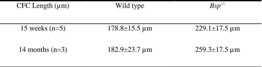
Table 3.1 CFC lengths of 15 week and 14 month old wild type and Bsp-/- QCT
xii
APPENDIX A: Statement of Permission for the Use of Animals for Experimental
xiii
Ala Alanine
BMP Bone Morphogenic Protein
BSP Bone Sialoprotein
CFC Calcified Fibrocartilage
CMV Cytomegalovirus
CT Critical Threshold
Da Dalton
DAB 3,3’-Diaminobenzidine
DMP1 Dentin Matrix Protein 1
DSPP Dentin Sialophosphoprotein
ECM Extracellular Matrix
EDTA Ethylenediaminetetraacetic acid
Glu Glutamic Acid
Gly Glycine
HA Hydroxyapatite
IHC Immunohistochemistry
IHH Indian Hedgehog
KAE Lysine-Alanine-Glutamic Acid
KD Dissociation Constant
kDa Kilodalton
LCM Laser Capture Microdissection
MALDI-TOF Matrix Assisted Laser Desorption/Ionization Time of Flight
MEPE Matrix Extracellular Phosphoglycoprotein
microCT Micro-Computed Tomography
mRNA Messenger Ribonucleic Acid
NMR Nuclear Magnetic Resonance
xiv
PBS-T Phosphate Buffed Saline-Tween 20
PCR Polymerase Chain Reaction
PDL Periodontal Ligament
PHEX Phosphate Regulating Endopeptidase Homolog, X-Linked
pI Isoelectric point
PMMA Polymethyl Methacrylate
Ptch Patched
PTHrP Parathyroid Hormone-Related Protein
QCT Quadriceps Femoris Tendon
qPCR Quantitative Real-Time Polymerase Chain Reaction
RGD Arginine-Glycine-Aspartic Acid
RNA Ribonucleic Acid
RNAseq Ribonucleic Acid Sequencing
ROI Region of Interest
Runx2 Runt-Related Transcription Factor 2
Scx Scleraxis
SIBLING Small Integrin-Binding Ligand, N-Linked Glycoproteins
Smo Smoothened
Sox Sex Determining Region on Y-box
SST Supraspinatus Tendon
TGFβ Transforming Growth Factor β
TGFβr Transforming Growth Factor β Receptor
UFC Uncalcified Fibrocartilage
CHAPTER ONE
1.1 Mineralized Tissues
Biomineralization refers to the processes by which organisms form minerals
(Lowenstam and Weiner, 1989). It requires the selective extraction and uptake of
elements from the local environment and their incorporation into functional structures
under strict biological control. The earliest fossil evidence of biomineralization dates
back 3.5 billion years (Lepot et al., 2008) and has since been observed in all five of the
biological kingdoms (Lowenstam and Weiner, 1989). Mineralized tissues are biological
matrices that have incorporated inorganic minerals such as calcium and phosphorus into
their structure. Mineralized tissues serve a variety of functions. They can act as protective
armor, such as a shell, they can be involved in cutting and grinding functions, such as
teeth, and can offer support and stability, as in the skeleton. Perhaps the most
recognizable, and by far the best characterized, mineralized tissue is that of mammalian
bone, of which a brief description follows.
Bone is a complex connective tissue composed of three main phases: the cellular
phase, the inorganic phase, and the organic phase. The major cell types in bone are
osteoblasts, osteocytes and osteoclasts. Osteoblasts are anabolic cells associated with the
secretion of matrix and the deposition of mineral. Osteocytes, derived from osteoblasts
that have become entombed within the matrix of the bone, are associated with its
regulation and homeostasis. Osteoclasts are catabolic cells involved in the resorption and
remodeling of bone, releasing mineral and breaking down the extracellular matrix. The
mineral content of bone, constituting the inorganic phase, is largely hydroxyapatite (HA),
a stable crystal of calcium phosphate with unit formula Ca10(PO4)6(OH)2. HA deposition
comprised of type I collagen. Roughly 10% of this matrix consists of non-collagenous
proteins which serve a variety of functions such as cell attachment and signaling,
regulation of mineral formation, which includes nucleation, binding and inhibition, as
well as structural functions.
Bones form through two major processes: intramembranous ossification and
endochondral ossification. Flat bones, such as the components of the skull and scapula
form through intramembranous ossification in which mesenchymal tissues become
mineralized without a cartilage intermediate. Long bones, such as the femur and
humerus, form through the process of endochondral ossification. Endochondral
ossification begins embryonically when cells of the mesenchyme condense in regions that
will eventually become the long bones of the skeleton. These cells differentiate into
chondrocytes and form a cartilaginous anlage rich in type II collagen. Cells in the central
region of this structure begin to exit the cell cycle and enter hypertrophy, in which they
swell in size and excrete extracellular matrix. Hypertrophic chondrocytes secrete
angiogenic factors which promotes the invasion of blood vessels into the tissue. These
blood vessels deliver precursor cells which differentiate into osteoblasts and osteoclasts,
as well as blood and bone marrow-forming hematopoietic cells, to the developing bone.
This creates a primary ossification center in the middle of the anlage from which the
mineralization process begins. Osteoblasts promote mineral deposition, while osteoclasts
remodel the mineralized cartilage, facilitating the formation of lamellar bone. At the
terminals of the developing long bones, the epiphysis, secondary ossification centers also
form, creating a sandwich of resting, proliferative and hypertrophic chondrocytes termed
chondrocytes of the growth plate drive the elongation and growth of the bone, until the
plates fuse at the end of puberty
1.2 Biomineralization
The exact mechanism by which HA is deposited into the type I collagen matrix of
bone is not fully understood. Serum concentrations of Ca2+ and PO43- are above the
solubility product constant of HA (Eidelman et al., 1987), however, they are kept from
precipitating in the body by a variety of inhibitors of crystal formation. In bone, type I
collagen fibrils are arranged in staggered arrays creating a periodic gap, call the hole
zone, every 67 nm (Katz and Li, 1973). It is within these hole zones that the initiation of
HA crystal formation takes place (Traub et al., 1992). It has been suggested that the
aqueous compartment created by the collagen hole zone is sufficiently small to exclude
large HA inhibitors, which leads to mineral deposition by the exclusion of inhibitors
(Nudelman et al., 2010; Price et al., 2009). Despite this evidence, type I collagen fibrils
themselves are not sufficient to nucleate HA formation (Blumenthal et al., 1990; Hunter
et al., 1986), indicating that some additional factor must be present to initiate HA crystal
formation. Acidic collagen binding proteins have long been speculated to fulfill this role
(Glimcher and Muir, 1984), of which members of the SIBLING protein family are the
prime examples.
1.3 SIBLINGs
Members of the Small Integrin Binding LIgand, N-linked Glycoprotein
The SIBLINGs have flexible structures and extensive post-translational modifications.
All members of the SIBLING family contain an RGD integrin-binding motif (Fisher et
al., 2001; George and Veis, 2008). The SIBLING genes are grouped in a 375 kbp cluster
on human chromosome 4 (Fisher and Fedarko, 2003) and mouse chromosome 5 (Crosby
et al., 1996). The family is comprised of 5 members: Osteopontin (OPN), Bone
Sialoprotein (BSP), Dentin Matrix Protein 1 (DMP1), Matrix Extracellular
Phosphoglycoprotein (MEPE) and Dentin Sialophosphoprotein (DSPP) (Fisher and
Fedarko, 2003; Fisher et al., 2001). Although the SIBLING proteins do not share a high
degree of sequence similarity, an examination of their exon structure reveals that the 5
genes are indeed related, likely resulting from gene duplication events and subsequent
divergences, facilitated by their flexible structure (Fisher and Fedarko, 2003).
All members of the SIBLING family seem to be involved with the regulation and
control of biological mineral deposition, although in different ways. The SIBLING
proteins are largely acidic, with isoelectric points ranging from 3.4-4.3, however MEPE
differs from the rest of the group as it is highly basic with a pI of 9.2 (Fisher and Fedarko,
2003) BSP, DMP1, DSPP are promoters of mineral deposition and are mostly found
within mineralized tissues, with greater concentrations of BSP found in bone, while
dentin contains greater amounts of DMP1 and DSPP (George and Veis, 2008). OPN and
MEPE are potent inhibitors of HA formation and are expressed ubiquitously, likely to
prevent ectopic calcification of soft tissues (George and Veis, 2008). Single knock-out
animals for each of the SIBLINGS have been created, with each displaying alterations in
the properties of their mineralized tissues, however the loss of a single SIBLING gene
suggests that the SIBLINGs work in concert with each other to control the process of
biomineralization in vivo and a great deal of functional redundancy is present within the
family. As the focus of this thesis is bone sialoprotein, an in depth discussion of BSP
follows.
1.4 Bone Sialoprotein
BSP is an intrinsically disordered and extensively post-translationally modified
phosphoprotein found in the mineralized tissues of bones and teeth. BSP was initially
isolated from bone in 1963 as a 23-kDa sialic acid-containing glycoprotein (Herring and
Kent, 1963)(Figure 1.1). By utilizing a dissociative extraction procedure, Fisher et al.
isolated a 70-80 kDa variant from calf bone, representing full length native BSP complete
with its post translational modifications (Fisher et al., 1983). The rat Bsp gene was first
sequenced in 1988 by Oldberg et al. (Oldberg et al., 1988). The amino acid sequences of
BSP have since been deduced in a variety of mammalian species and show a high level of
conservation, with specific domains showing identity approaching 90% (Goldberg and
Hunter, 2012).
1.4.1 Structure
Mammalian BSP contains an average of 300 amino acids, 20% of which are Glu
and 11% are Gly. Additionally a 16-residue signal sequence is present at the N-terminus.
Unmodified BSP has a pI of 3.9. BSP’s calculated mass based on amino acid composition
Figure 1.1 Schematic of bone sialoprotein. BSP is approximately 300 amino acids long.
The collagen-binding sequence (yellow) spans residues 18-45. There are two
poly-glutamic acid sequences (red) responsible for HA nucleation. Finally, there is an
mass spectroscopy studies of BSP found mean masses of 52.5 and 49.0 kDa (Wuttke et
al., 2001; Zaia et al., 2001). Broad mass peaks were observed in both these studies,
reflecting the significant heterogeneity of post-translation modifications on individual
BSP molecules. Oligosaccharides comprise approximately 30% of BSP’s weight and
BSP contains 23 sialic acid residues (Zaia et al., 2001). BSP (bovine) also contains 11
identifiedphosphorylation sites, however not all of these sites appear to be
phosphorylated on each molecule. MALDI-TOF mass spectroscopy studies on proteolytic
digests of BSP indicate that on average BSP molecules contain 5.8 phosphorylated serine
residues (Salih and Flückiger, 2004; Salih et al., 2002). BSP also contains several
sulfated tyrosine residues located in the C-terminal region of the protein. Structural
analyses of BSP utilizing circular dichroism spectropolarimetry, NMR and small-angle
X-ray scattering indicate the loose, flexible structure of an intrinsically disordered protein
(Fisher et al., 2001; Tye et al., 2003; Wuttke et al., 2001).
1.4.2 Collagen Binding
BSP binds to type I collagen fibrils with a high affinity (KD =12.1 nM) (Tye et al.,
2005). The highly conserved collagen-binding domain of BSP encompasses residues
18-45 and is rich in hydrophobic and basic amino acids. The KD of BSP18-45 (150 nM) is
weaker than that of native, full-length BSP however, indicating that other regions of the
molecule are involved in stabilizing the interaction with collagen (Baht et al., 2008).
Intermolecular forces involved in this binding appear to be comprised mostly of
hydrophobic interactions, as elution of BSP with 1.0 M NaCl yielded little protein;
the full release of BSP from a type I collagen affinity column (Baht et al., 2008). The
exact location that BSP binds to on the collagen molecule remains unknown, however
BSP appears to localize to the hole regions of type I collagen fibrils (Fujisawa et al.,
1995), which as previously mentioned are associated with early mineral formation
(Fratzl et al., 1991; Traub et al., 1992).
1.4.3 Hydroxyapatite Binding and Nucleation
BSP’s many negatively charged groups confer HA binding properties. At
physiological salt concentrations, native rat BSP bind ~60 Ca2+ ions per molecule with
low affinity (Chen et al., 1992), however bone-derived porcine BSP’s affinity for HA is
higher with a KD of ~0.9 μM (Goldberg et al., 2001). This affinity is largely attributed to
the 2-3 contiguous Glu sequences present in the central region of the protein, as
competitive binding studies using synthetic poly-Asp and poly-Glu peptides reduced
BSP’s affinity for HA by 90% and 68% respectively (Goldberg et al., 2001).
Additionally, mutation of these poly-Glu sequences to poly-Ala sequences reduced in
vivo HA adsorption 4-fold (Wazen et al., 2007).
BSP is also a potent nucleator of HA formation as demonstrated by Hunter and
Goldberg using a steady-state agarose gel system (Hunter and Goldberg, 1993). This
group went on to show that BSP’s poly-Glu sequences are largely responsible for HA
formation (Goldberg et al., 1996). Synthetic peptides spanning BSP poly-Glu sequences
can mediate HA formation on its own, and a 2.5 fold increase in nucleation activity is
unmodified full length BSP also nucleates hydroxyapatite formation, however native BSP
with its post translational modifications is ~100 fold more potent (Goldberg et al., 1996).
1.4.4 Cell Attachment and Signaling
Akin to its other SIBLING members, BSP contains an RGD integrin-binding
motif, which is located towards its C-terminus. BSP’s RGD sequence is recognized by
the αvβ3 integrin (Oldberg et al., 1988), a receptor identified on the surfaces of osteoblasts
(Prince et al., 1991) and osteoclasts (Miyauchi et al., 1991; Ross et al., 1993)
BSP-mediated attachment of these cell types has been demonstrated respectively by
Somerman et al. and Helfrich et al. (Helfrich et al., 1992; Somerman et al., 1988). Cell
attachment of osteoblasts, fibroblasts, and chondrocytes is abolished by alteration to a
KAE sequence (Gill et al., 2008; Gordon et al., 2007; Harris et al., 2000). Eukaryotic cell
binding via an RGD-independent mechanism has also been described (Mintz et al.,
1993).
BSP has been shown to promote osteoblast differentiation and mineral formation
in cell culture (Cooper et al., 1998; Zhou et al., 1995). Furthermore, a reduction in
mineralized nodules was observed when osteoblast-like cell cultures were treated with an
anti-BSP antibody (Boskey et al., 2008). Overexpression of BSP in primary rat calvarial
cells increased mineral formation and markers of osteoblast differentiation (Gordon et al.,
2007). Treatment with BSP and osteogenic media of human bone marrow-derived cells
resulted in a decrease in cell proliferation and promoted differentiation into osteoblasts
BSP mediates Ca2+ signaling in osteoclasts through the αvβ3 integrin receptor
(Paniccia et al., 1995, 1993) and can modulate their activity. BSP is involved in the
generation of osteoclasts (Valverde et al., 2005, 2008) and stimulates in vitro bone
resorption (Raynal et al., 1996), however it is not critical for this process (Malaval et al.,
2008).
1.4.5 Tissue Distribution and Expression
BSP expression is largely restricted to mineralized tissue. BSP has long been
known to be expressed in osteoid tissue (Ganss et al., 1999) and has particularly strong
expression in developing bone (Bianco et al., 1991). BSP is relatively uniform in its
distribution throughout the bone, where it is likely bound to and entrapped by HA
(Bianco et al., 1993; Chen et al., 1993; Kasugai et al., 1992; Riminucci et al., 1995)
although BSP protein is particularly concentrated at the mineralization front (Bianco et
al., 1993; Riminucci et al., 1995; Shapiro et al., 1993). Chen et al. showed BSP mRNA in
rat tissue is abundant during times of de novo bone synthesis and mineralization in bone,
dentin and cementum using northern blotting and in situ hybridization, however
expression declines as the animals matured, with little mRNA detected in 100 day old
rats (Chen et al., 1992). Thus, BSP’s expression and localization coincides with the
formation of a mineralized matrix. BSP mRNA is abundant and easily detected in mature
osteoblasts and osteocytes, however it is not present in pre-osteoblastic cells, indicating
that BSP expression is induced upon differentiation (Bianco et al., 1991; Chen et al.,
1991). In line with its proposed role in de novo bone synthesis, BSP is also expressed by
BSP expression has been detected in osteoclasts (Arai et al., 1995; Bianco et al., 1991),
suggesting that it is involved in the modulation of mineral resorption.
BSP is expressed in a variety of dental tissues, and may have a role in the
formation and maintenance of teeth. BSP protein and mRNA have been detected in both
dentin and cementum where it is expressed by odontoblasts and cementoblasts,
respectively (Chen et al., 1993; Somerman et al., 1991). Cementum in particular is rich in
BSP and BSP appears to be critical to the formation of acellular cementum (Foster et al.,
2013). Low levels of BSP have been detected in enamel and ameloblasts (Chen et al.,
1998). BSP has also been detected in three non-mineralized tissues; the trophoblastic
cells of the human placenta (Bianco et al., 1991), salivary glands (Ogbureke and Fisher,
2004) as well as platelets where it is probably endocytosed from the serum (Chenu and
Delmas, 1992).
1.4.6 In Vivo Function
Due to its tissue distribution, expressional patterns and biochemical properties,
BSP is thought to be involved in de novo bone synthesis. Specifically, BSP is believed to
bind to collagen fibers and nucleate HA formation, while also recruiting osteogenic cells
to sites of mineral formation (Ganss et al., 1999). A variety of in vivo studies support this
hypothesis. When BSP and type I collagen are implanted into calvarial defects of rats,
cell proliferation, osteoblast differentiation and mineral deposition are observed (Wang et
al., 2006; Xu et al., 2007). BSP implantation into the pulp of rat molars may also
Mice lacking the Bsp gene (Bsp-/-) demonstrate reduced body size and weight with
undermineralized fetal bones compared to wild-type littermates. At 4 months of age Bsp-/-
mice display thinner cortical bones than wild type mice and have increased trabecular
bone volume, however at 12 months few difference are apparent (Malaval et al., 2008).
This suggests that the defects are transient, and mutant mice eventually "catch up" with
their wild-type counterparts. Interestingly, mice which overexpress BSP via a CMV
promoter also display thinner cortical bone and mild dwarfism, although trabecular bone
volume is decreased in these animals (Valverde et al., 2008). Bone formation rate is
decreased in Bsp-/- mice, as is bone resorption and osteoclastogenesis (Boudiffa et al.,
2010; Malaval et al., 2008). Conversely, BSP overexpressing mice display increased
osteoclast activity and bone resorption (Valverde et al., 2008). Osteoblasts isolated from
Bsp-/- mice form fewer mineralizing colonies and express decreased levels of osteoblastic
cell markers (Malaval et al., 2008); however BSP overexpressing mice also display
decreased osteoblast populations and differentiation markers (Valverde et al., 2008).
Repair of cortical defects drilled into the femur of Bsp-/- mice is slowed compared
to wild type animals (Malaval et al., 2009), and marrow ablation models indicate primary
bone formation is also delayed (Wade-Gueye et al., 2012). Embryonic mice at 15.5 days
gestation display a delay in primary ossification (Holm et al., 2015). In newborn Bsp-/-
mice, microCT analysis of the skull revealed wider cranial sutures (Bouleftour et al.,
2014), suggesting a delay in membranous ossification.
Recent studies suggest that there are alterations in the growth plates of these
animals as well. Bouleftour et al. reported a thinner hypertrophic zone in the epiphyseal
zone at 3 weeks of age (Bouleftour et al., 2014). Holm et al. also report growth plate
abnormalities in the Bsp-/- mouse however this group observed an increase in the size of
the resting zone with a reciprocal decrease in the size of the proliferative zone in newborn
animals (Holm et al., 2015).
A significant periodontal phenotype has recently been uncovered in the Bsp-/-
mouse. The alveolar bone, into which the teeth are set, displays significant resorption in
Bsp-/- mice (Foster et al., 2013). Briefly mentioned previously, acellular cementum is a
thin mineralized tissue found along the apical portion of the root of the tooth, into which
the periodontal ligament (PDL) inserts and anchors the tooth into alveolar bone.
Immunohistochemical staining has demonstrated that acellular cementum is rich in BSP,
and this tissue is significantly reduced in Bsp-/- mice (Foster et al., 2013). Additionally,
the PDL in Bsp-/- mice is disorganized, with poorly aligned collagen fibers that do not
properly insert into the tooth root (Foster et al., 2013). This phenotype spurred us to
investigate other junctions between hard and soft tissues.
1.5 The Enthesis
Tendons and ligaments attach to bones through a transitional structure known as
the enthesis, however authors have given the structure several other names such as
osteotendious junction or simply the tendon-bone insertion (Benjamin and McGonagle,
2001; Benjamin et al., 2002). Entheses are vital to locomotion and movement as they
transmit the contractile force of skeletal muscles to their corresponding structures of the
differences in the mechanical properties of the two tissues they connect, entheses possess
unique mechanics and biology.
Entheses fall under two district categories which again have been called a variety
of different names including periosteal-diaphyseal and chondroapophyseal attachments as
well as indirect and direct attachments. For the purposes of this thesis we shall use the
terms fibrous enthesis and fibrocartilaginous enthesis, respectively, for these structures
since these are the consensus terms in the literature (Benjamin and McGonagle, 2001).
1.5.1 Fibrous Entheses
Fibrous insertions originating from tendon/ligaments usually occur over large
areas where they connect either directly with the osteoid tissue of the bone or indirectly
through the periosteum via perforating mineralized collagen fibers. When they attach to
long bones, fibrous entheses usually occur at the diaphysis (Benjamin et al., 2002). Two
examples of fibrous entheses are the tibial attachment of the medial collateral ligament of
the knee and the deltoid attachment to the humerus in the shoulder. Fibrous entheses have
received little research attention when compared to the fibrocartilaginous enthesis, which
is likely due to the latter’s clinical relevance. Indeed, the focus of this thesis too, is the
fibrocartilaginous enthesis.
1.5.2 Fibrocartilaginous Entheses
The far more common fibrocartilaginous enthesis is distinguished from the
fibrous enthesis by the presence of fibrocartilage in the transition from tendon/ligament to
in tears of the rotator cuff and ligaments of the knee. Examples of fibrocartilaginous
entheses include the humeral attachment of the supraspinatus tendon (SST) and the
insertion of the quadriceps femoris tendon (QCT) into the back of the patella (Figure 1.2).
The fibrocartilaginous enthesis has traditionally been described as having four
transitional zones: dense fibrous connective tissue of the tendon, uncalcified
fibrocartilage, calcified fibrocartilage, and bone (Benjamin and McGonagle,
2001)(Figure 1.3).
The first zone of the fibrocartilaginous entheses is pure dense connective tissue of
the tendon/ligament. This zone is primarily composed of linear type I collagen fibers with
additional type III collagen, as well as small amounts of elastin and the proteoglycans
biglycan and decorin. The major cells of this tissue are the elongated spindle-shaped
fibroblasts which are responsible for the creation and maintenance of the tissue
(Benjamin and Ralphs, 2000).
Next follows a region of fibrocartilage, part of which is mineralized and part of
which is not, the calcified fibrocartilage (CFC) and uncalcified fibrocartilage (UFC).
Tendon gradually gives way to the UFC, which is composed mainly of type II collagen,
although type I and III collagen are also present. Proteoglycans that have been identified
in the UFC include decorin, biglycan, lumican, versican, and aggrecan and the cells that
populate this region referred to as fibrochondrocytes (Fukuta et al., 1998; Kumagai et al.,
1994; Waggett et al., 1998). Fibrochondrocytes are isolated from one another and do not
communicate with each other via gap junctions as the cells of the tendon and bone do,
Figure 1.2 Anatomical location of selected fibrocartilaginous entheses. (A) Anterior
view of the bones, muscles, tendons and ligaments of the knee. Insertion of the
quadriceps femoris tendon into the back of the patella indicate (red arrow). (B) Posterior
view of the muscles and bones of the shoulder. Insertion of the supraspinatus tendon into
Figure 1.3 The transitional zones of the fibrocartilaginous enthesis.
Fibrocartilaginous entheses have been classically divided into four zones: tendon,
uncalcified fibrocartilage (UFC), calcified fibrocartilage (CFC), and bone. The tendon
proper, made mostly of type I collagen, a region of fibrocartilage composed of type II
collagen that is divided into an uncalcified and calcified region. The boundary is called
the tidemark and indicates the edge of the mineralized tissue. The calcified fibrocartilage
gives way to bone. Bar = 100 µm
(Ralphs et al., 1998). The UFC is more pliable than either the tendon or the CFC and it is
thought that its primary function is stress dissipation and force dampening. This is
evidenced by larger regions of UFC in entheses which vary more in their insertion angles
during joint movement (Benjamin and McGonagle, 2001; Benjamin and Ralphs, 1998).
A sharp boundary exists between the UFC and the CFC, termed the tidemark, and
represents the maximum extent of the mineralized zone of the fibrocartilaginous enthesis.
Using Raman spectroprobe analysis Schwartz et al. characterized the mineral gradient
that occurs between the UFC and CFC at the tidemark in the mouse supraspinatus tendon
insertion (Schwartz et al., 2012). Mineral increases from zero to maximal levels over a
length of approximately 20 µm, a relatively narrow band as the total length of the
calcified fibrocartilage of the murine supraspinatus tendon enthesis is roughly 100 µm.
Furthermore, although the length of the CFC increases as the animal ages, the length of
the mineral gradient remains the same. A separate study on the rat supraspinatus tendon
enthesis found a gradient with length of roughly 100 µm (Wopenka et al., 2008), however
it is not clear how the span of the gradient scales with the size of the animal.
The mineral content of the CFC beyond the gradient zone is constant across its
length (Schwartz et al., 2012), and the CFC has a slightly different matrix composition
than the UFC. Type II collagen is much more abundant here, although type I collagen can
still be detected. Furthermore, type X collagen is present in small amounts and its
expression is not transient, as it is in the growth plate (Fujioka et al., 1997).
The major proteoglycan of the CFC is aggrecan, and notably this is the only
region of the fibrocartilaginous enthesis where decorin is absent (Waggett et al., 1998).
Liu et al. observed asporin mRNA in the developing insertion of the patellar ligament to
the tibia (Liu et al., 2013). Asporin is a member of the leucine rich repeat family of
proteins, which includes the proteoglycans decorin and biglycan with collagen binding
and HA binding properties (Kalamajski et al., 2009; Lorenzo et al., 2001). The CFC is
intermediate in stiffness between tendon and bone and this characteristic is also thought
to assist in stress dissipation during movement and loading (Benjamin et al., 2002;
Fujioka et al., 1997).
The final zone of the fibrocartilaginous enthesis is bone, which has been
described previously. The junction between the CFC and the lamellae of the bone is
highly irregular, in contrast to the smooth straight line of the tidemark. This
interdigitating of bone and CFC is thought to create a firm joining of these tissues,
ultimately anchoring the tendon/ligament to the bone.
1.5.3 Development
The developmental pathways of the fibrocartilaginous enthesis are still being
explored. The structure can first be identified in mice at embryonic day 13.5 while an
apparently mature enthesis can be observed histologically as early as 21 days post-natal
(Zelzer et al., 2014). As discussed previously, the fibrocartilaginous enthesis is a
transitional tissue between bone and tendon, and as such, the origin of the enthesis is
likely to include developmental components from the two tissues it eventually unites.
Mentioned above, bone initially develops as a cartilaginous anlage composed of a
condensation of mesenchymal cells that differentiate into chondrocytes. A key regulator
expressed by all chondroprogenitors and chondrocytes during chondrogenesis (Akiyama
et al., 2002; Dy et al., 2012; Ng et al., 1997; Zhao et al., 1997). Tenocyte differentiation
is largely controlled by a basic helix loop helix transcription factor known as scleraxis
(Scx) (Cserjesi et al., 1995; Murchison et al., 2007; Schweitzer et al., 2001).
Generally tendons and ligaments insert into bone at boney eminences or
protrusions, thus initial studies of the development of fibrocartilaginous entheses began
with these structures. Intriguingly, cell lineage tracing indicates that bone eminences are
not derived from the cartilaginous anlage of the bone but rather arise as a distinct unit
derived from a pool of progenitors which are appended onto the developing bone (Blitz et
al., 2013). Gene expression analysis of these progenitors reveals that they are positive for
both Sox9 and Scx (Blitz et al., 2013; Sugimoto et al., 2013). This observation has led
Zelzer et al. to propose the segregation model of enthesis development and cell
differentiation (Zelzer et al., 2014). In this model, the bone eminence, Sox9 Scx double
positive, progenitor cells differentiate and segregate into either the Sox9 positive cells of
the enthesis fibrocartilage, or the Scx positive cells of the incoming tendon, uniting the
two tissues without the need for a complex homing mechanism to direct a tendon to its
proper attachment point.
Other molecules have been identified to participate in the development of
entheses. Transforming growth factor β (TGFβ) is necessary for tenocyte differentiation
as evidenced by a lack of all tendinous tissue in mice which lack either the genes for
Tgfβ1 andTgfβ2, or its receptor TgfβrII(Pryce et al., 2009). The role of TGFβ in bone is
poorly understood, as overexpression triggers chondrogenesis (Carrington et al., 1991;
Merino et al., 1998; Verrecchia and Mauviel, 2002) while lack of expression results in
mild phenotypes (Seo and Serra, 2007; Spagnoli et al., 2007; Verrecchia and Mauviel,
2002). At the developing enthesis, deletion of TgfβrII results in mice which lack bone
eminence progenitor cells (Blitz et al., 2013), indicating that the TGFβ signaling pathway
is required to commit mesenchymal cells to the enthesis development pathway. In
contrast, the Scx/bone morphogenic protein 4 (BMP4)pathway does not seem to be
involved in the specification of cells to enthesis development, as mice that lack these
genes still possess Sox9 positive bone eminence progenitors. However, this pathway is
involved in their differentiation as the bone eminence progenitors in both Scx-/-and Bmp4
-/-mice fail to differentiate and do not express type II collagen (Blitz et al., 2013). Other
BMPs may be involved in enthesis development as well, as loss of the receptor for BMP2
and BMP7 results in the failure of bone eminences of the humerus to form (Ovchinnikov
et al., 2006). Dominant negative mutations in Bmp5 also cause alterations in bone
eminence formation (Ho et al., 2008).
The mechanism by which the developing enthesis mineralizes is largely unknown
however the process is speculated to be similar to endochondral ossification of the long
bones. Endochondral ossification is regulated in part by the Indian hedgehog (IHH)
pathway, which also seems to play a role in enthesis development. IHH is expressed by
hypertrophic chondrocytes as they terminally differentiate. IHH stimulates further
chondrocyte proliferation by binding to the membrane receptor Patched (Ptch) which
activates the membrane receptor Smoothened (Smo), resulting in the expression of
parathyroid hormone-related protein (PTHrP). PTHrP inhibits chondrocyte maturation
which chondrocyte proliferation and hypertrophy can be finely controlled, based on the
distance between the hypertrophic and proliferating chondrocytes. Expression of PTHrP
and IHH has been detected in the developing enthesis, suggesting a developmental role
for this pathway (Chen et al., 2006; Liu et al., 2012). Deletion of PTHrP in Scx
expressing cells appears to deleteriously effect the ability of osteoclasts to excavate
insertions for fibrous entheses (Wang et al., 2013). Constitutive expression of IHHin
Scx-expressing cells leads to enthesis marker expression in the central region of the
tendon (tendon midsubstance), such as type II collagen, where it is not normally
expressed(Liu et al., 2013). Deletion of Smo in Scx-expressing cells results in
chondrocyte impairment such that they fail to form fibrocartilage at the insertion.
Furthermore, adult animals lacking Smo in these cells lack a tidemark, indicating
mineralization is impaired (Liu et al., 2013). Although several studies have investigated
the role of classical endochondral ossification signaling pathways in enthesis
development, to date, protein regulators of mineralization remain wholly uncharacterized
in the development and function fibrocartilaginous enthesis.
Mechanical load has a significant influence on enthesis development. The role of
physical cues in the development of the musculoskeletal system has been extensively
studied (Galloway et al., 2013; Gkiatas and Lykissas, 2015). In the absence of muscle
forces, defects in bone size, shape and mineralization can occur (Gomez et al., 2007;
Mikic et al., 2000; Osborne et al., 2002; Sharir et al., 2011). Bone eminences too require
mechanical force to fully mature as evidenced by transplantation experiments
(Hamburger and Waugh, 1940; Hamburger, 1939, 1938). However, force is not required
stunted versions of these structures. It appears that instead, mechanical force drives
growth and cell proliferation (Blitz et al., 2009; Kahn et al., 2009), and as a result,
maturation of these structures in the absence of muscle force is delayed, with little or no
fibrocartilage present at 8 weeks of age (Das et al., 2011; Schwartz et al., 2013;
Thomopoulos et al., 2007). Disorganization of enthesis collagen fibres was also apparent
at 8 weeks of age (Schwartz et al., 2013). Mineralization of the enthesis is also severely
impaired by a loss of mechanical loading, causing an overall reduction in mineral content
as well as altering the characteristics of the mineral crystals (Thomopoulos et al., 2007).
Together these defects ultimately lead to a mechanically inferior tissue that failed under
lower loads (Schwartz et al., 2013).
1.5.4 Tissue Injury and Healing
Tearing in tendons and ligaments is a common orthopedic injury, however, when
the damage sustained involves entheseal tissue significant morbidity can occur. The
muscles involved in the movement of the shoulder are collectively termed the rotator cuff
and tears in these structures due to work and sports related injuries and aging are
common (Ricchetti et al., 2012; Rothrauff and Tuan, 2014). Between 30,000 and 75,000
rotator cuff repairs are performed annually in the United States (relevant Canadian
statistics could not be identified) however the outcome of these surgeries remains poor
(Ricchetti et al., 2012). Post-surgical repair failure rates for minor tears are as high as
20% while the re-tear rate for massive rotator cuff tears are reported to reach a staggering
94% (Derwin et al., 2012; Galatz et al., 2004). Repair failure is due in large part to a lack
Various studies using animal models have demonstrated that reattachment of a
torn tendon/ligament to bone does not result in the regeneration of an enthesis, but rather
the formation of a fibrovascular scar (Aoki et al., 2001; Fujioka et al., 1998; Rodeo et al.,
1993; Thomopoulos and Williams, 2003). Although these therapies elicited a robust
healing response, the resulting scar tissue did not have the same mechanical integrity as a
true enthesis (Pierre et al., 1995; Thomopoulos and Williams, 2003). A detailed study
using a rat rotator cuff injury model by Thomopoulos and Williamsshowed that a sharp
boundary forms between the hard and soft tissues, and the continuous collagen fibers
spanning the length of the native enthesis were absent and replaced by a disorganized
matrix rich in type III collagen. Also absent was the gradation of mineral between soft
tissue and bone, thought to be so crucial to stress dissipation. The authors report a tenfold
reduction in the mechanical properties of this tissue (Thomopoulos and Williams, 2003).
Numerous therapies have been developed to overcome this problem, yet none so
far have succeeded in creating a tissue with the same or greater mechanical properties of
the native fibrocartilaginous enthesis. Further study of the composition and development
of this structure, especially in regards to its mineralized portions, are required to develop
1.6 Purpose of Thesis
A common joint injury is the avulsion or tear of a tendon or ligament from its
attachment point on the skeleton, the fibrocartilaginous enthesis. These injuries cause
significant morbidity, yet surgical and regenerative techniques fail to restore the full
biomechanical properties of the original tissue. Our knowledge of the enthesis is
incomplete, with the specifics of its composition and development still being elucidated.
The calcified fibrocartilage in particular is poorly characterized. Although the presence
and distribution of mineral in the CFC is vital to the mechanical properties and function
of the tissue, a great void in our knowledge exists with regards to the mechanism of
mineral deposition and the regulation of this process within it. Rationale for studying
bone sialoprotein in the fibrocartilaginous enthesis is abundant. BSP’s presence in
mineralizing tissues and its abilities to bind collagen, nucleate HA formation and interact
with osteogenic cells are applicable and transferrable to the development of the
fibrocartilaginous enthesis. In fact, numerous researchers have already highlighted the
similarities and parallels present in the development of growth plates and
fibrocartilaginous entheses. Furthermore, a significant defect caused by the loss of BSP
has been identified at the site where the periodontal ligament attaches to the root of the
tooth. The purpose of this thesis is to establish the presence of BSP in the
fibrocartilaginous enthesis and to characterize the phenotype of this enthesis in the Bsp-/-
mouse. It is hoped that a better understanding of the role BSP plays in the mineralization
of this structure will further our understanding of its development and ultimately lead to
more effective therapeutic and biomimetic techniques in the regeneration of the
1.7 References
Akiyama, H., Chaboissier, M.-C.C., Martin, J.F., Schedl, A., Crombrugghe, B. de, 2002. The transcription factor Sox9 has essential roles in successive steps of the chondrocyte differentiation pathway and is required for expression of Sox5 and Sox6. Genes Dev. 16, 2813–28.
Aoki, M., Oguma, H., Fukushima, S., Ishii, S., Ohtani, S., 2001. Fibrous connection to bone after immediate repair of the canine infraspinatus: the most effective bony surface for tendon attachment. J. Shoulder Elbow Surg. 10, 123-8.
Arai, N., Ohya, K., Kasugai, S., 1995. Expression of bone sialoprotein mRNA during bone formation and resorption induced by colchicine in rat tibial bone marrow cavity. J. Bone Miner. Res. 10, 1209-17.
Baht, G.S., O’Young, J., Borovina, A., Chen, H., Tye, C.E., Karttunen, M., Lajoie, G.A., Hunter, G.K., Goldberg, H.A., 2010. Phosphorylation of Ser136 is critical for potent bone sialoprotein-mediated nucleation of hydroxyapatite crystals. Biochem. J. 428, 385–95.
Baht, G.S., Hunter, G.K., Goldberg, HA, 2008. Bone sialoprotein–collagen interaction promotes hydroxyapatite nucleation. Matrix Biol. 27, 600–608.
Benjamin, M., McGonagle, D., 2001. The anatomical basis for disease localisation in seronegative spondyloarthropathy at entheses and related sites. J. Anat. 199, 503-26.
Benjamin, M., Ralphs, J.R., 2000. The cell and developmental biology of tendons and ligaments. International review of cytology. 196, 85-130.
Benjamin, M., Kumai, T., Milz, S., Boszczyk, B.M., Boszczyk, A.A., Ralphs, J.R., 2002. The skeletal attachment of tendons--tendon “entheses”. Comp. Biochem. Physiol., Part A Mol. Integr. Physiol. 133, 931–45.
Benjamin, M., Ralphs, J.R., 1998. Fibrocartilage in tendons and ligaments--an adaptation to compressive load. J. Anat. 193( Pt 4), 481–94.
Bianco, P., Fisher, L.W., Young, M.F., Termine, J.D., 1991. Expression of bone sialoprotein (BSP) in developing human tissues. Calcif. Tissue Int. 49, 421-26.
Bianco, P., Riminucci, M., Bonucci, E., 1993. Bone sialoprotein (BSP) secretion and osteoblast differentiation: relationship to bromodeoxyuridine incorporation, alkaline phosphatase, and matrix deposition. J. Histochem. Cytochem. 41(2), 183-91.
Blitz, E., Viukov, S., Sharir, A., Shwartz, Y., Galloway, J., Pryce, B., Johnson, R., Tabin, C., Schweitzer, R., Zelzer, E., 2009. Bone Ridge Patterning during Musculoskeletal Assembly Is Mediated through SCX Regulation of Bmp4 at the Tendon-Skeleton Junction. Developmental Cell 17, 861-73.
Blumenthal, N., Cosma, V., Gomes, E., 1990. Regulation of hydroxyapatite formation by gelatin and type I collagen gels. Calcif. Tissue Int. 48, 440–442.
Boskey, A.L., Doty, S.B., Kudryashov, V., Mayer-Kuckuk, P, 2008. Modulation of extracellular matrix protein phosphorylation alters mineralization in differentiating chick limb-bud mesenchymal cell micromass cultures. Bone 42, 1061-71.
Boudiffa, M., Wade‐ Gueye, N., Guignandon, A., Vanden‐ Bossche, A., Sabido, O., Aubin, J., Jurdic, P., Vico, L., Lafage‐ Proust, M., Malaval, L., 2010. Bone sialoprotein deficiency impairs osteoclastogenesis and mineral resorption in vitro. J. Bone Miner. Res. 25, 2669–2679.
Bouleftour, W., Boudiffa, M., Wade-Gueye, N., Bouët, G., Cardelli, M., Laroche, N., Vanden-Bossche, A., Thomas, M., Bonnelye, E., Aubin, J., 2014. Skeletal Development of Mice Lacking Bone Sialoprotein (BSP)-Impairment of Long Bone Growth and Progressive Establishment of High Trabecular Bone Mass. PloS one 9(5), e95144.
Carrington, J.L., Chen, P., Yanagishita, M., Reddi, A.H., 1991. Osteogenin (bone
morphogenetic protein-3) stimulates cartilage formation by chick limb bud cells in vitro. Dev. Biol. 146, 406–15.
Chen, J., McCulloch, C., Sodek, J., 1993. Bone sialoprotein in developing porcine dental tissues: cellular expression and comparison of tissue localization with osteopontin and osteonectin. Arch. Oral. Biol. 38(3), 241-9.
Chen, J., Sasaguri, K., Sodek, J., 1998. Enamel epithelium expresses bone sialoprotein (BSP). Eur. J. Oral. Sci. 106(Supp 1), 331-6.
Chen, J., Shapiro, H.S., Wrana, J.L., Reimers, S., 1991. Localization of bone
sialoprotein (BSP) expression to sites of mineralized tissue formation in fetal rat tissues by in situ hybridization. Matrix. 11(2), 133-43.
Chen, J., Shapiro, H., Sodek, J., 1992. Developmental expression of bone sialoprotein mRNA in rat mineralized connective tissues. Journal of Bone and Mineral Research 7, 987–997.
Chenu, C., Delmas, P.D., 1992. Platelets contribute to circulating levels of bone sialoprotein in human. J. Bone. Miner. Res. 7(8), 987-97.
Chimal-Monroy, J., Díaz de León, L., 1997. Differential effects of transforming growth factors beta 1, beta 2, beta 3 and beta 5 on chondrogenesis in mouse limb bud
mesenchymal cells. Int. J. Dev. Biol. 41, 91–102.
Cooper, L.F., Yliheikkilä, P.K., Felton, D.A., 1998. Spatiotemporal assessment of fetal bovine osteoblast culture differentiation indicates a role for BSP in promoting
differentiation. J. Bone. Miner. Res. 13(4), 620-32.
Crosby, A.H., Lyu, M.S., Lin, K., McBride, O.W., Kerr, J.M., Aplin, H.M., Fisher, L.W., Young, M.F., Kozak, C.A., Dixon, M.J., 1996. Mapping of the human and mouse bone sialoprotein and osteopontin loci. Mammalian Genome 7, 149–151.
Cserjesi, P., Brown, D., Ligon, K.L., Lyons, G.E., Copeland, N.G., Gilbert, D.J., Jenkins, N.A., Olson, E.N., 1995. Scleraxis: a basic helix-loop-helix protein that prefigures
skeletal formation during mouse embryogenesis. Development 121, 1099–110.
Das, R., Rich, J., Kim, M., McAlinden, A., Thomopoulos, S., 2011. Effects of botulinum toxin-induced paralysis on postnatal development of the supraspinatus muscle. J. Orthop. Res. 29, 281–288.
Decup, F., Six, N., Palmier, B., Buch, D., Lasfargues, J.J., Salih, E., Goldberg, M., 2000. Bone sialoprotein-induced reparative dentinogenesis in the pulp of rat’s molar. Clin. Oral. Investig. 4, 110–119.
Derwin, K.A., Milks, R.A., Davidson, I., Iannotti, J.P., 2012. Low-dose CT imaging of radio-opaque markers for assessing human rotator cuff repair: Accuracy, repeatability and the effect of arm position. J. Biochem. 45(3), 614-8.
Dy, P., Wang, W., Bhattaram, P., Wang, Q., Wang, L., Ballock, T., Lefebvre, V., 2012. Sox9 Directs Hypertrophic Maturation and Blocks Osteoblast Differentiation of Growth Plate Chondrocytes. Developmental Cell 22, 597–609.
Eidelman, N., Chow, L.C.., Brown, W.E. 1987. Calcium phosphate saturation levels in ultrafiltered serum. Calcif. Tissue Int. 41(1), 18-26.
Fisher, L., Fedarko, N., 2003. Six Genes Expressed in Bones and Teeth Encode the Current Members of the SIBLING Family of Proteins. Connect. Tissue Res. 44, 3340.
Fisher, L.W., Termine, J.D., Dejter, S.W., Whitson, S.W.,Yanagishita M., Kimura J.H., Hascall V.C., Kleinman H.K., Hassell J.R., Nilsson B., 1983., Proteoglycans of
developing bone. J. Biol. Chem. 258(10), 6588-94.
Foster, B.L., Soenjaya, Nociti, F.H., Holm, Zerfas, P.M., Wimer, H.F., Holdsworth, D.W., Aubin, J.E., Hunter, G.K., Goldberg, H.A., Somerman, M.J., 2013. Deficiency in Acellular Cementum and Periodontal Attachment in Bsp Null Mice. J. Dent. Res. 92, 166–72.
Fratzl P., Fratzl-Zelman N., Klaushofer K., Vogl G., Koller K.,1991. Nucleation and growth of mineral crystals in bone studied by small-angle X-ray scattering. Calcif. Tissue Int. 48(6), 407-13.
Fujioka, H., Thakur, R., Wang, G.J., 1998. Comparison of surgically attached and non-attached repair of the rat Achilles tendon-bone interface. Cellular organization and type X collagen expression. Connect. Tissue Res. 37(3-4), 205-18.
Fujioka, H., Wang, G., Mizuno, K., Balian, G., Hurwitz, S., 1997. Changes in the expression of type‐ X collagen in the fibrocartilage of rat Achilles tendon attachment during development. J. Orthop. Res. 15(5): 675-81.
Fujisawa R., Nodasaka Y., Kuboki Y., 1995. Further characterization of interaction between bone sialoprotein (BSP) and collagen. Calcif. Tissue Int. 56, 140–144.
Fukuta, S., Oyama, M., Kavalkovich, K., Fu, F.H., Niyibizi, C., 1998. Identification of types II, IX and X collagens at the insertion site of the bovine achilles tendon. Matrix Biol. 17(1), 65-73.
Galatz, L.M., Ball, C.M., Teefey, S.A., Middleton, W.D., 2004. The outcome and repair integrity of completely arthroscopically repaired large and massive rotator cuff tears. J. Bone Joint Surg. Am. 86-A(2), 219-24.
Galloway, M.T., Lalley, A.L., Shearn, J.T., 2013. The role of mechanical loading in tendon development, maintenance, injury, and repair. J. Bone Joint Surg. Am. 95, 1620– 8.
Ganss, B., Kim, R.H., Sodek, J.S., 1999. Bone Sialoprotein. Crit. Rev. Oral Biol. Med. 10, 79–98.
George, A., Veis, A., 2008. Phosphorylated proteins and control over apatite nucleation, crystal growth, and inhibition. Chemical Reviews 108, 4670–93.
Gkiatas, I., Lykissas, M., 2015. Factors Affecting Bone Growth. Am. J. Orthop. 44(2), 61-7.
Glimcher, M.J., Muir, H., 1984. Recent Studies of the Mineral Phase in Bone and Its Possible Linkage to the Organic Matrix by Protein-Bound Phosphate Bonds. Phil. Trans. R. Soc. Lond. 304, 479-508.
Goldberg, H.A., Warner, K.J., Li, M.C., Hunter, G.K., 2001. Binding of bone
sialoprotein, osteopontin and synthetic polypeptides to hydroxyapatite. Connect. Tissue Res. 42(1), 25-37.
Goldberg, H.A., Warner, K.J., Stillman, M.J., Hunter, G.K., 1996. Determination of the hydroxyapatite-nucleating region of bone sialoprotein. Connect. Tissue Res. 35(1-4), 385-92.
Goldberg, H.A., Hunter, G.K., 2012. Functional domains of bone sialoprotein. In: Goldberg M., ed. Phosphorylated extracellular matrix proteins of bone and dentin. Vol. 2. Oak Park (IL): Bentham Science Publishers. p. 266–282.
Gomez, C., David, V., Peet, N., Vico, L., Chenu, C., Malaval, L., Skerry, T., 2007. Absence of mechanical loading in utero influences bone mass and architecture but not innervation in Myod-Myf5-deficient mice. J. Anat. 210, 259–271.
Gordon, J., Tye, C., Sampaio, A., Underhill, M., Hunter, G., Goldberg, H., 2007. Bone sialoprotein expression enhances osteoblast differentiation and matrix mineralization in vitro. Bone 41, 462473.
Hamburger, V., Waugh, M., 1940. The primary development of the skeleton in
nerveless and poorly innervated limb transplants of chick embryos. Physiol. Zool. 13(4), 367-82.
Hamburger, V., 1938. Morphogenetic and axial self-differentiation of transplanted limb primordia of 2-day chick embryos. Journal of Experimental Zoology 77, 379–399.
Hamburger, V., 1939. The development and innervation of transplanted limb primordia of chick embryos. J. Exp. Zool.80, 347–389.
Harris, N.L., Rattray, K.R., Tye, C.E., Underhill, T.M., 2000. Functional analysis of bone sialoprotein: identification of the hydroxyapatite-nucleating and cell-binding domains by recombinant peptide expression and site-directed mutagenesis. Bone 27(6), 795-802.
Herring, G.M., Kent, P.W., 1963. Some studies on mucosubstances of bovine cortical bone. Biochem. J. 89, 405-14.
Ho, A.M., Marker, P.C., Peng, H., Quintero, A.J., Kingsley, D.M., Huard, J., 2008. Dominant negative Bmp5 mutation reveals key role of BMPs in skeletal response to mechanical stimulation. BMC Dev. Biol. 8, 35.
Holm, E., Aubin, J.E., Hunter, G.K., Beier, F., Goldberg, H.A., 2015. Loss of bone sialoprotein leads to impaired endochondral bone development and mineralization. Bone 71, 145-54.
Hunter, G.K., Nyburg, S.C., Pritzker, K., 1986. Hydroxyapatite formation in collagen, gelatin, and agarose gels. Coll. Relat. Res. 6(3), 229-38.
Hunter, G.K., Goldberg, H.A., 1993. Nucleation of hydroxyapatite by bone sialoprotein. Proc. Natl. Acad. Sci. U.S.A. 90, 8562–5.
Kahn, J., Shwartz, Y., Blitz, E., Krief, S., Sharir, A., Breitel, D.A., Rattenbach, R., Relaix, F., Maire, P., Rountree, R.B., Kingsley, D.M., Zelzer, E., 2009. Muscle contraction is necessary to maintain joint progenitor cell fate. Dev. Cell 16, 734–43.
Kalamajski, S., Aspberg, A., Lindblom, K., Heinegård, D., Oldberg, Å., 2009. Asporin competes with decorin for collagen binding, binds calcium and promotes osteoblast collagen mineralization. Biochem. J. 423(1), 53-9.
Kasugai, S., Nagata, T., Sodek, J., 1992. Temporal studies on the tissue
compartmentalization of bone sialoprotein (BSP), osteopontin (OPN), and SPARC protein during bone formation in vitro. J. Cell Physiol. 152(3), 467-77.
Katz, E.P., Li, S.T., 1973. The intermolecular space of reconstituted collagen fibrils. J. Mol. Biol. 80(1), 1-15.
Kulyk, W.M., Rodgers, B.J., Greer, K., Kosher, R.A., 1989. Promotion of embryonic chick limb cartilage differentiation by transforming growth factor-beta. Dev. Biol. 135, 424–30.
Kumagai, J., Sarkar, K., Uhthoff., HK, Okawara., Y., 1994. Immunohistochemical distribution of type I, II and III collagens in the rabbit supraspinatus tendon insertion. J. Anat. 185(Pt 2), 279-84.
Lepot, K., Benzerara, K., Brown, G.E., Philippot, P., 2008. Microbially influenced formation of 2,724-million-year-old stromatolites. Nature Geoscience 1, 118–121.
Liu, C.F., Aschbacher-Smith, L., Barthelery, N., Dyment, N., Butler, D., Wylie, C., 2012. Spatial and Temporal Expression of Molecular Markers and Cell Signals During Normal Development of the Mouse Patellar Tendon. Tissue Eng. Part A 18, 598–608.
Liu, C.F., Breidenbach, A., Aschbacher-Smith, L., Butler, D., Wylie, C., 2013. A Role for Hedgehog Signaling in the Differentiation of the Insertion Site of the Patellar Tendon in the Mouse. PLoS ONE 8, e65411.
Lorenzo, P., Aspberg, A., Önnerfjord, P., Bayliss, M., Neame, P., Heinegård, D., 2001. Identification and Characterization of Asporin. A novel mmeber of the leucine-rich repeat protein family closely related to decorin and biglycan.J. Biol. Chem. 276, 12201–11.
Lowenstam, H.A., Weiner, S., 1989. On Biomineralization. Oxford University Press, Oxford.
Malaval, L., Monfoulet, L., Fabre, T., Pothuaud, L., Bareille, R., Miraux, S., Thiaudiere, E., Raffard, G., Franconi, J.-M., Lafage-Proust, M.-H., Aubin, J., Vico, L., Amédée, J., 2009. Absence of bone sialoprotein (BSP) impairs cortical defect repair in mouse long bone. Bone 45(5), 853-61.
Malaval, L., Wade-Guéye, N., Boudiffa, M., Fei, J., Zirngibl, R., Chen, F., Laroche, N., Roux, J.-P., Burt-Pichat, B., Duboeuf, F., Boivin, G., Jurdic, P., Lafage-Proust, M.-H., Amédée, J., Vico, L., Rossant, J., Aubin, J., 2008. Bone sialoprotein plays a functional role in bone formation and osteoclastogenesis. J. Exp. Med. 205(5), 1145-53.
Merino, R., Gañan, Y., Macias, D., Economides, A.N., Sampath, K.T., Hurle, J.M., 1998. Morphogenesis of digits in the avian limb is controlled by FGFs, TGFbetas, and noggin through BMP signaling. Dev. Biol. 200, 35–45.
Mikic, B., Johnson, T.L., Chhabra, A.B., 2000. Differential effects of embryonic immobilization on the development of fibrocartilaginous skeletal elements. J. Rehabil. Res. Dev. 37(2), 127-33.
Mintz, K.P., Grzesik, W.J., Midura, R.J., 1993. Purification and fragmentation of nondenatured bone sialoprotein: Evidence for a cryptic, RGD‐ resistant cell attachment domain. J. Bone Miner. Res. 8(8), 985-95.