Western University Western University
Scholarship@Western
Scholarship@Western
Electronic Thesis and Dissertation Repository
6-21-2016 12:00 AM
Structure and Function Relationships between ATPase Family,
Structure and Function Relationships between ATPase Family,
AAA Domain Containing Protein 5, Proliferating Cell Nuclear
AAA Domain Containing Protein 5, Proliferating Cell Nuclear
Antigen, and USP1-Associated Factor 1
Antigen, and USP1-Associated Factor 1
Tam T. Bui
The University of Western Ontario
Supervisor Dr. Hong Ling
The University of Western Ontario
Graduate Program in Biochemistry
A thesis submitted in partial fulfillment of the requirements for the degree in Master of Science © Tam T. Bui 2016
Follow this and additional works at: https://ir.lib.uwo.ca/etd
Part of the Biochemistry Commons, and the Structural Biology Commons
Recommended Citation Recommended Citation
Bui, Tam T., "Structure and Function Relationships between ATPase Family, AAA Domain Containing Protein 5, Proliferating Cell Nuclear Antigen, and USP1-Associated Factor 1" (2016). Electronic Thesis and Dissertation Repository. 4020.
https://ir.lib.uwo.ca/etd/4020
This Dissertation/Thesis is brought to you for free and open access by Scholarship@Western. It has been accepted for inclusion in Electronic Thesis and Dissertation Repository by an authorized administrator of
The genome is constantly damaged by intracellular and extracellular factors. At
sites of DNA damage, replication forks are stalled, leading to monoubiquitination of
proliferating cell nuclear antigen (PCNA). Monoubiquitination of PCNA promotes the
switch from regular high-fidelity polymerases to Y-family polymerases for bypassing
damaged DNA. Prolonged replication by these polymerases may lead to increased
mutagenesis, so tight regulation of this process is required. ATAD5 recruits a
deubiquitinase complex consisting of ubiquitin-specific protease 1 (USP1) and
USP1-associated factor 1 (UAF1) to control PCNA monoubiquitination. The mechanism by
which ATAD5 and PCNA interact has been previously unexplored. We show through
biochemical and structural studies that ATAD5 contains a non-canonical
PCNA-interacting protein motif that interacts with PCNA in the low µM range. Our structural
studies indicate that the binding of ATAD5’s PIP Box to PCNA is topologically conserved
with respect to canonical PIP Boxes from other proteins. Furthermore, we detected we
interactions between ATAD5’s and UAF1’s protein interacting motifs. This suggests that
ATAD5 acts as an adapter between PCNA and the UAF1-USP1 deubiquitinase complex.
Characterization of these interactions will increase our understanding of DNA damage
tolerance and may lead to the design of better cancer therapeutics.
ii
ACKNOWLEDGEMENTS
I would like to thank my supervisor, Dr. Hong Ling for providing me with the
opportunity to be a part of this project, as well as for her guidance and support throughout
my time as her student. I have gained invaluable experience within and out of the lab that
will surely be useful in my future endeavours. I would also like to thank my advisory
committee members, Dr. Brian Shilton and Dr. Chris Brandl for their continued support,
feedback, and advice during meetings. I would also like to thank Dr. Chris Brandl for the
yeast strain and plasmids used in the two-hybrid experiments, sharing his expertise in yeast,
and space in his lab for me to work.
To Dr. Guangxing Xing, for offering his aid and advice in troubleshooting
experiments and maintaining the lab. A special thanks to Dr. Vikash Jha for all of his help
throughout the crystallization process and structure building and refinement.
To the past and current members of the lab, Dr. Chuanbing Bian, Lizhen Guo, Andy
Liu, Ryan Martin, Rebecca Earnshaw, Josh Brar, Sam Chu, Ian Wei, and all of the other
undergraduate students, you have made my graduate experience a special one. I would also
like to thank Lee-Ann Briere for your training and advice, I always look forward to your
stories.
To my friends from graduate school and the University of Guelph, especially
Michelle Dubinsky, Dee Laxton and Erin Garbett, for always lending me an ear to talk to
and a shoulder to lean on. Finally, I would like to thank my parents for their support and
iii
TABLE OF CONTENTS
ABSTRACT ... i
ACKNOWLEDGEMENTS ... ii
TABLE OF CONTENTS ... iii
LIST OF TABLES ... vi
LIST OF FIGURES ... vii
LIST OF ABBREVIATIONS AND ACRONYMS ... ix
CHAPTER 1: INTRODUCTION ... 1
1.1 Translesion DNA Synthesis ... 1
1.2 Proliferating Cell Nuclear Antigen ... 2
1.2.1 Human PCNA Structure ... 3
1.2.2 PCNA-interacting protein motif (PIP box) ... 5
1.2.3 Post-translational modification of PCNA ... 7
1.3 Ubiquitin-Specific Protease 1 (USP1) and USP1 Associated Factor (UAF1) Complex ... 9
1.4 ATPase family, AAA domain-containing protein 5 (ATAD5) ... 10
1.5 Scope of thesis ... 13
1.6 Hypothesis... 13
CHAPTER 2: MATERIALS AND METHODS ... 16
2.1.1 Bacterial strains and plasmids ... 16
2.1.2 Yeast strains and plasmids ... 16
2.1.3 List of buffers ... 16
2.1.4 Cloning of constructs used in yeast two-hybrid assay ... 18
2.1.5 Cloning of constructs used for protein expression ... 20
2.2 Expression and purification ... 23
2.2.1 Expression and purification of PCNA ... 23
2.2.2 Expression and purification of recombinant proteins ... 24
2.2.3 Purification of p21, ATAD5_PIP, ATAD5_PIPM, and GST-ATAD_SIM ... 25
2.2.4 Purification of GST, UAF1_SLD2, UAF1_SLD2M, and ATAD5_SIM ... 25
iv
2.4 Isothermal titration calorimetry ... 27
2.4 Yeast two-hybrid assays ... 28
2.5 Crystallization ... 29
2.5.1 Crystal screening and optimization ... 29
CHAPTER 3: RESULTS I INTERACTION BETWEEN ATAD5 AND PCNA ... 31
3.1 Cloning of plasmids ... 31
3.2 Purification of proteins ... 32
3.2.1 Purification of PCNA ... 32
3.2.2 Purification of GST-fusion proteins... 34
3.2 GST Pull-down assays ... 37
3.3 Yeast two-hybrid assays ... 39
3.4 Isothermal titration calorimetry ... 40
3.5 Crystallization of PCNA:ATAD5 peptide complex ... 44
3.5.1 Screening and optimization... 44
3.5.2 Structure determination of PCNA:ATAD5 peptide complex ... 48
3.5.3 PCNA:ATAD5 peptide complex structure ... 50
CHAPTER 4: RESULTS II UAF1-ATAD5 INTERACTION ... 55
4.1 Expression and purification of ATAD5_SIM, UAF1_SLD2 and UAF1_SDL2M 55 4.2 GST pull-down assay ... 57
4.3 Isothermal titration calorimetry ... 59
4.2 Crystallography ... 62
CHAPTER 5: DISCUSSION ... 64
5.1 ATAD5 contains a PIP box motif ... 64
5.2 Comparing ATAD5’s PIP box to other known structures ... 65
5.2.1 ATAD5 does not possess a canonical PIP box ... 65
5.2.2 ATAD5’s PIP box binds PCNA in a topologically conserved manner ... 66
5.2.3 ATAD5 peptide’s interaction to the IDCL is limited ... 68
5.3 UAF1 interacts with ATAD5 ... 69
5.3.1 UAF1_SLD2/ATAD5_SIM interaction was not detectable in affinity pull-down assay ... 70
v
5.4 Functional implications ... 71
5.5 Conclusion and future directions ... 74
REFERENCES ... 77
APPENDIX ... 85
vi
LIST OF TABLES
Table 1. Summary of buffers used in this study ... 17
Table 2. Summary of constructs used for yeast two-hybrid experiments ... 19
Table 3. Summary of constructs used for protein expression and purification ... 22
Table 4. Data collection and refinement statistics for the structure of PCNA with a
synthetic peptide derived from ATAD5 ... 49
Table 5. Summary of PIP box-containing peptides from crystal structures available from
vii
LIST OF FIGURES
Figure 1. Structure of native human PCNA (PDB ID: 1W60) ... 4
Figure 2. Sequence alignment of human proteins containing PIP boxes ... 6
Figure 3. PCNA ubiquitination is regulated by the RAD6 pathway and the UAF1-USP1 8
Figure 4. Schematic of ATAD5 and UAF1 ... 11
Figure 5. Schematic diagram of ATAD5 acting as an adaptor protein between
monoubiquitinated human PCNA and the UAF1-USP1 DUB complex ... 15
Figure 6. Purification of human proliferating cell nuclear antigen ... 33
Figure 7. Purification of GST-fusion proteins ... 36
Figure 8. Pull-down assay showing the interaction between ATAD’s PIP box and PCNA
... 38
Figure 9. Interaction of ATAD5’s N-terminal 250 amino acids with native PCNA in a
yeast two-hybrid system ... 41
Figure 10. Determination of the thermodynamic parameters of the binding between
ATAD5’s PIP box and PCNA ... 43
Figure 11. Hits from crystallization trials for complex of PCNA and a synthetic peptide
derived from ATAD5 ... 45
Figure 12. Optimization of the crystallization condition for the crystallization of
PCNA/Peptide complex ... 47
Figure 13. Structure of PCNA co-crystalized with a synthetic peptide derived from
ATAD5 ... 51
Figure 14. Binding of peptide does not induce significant conformational changes in
PCNA ... 52
Figure 15. ATAD5 binds to the hydrophobic pocket of PCNA ... 54
Figure 16. Representative purification of tagged protein and subsequent tag cleavage by
TEV protease ... 56
Figure 17. Interaction between ATAD5’s SIM and UAF1’s SLD2 cannot be detected by
GST pull-down assay ... 58
Figure 18. Determination of the thermodynamic parameters of the binding between
ATAD5_SIM and UAF1_SLD2 ... 61
viii
ix
LIST OF ABBREVIATIONS AND ACRONYMS
Amino Acids
Ala (A) Alanine
Arg (R) Arginine
Asn (N) Asparagine
Asp (D) Aspartic acid
Cys (C) Cysteine
Gln (Q) Glutamine
Glu (E) Glutamate
Gly (G) Glycine
His (H) Histidine
Ile (I) Isoleucine
Leu (L) Leucine
Lys (K) Lysine
Met (M) Methionine
Phe (F) Phenylalanine
Pro (P) Proline
Ser (S) Serine
Thr (T) Threonine
Trp (W) Tryptophan
Tyr (Y) Tyrosine
Val (V) Valine
AD Activating domain
ATAD5 ATPase family, AAA domain-containing protein 5
BD Binding domain
DDT DNA damage tolerance
DNA Deoxyribonucleic acids
dNTP Nucleoside triphosphate
DTT Dithiothreitol
x
E. coli Escherichia coli
EDTA Ethylenediaminetetraacetic
g Times gravity
GST Glutathione S-transferase
IDCL Interdomain connecting loop
IMAC Immobilized metal affinity chromatography
IPTG Isopropyl β-D-1-thiogalactopyranoside
ITC Isothermal titration calorimetry
Kd Dissociation constant
kDa KiloDalton
M-PIPE Mutagenic primer incomplete polymerase extension
MBP Maltose binding protein
MgCl2 Magnesium chloride
MW Molecular Weight
NaCl Sodium chloride
PCNA Proliferating cell nuclear antigen
PCR Polymerase chain reaction
PEG Polyethylene Glycol
PIP Box PCNA-interacting protein motif
PIPE Primer incomplete polymerase extension
PMSF Phenylmethylsulfonyl fluoride
Pol Polymerase
RFC Replication factor C
rpm Rotations per minute
SDS-PAGE Sodium dodecyl sulfate poly acrylamide gel electrophoresis
SIM SUMO-interacting motif
SLD SUMO-like domain
SUMO Small ubiquitin-like modifier
TLS Translesion synthesis
UAF1 USP1-associated factor 1
xi
UBM Ubiquitin binding motif
ubPCNA Monoubiquitinated PCNA
UBZ Ubiquitin binding zinc finger motif
USP1 Ubiquiti specific protease 1
β-me β-mercaptoethanol
1
CHAPTER 1: INTRODUCTION
1.1 Translesion DNA Synthesis
Faithful replication of a cell’s genetic material during replication is essential in
order to pass genetic material from one generation to the next; however, DNA-damaging
agents constantly injure the genome. High fidelity replication of the cell’s genetic material
is normally carried out through the combination of accurate DNA polymerases and DNA
mismatch repair (1). These high-fidelity DNA polymerases have evolved mechanisms to
strongly favor correct dNTP incorporation opposite to the template strand during
replication (1). However, these polymerases are unable to replicate past damaged DNA,
and are stalled at these sites (2). Stalled replication forks are dangerous as they can collapse,
causing chromosomal breaks and genomic instability (3).
Cells belonging to all branches of life possess DNA damage tolerance (DDT)
pathways in order to survive in the presence of DNA-polymerase blocking lesions and to
restart stalled replication forks (4). One of the DDT pathways is the translesion synthesis
(TLS) pathway, which involves the use of specialized DNA polymerases, most of which
belong to the Y-family (5). Y-family polymerases differ from other polymerases in that
they are able to replicate past DNA lesions and have reduced fidelity on undamaged
templates (6). Prolonged replication by these low-fidelity polymerases could have
deleterious consequences arising from mutations, so it is crucial that their activity is strictly
regulated (6). The specific mechanism in which different polymerases are loaded on and
off of the replication fork involves post-translational modification of proliferating cell
1.2 Proliferating Cell Nuclear Antigen
PCNA is a DNA sliding clamp with multiple functions in DNA replication and
repair. Two separate groups first identified PCNA independently from each other. The first
group, Miyachi et al.(1978), observed an auto-antigen in the nucleus of dividing cells from
patients with systemic lupus erythematosus and named it PCNA (8). The second group,
Bravo and Celis (1980), identified a protein that was synthesized during the S-phase of the
cell cycle using two-dimensional gel electrophoresis and named it cyclin (9). Work by
Mathews et al.(1984) later demonstrated that the two proteins discovered by these groups
were the same (10). Today the protein is called PCNA, as the term cyclin is now reserved
for a family of proteins involved in cell cycle regulation (11).
PCNA has functions at various levels of DNA metabolic pathways while unbound
or bound to DNA. When unbound to DNA, PCNA promotes the localization of replication
factors with PCNA-binding domains to replication factories (12). When bound to DNA,
PCNA directs proteins involved in DNA replication, repair, modification, and chromatin
modeling (12). PCNA is the processivity factor of Polδ and is therefore the functional
homologue of other processivity factors such as the β subunit of E. coli DNA polymerase
III holoenzyme (Pol III) and the gene-45 product of bacteriophage-T4 (11). Stukenberg et
al. (1991) first established the concept that a processivity factor functions by encirculating
DNA through experiments on the β subunit of E. coli Pol III (13). They demonstrated that
the β subunit bound tightly to nicked circular plasmid, however, the clamp dissociates off
of DNA by sliding over the ends upon linearization of the plasmid (13). The determination
of the crystal structure of the β subunit confirmed these studies and revealed that it is a
3
1.2.1 Human PCNA Structure
The ortholog of the β subunit of Pol III in eukaryotes is PCNA. Although the
sequence similarity between these two proteins is limited, they are structurally similar.
Human PCNA consists of three identical monomers instead of two (Figure 1A) (15). Each
monomer of human PCNA contains two similar domains, giving the complete protein a
six-fold pseudo symmetry (Figure 1C) (12). Each of the domains is composed of a series
of antiparallel β strands forming curved β sheets. Six curved β sheets make up a donut-like
scaffold. Two alpha helices from each domain face inward approximately perpendicular to
the face of the ring to form a cylindrical inner cavity with a diameter of about 34 Å. This
is much larger than the diameter of double stranded B DNA (20 Å). Although PCNA has
a net negative charge, the inner cavity is electrostatically positive. Up to 10 positively
charged amino acid side chains face toward the center from the alpha helices, however,
there are only three negatively charged amino acids to counteract the charge. The positively
charged ring forms favourable interactions through the solvent to the negatively charged
phosphodiester backbone of DNA (12).
PCNA has a distinct back and front face (Figure 1B). The front face of PCNA is
oriented in the direction of DNA synthesis and contains structural elements that are
involved in many protein interactions (16). The C-terminal tails of the monomers face
frontward along with an interdomain connecting loop (IDCL) which links the N- and C-
terminals of each monomer (12). The IDCL covers a hydrophobic pocket, which can be
Figure 1. Structure of native human PCNA (PDB ID: 1W60). Each monomer is coloured differently. A) Cartoon representation with transparent surface of the front (top) and back (bottom) view of PCNA showing the diameter of the inner ring. B) Side view of PCNA with the front and back labelled. C) Front view of PCNA monomer. The two similar globular domains have been divided by a dotted line. N- and C- terminals are indicated in bold.
A
B
C
34Å
Front
Back
Front
Back
5
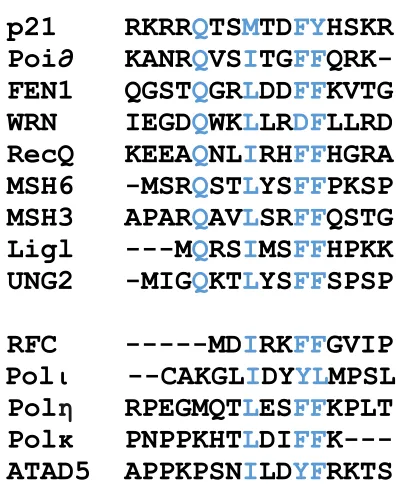
1.2.2 PCNA-interacting protein motif (PIP box)
Virtually all proteins that interact with PCNA contain a PCNA-interacting protein
motif termed the “PIP box”. The consensus binding motif of the PIP box is:
Qxx[Ψ]xx[ϑ][ϑ] (x being any amino acid, ψ being the hydrophobic residues L, M, or I and
ϑ being the aromatic residues F or Y (17). In some cases, instead of being C-terminally
flanked by xx[ϑ][ϑ], the Qxx[Ψ] motif can be flanked by KAx instead (18). A sequence
alignment of human proteins that interact with PCNA is shown in Figure 2. Gulbis et al.
(1996) first published a crystal structure that revealed how the PIP box interacts with
PCNA using a peptide derived from p21WAF1/CIP1 (p21), an inhibitor of the
cyclin-dependent kinases that control the initiation of S phase and DNA replication (19). This
peptide binds PCNA with high affinity, the interaction has a Kd in the low nanomolar range
(15, 20). The PIP box of this peptide was shown to form an alpha helix that acts as a
hydrophobic plug, docking into the hydrophobic pocket on PCNA. Furthermore, the
C-terminal of this peptide forms a β sheet structure that makes extensive contacts with the
IDCL (15). Proteins that interact with PCNA through a PIP box may also use additional
regions for interaction (21). Because many proteins that contain PIP boxes bind to PCNA
in the same region, the binding is often mutually exclusive and competitive (21).
Many of the pathways which PCNA is part of involves the transfer of DNA from
one enzyme to another. For example, during Okazaki fragment processing, FEN1 must
cleave the 5’ flap on DNA before DNA ligase can patch the nick (22). One possibility of
how this occurs is that PCNA acts as a “toolbelt”, binding all the necessary enzymes
Consensus
QxxΨxxϑϑ
p21 RKRRQTSMTDFYHSKR
Poi∂ KANRQVSITGFFQRK-
FEN1 QGSTQGRLDDFFKVTG
WRN IEGDQWKLLRDFLLRD
RecQ KEEAQNLIRHFFHGRA
MSH6 -MSRQSTLYSFFPKSP
MSH3 APARQAVLSRFFQSTG
Ligl ---MQRSIMSFFHPKK
UNG2 -MIGQKTLYSFFSPSP
RFC ---MDIRKFFGVIP
Polι --CAKGLIDYYLMPSL
Polη RPEGMQTLESFFKPLT
Polκ PNPPKHTLDIFFK---
ATAD5 APPKPSNILDYFRKTS
7
S. solfataricus where a PCNA heterotrimer simultaneously binds to DNA polymerase,
FEN1, and DNA ligase (23). However, There is currently no clear evidence that suggests
eukaryotic PCNA functions as a toolbelt, however, it is likely (22).
1.2.3 Post-translational modification of PCNA
PCNA is post-translationally modified in response to stalling of the replication fork
via sumoylation or monoubiquitination (24). As previously mentioned, prolonged stalling
of the replication fork during DNA replication is dangerous as it can collapse. This causes
double-stranded breaks and gross chromosomal rearrangements which may lead to cell
death (3). Restart of the replication fork is possible through post-translational modification
of PCNA and subsequent activation of one of two DDT pathways. These pathways include
the homologous recombination repair pathway through sumoylation of K164 or the
post-replication repair pathway via monoubiquitination of the same lysine residue (25, 26). As
seen in Figure 3A, monoubiquitination of K164 is carried out by E2 ubiquitin conjugating
enzyme Rad6 and E3 ubiquitin ligase Rad18 (Rad6/Rad18 complex) in the RAD6 pathway
(25). Rad18 recruits the ubiquitination machinery to DNA-bound PCNA through
interactions with Rad6, PCNA, and DNA (27). The structure of mono-ubiquitinated PCNA
is shown in Figure 3B.
Monoubiquitinated PCNA (ubPCNA) has been proven to promote TLS carried out
by Y-family polymerases. In addition to the PIP box motif, these polymerases also contain
ubiquitin-binding motifs (UBMs) or ubiquitin-binding zinc fingers (UBZ) which are able
to interact with the ubiquitin moiety and increase its affinity to ubPCNA (24). Tight
Figure 3. PCNA ubiquitination is regulated by the RAD6 pathway and the UAF1-USP1. A) PCNA is monoubiquitinated through the Rad6 pathway and deubiquitinated through the recruitment of the UAF1-USP1 deubiquitinase (DUB) complex. B) Structure of monoubiquitinated PCNA (PDB ID: 3TBL). Ubiquitin molecules (orange) are covalently linked to K164 of PCNA (blue) on two of three monomers.
Rad6/ Rad18
UAF1-USP1
A
B
9
polymerases may lead to increased mutagenesis. Zhuang et al. (2008) demonstrated that
the switching from the TLS polymerase η (Polη) to the normal high-fidelity Polδ is
inhibited by the ubiquitin moiety on PCNA (28). This suggests that deubiquitination is
required for the normal high-fidelity polymerase to switch back to DNA synthesis after
lesion bypass. Ubiquitin specific protease 1 (USP1) was identified in a siRNA screen as a
putative deubiquitinase (DUB) for PCNA (29).
1.3 Ubiquitin-Specific Protease 1 (USP1) and USP1 Associated Factor (UAF1)
Complex
DUB enzymes control deubiquitination of human proteins. Although 95 distinct
proteins belong to this family of proteins, many of their functions are unknown (30). DUBs
are cysteine proteases that remove ubiquitin from their substrate, either from mono- or
poly- ubiquitinated proteins or from linear ubiquitin polypeptides (29).
USP1 is a DUB that negatively regulates PCNA monoubiquitination in vivo and in
vitro (29). Upon DNA damage, transcription of USP1 is immediately turned off leading to
a rapid decay of USP1. This allows for PCNA to become monoubiquitinated, which is
essential for translesion synthesis (31). Knockdown of USP1 increases mutation frequency
during DNA replication and causes increased levels of ubPCNA (29). USP1 is activated
through interaction with USP1 associated factor 1 (UAF1). UAF1 stabilizes USP1 and is
essential for its deubiquitinating activity. UAF1 is also part of the Fanconi Anemia pathway
in charge of FANCD2 deubiquitination (30). USP1 alone has very low enzymatic activity,
however when it is part of a complex with UAF1, its activity increases 35-fold. This
proteins affinity to its substrate (31). The UAF1-USP1 DUB complex is likely recruited to
ubiquitinated PCNA through its interaction with ATAD5 (32).
UAF1 has an N-terminal WD40 domain containing eight WD40 propeller
sequences and two consecutive SUMO-like domains (SLD1 and SLD2) in its C-terminal
(Figure 4). The N-terminal binds and stimulates USP1’s ubiquitin protease activity (33).
Of the two SLDs in the C-terminal of UAF1, only the second SLD interacts with a
SUMO-interacting motif (SIM) on ATAD5.
1.4 ATPase family, AAA domain-containing protein 5 (ATAD5)
ATPase family, AAA domain-containing protein 5 (ATAD5) is the human
homolog of the yeast protein enhanced levels of genomic stability 1 (Elg1). Elg1 is divided
into three main domains: an extended N-terminal domain, a central AAA+ ATPase domain,
and a C-terminal domain (Figure 4) (34). Elg1 was originally identified in a screen for
suppressors of chromosomal instability and is a protein which participates in telomere
maintenance in yeast (35, 36). Elg1 has also been suggested to form an RFC-like complex,
which functions in stabilization of the DNA replication fork. This RFC-like complex
unloads PCNA from DNA following Okazaki fragment ligation in DNA lagging strand
synthesis (37, 38). Knockdown of ATAD5 in mammalian cells leads to spontaneous
chromosomal breaks, increased levels of recombination and aberrations in the chromosome
11
Figure 4. Schematic of ATAD5 and UAF1. A) Schematic diagram of ATAD5 with PCNA-interacting Protein (PIP) Box and sumo-PCNA-interacting protein motif (SIM) coloured green and yellow respectively. B) Schematic diagram of UAF1 with WD domains coloured orange and sumo-like domains coloured blue.
A
Knockdown of ATAD5 also causes PCNA and ubPCNA accumulate on DNA (41).
Consequently, the lifespan of replication factories (that use PCNA as a scaffold) is
extended, and remains well into G2 phase of cell division (41). The increase in PCNA
suggests that ATAD5 may interact with the core subunits of the RFC (rfc2-5) to form an
RFC-like complex, which is responsible for unloading PCNA similar to its yeast
homologue (41). Any binding of ATAD5 to PCNA has yet to be investigated, however, a
non-canonical PIP box motif has been shown to interact with PCNA in yeast (26, 34). The
accumulation of ubPCNA on DNA is due to ATAD5’s role in recruiting the UAF1-USP1
DUB complex.
ATAD5 interacts with UAF1 through a interaction similar to how the small
ubiquitin-like molecule (SUMO) interacts with a SIM: a SIM located in the N-terminal of
ATAD5 interacts with a SUMO-like domain (SLD) of UAF1 (32). The SIM consists of a
hydrophobic core, consisting of 3-4 aliphatic residues, juxtaposed to a negatively charged
cluster of acidic residues (42). The binding site of ATAD5 (KSNVVIQEEELELAVLE)
conforms to the consensus sequence of a SIM, and it is highly conserved among species
(43).
We have previously identified a putative PIP box motif in our lab in the N-terminal
of ATAD5 (Figure 2). Work done by Haley McConkey showed that a short peptide
containing this putative motif fused to GST was able to interact with PCNA in an affinity
pull-down assay. Furthermore, she showed that mutations to the two hydrophobic residues
13
1.5 Scope of thesis
Although it is proposed that ATAD5 functions to unload PCNA and recruit the
UAF1-USP1 DUB complex to ubPCNA, little is known about the interaction between
ATAD5 and PCNA. In yeast, Elg1 binds to PCNA through a non-canonical PIP box similar
to that of rfc1, a subunit of the RFC complex. PCNA has many binding partners, and
regulation of which protein binds at a given time is essential for certain cellular processes
such as the post replication repair or DDT pathways and DNA replication to occur.
Elucidation of the structural basis in which ATAD5 binds to PCNA and UAF1 can then be
applied to other proteins that bind to it.
The goal of this study is to elucidate the physical and functional relationships
between the binding of the human proteins: ATAD5, PCNA and UAF1, through structural
and biochemical methods. The ultimate objective of this study is to contribute
to our understanding of how the cell is able to recruit Y-family polymerases to stalled
replication forks arising from DNA lesions and facilitate switching from high- to low- and
back to high-fidelity polymerases.
1.6 Hypothesis
As seen in the sequence alignment of ATAD5’s PIP box with other human
PCNA-binding proteins, ATAD5 contains a putative PIP box motif close to its N-terminal that
resembles those of the Y-family polymerases and rfc1 (Figure 2). Furthermore, ATAD5
contains a SIM-like motif that interacts with a SLD on UAF1. Therefore, we propose that
ATAD5 serves as an adaptor protein (Figure 5) that is able to bind both PCNA and UAF1.
N-terminal of ATAD5 with PCNA and UAF1 through various biochemical methods.
Finally, we aim to elucidate the structural features of the binding between these proteins
15
CHAPTER 2: MATERIALS AND METHODS
2.1.1 Bacterial strains and plasmids
The plasmid expressing full-length wild-type PCNA was previously subcloned
from pAVR38, a generous gift from Dr. Roger Woodgate of the National Institutes of
Health (Maryland, USA), into pET22b. The pMCSG9, pMCSG10, pBluescriptR_UAF1
and pANT7-cGST-ATAD5 plasmids were purchased from the PlasmID Repository
(Harvard Medical School, Massachusetts, USA). Bacterial strain BL21(DE3) was
commercially purchased from Novagen. The maps of the vectors used are available in the
appendix (Figure I-II). A schematic diagram of the various domains from ATAD5 and
UAF1 used in this study are depicted in Figure 4.
2.1.2 Yeast strains and plasmids
The PJ694A yeast strain, and pASI and pACT2 vectors were generously provided
by Dr. Chris Brandl of the Schulich School of Medicine and Dentistry (Western University,
Ontario, Canada).
2.1.3 List of buffers
Buffers used in the purification of proteins and experiments have been compiled in
17
Table 1. Summary of buffers used in this study.*
Buffer Used In Components
Buffer A PCNA purification 25 mM Tris pH 7.5, 1 mM EDTA, 7 mM β-Me, 0.01 % NP-40
Buffer B PCNA purification 25 mM Tris pH 7.5, 1 mM EDTA, 7 mM β-Me, 0.01 % NP-40, 1 M NaCl Buffer C PCNA purification 50 mM NaAc pH 5.5, 10 % glycerol, 7 mM β-Me, 0.01 % NP-40
Buffer D PCNA purification 50 mM NaAc pH 5.5, 10 % glycerol, 7 mM β-Me, 0.01 % NP-40, 1 M NaCl Buffer E PCNA purification 25 mM KH2PO4/K2HPO4 pH 7.0, 10 % glycerol, 1 mM DTT
Buffer F PCNA purification 300 mM KH2PO4/K2HPO4 pH 7.0, 10 % glycerol, 1 mM DTT
Buffer G PCNA purification 25 mM Tris pH 7.5, 150 mM NaCl, 10 % Glycerol, 1 mM EDTA, 1 mM DTT ITC Buffer ITC 25 mM HEPES pH 7.5, 150 mM NaCl, 5 mM β-Me
HIS Buffer A MBP/GST-tagged Protein purification
25 mM Tris pH 7.5, 500 mM NaCl, 1 mM EDTA, 7 mM β-Me
HIS Buffer B MBP/GST-tagged protein purification
25 mM Tris pH 7.5, 500 mM NaCl, 1 mM EDTA, 7 mM β-Me, 300 mM imidazole
Q Buffer A MBP/GST-tagged Protein purification
25 mM Tris pH 7.5, 1 mM EDTA, 7 mM β-Me
Q Buffer B MBP/GST-tagged Protein purification
25 mM Tris pH 7.5, 1 M NaCl, 1 mM EDTA, 7 mM β-Me
Pull-Down Buffer Pull-down assays 25 mM HEPES pH 7.5, 150 mM NaCl, 1 mM EDTA, 5 mM β-Me
GST Elution Buffer GST Purification 25 mM Tris pH 7.5, 200 mM NaCl, 1 mM EDTA, 7 mM β-Me, 10 mM reduced glutathione
MBP Elution Buffer MBP Purification 25 mM Tris pH 7.5, 200 mM NaCl, 1 mM EDTA, 7 mM β-Me, 10 mM maltose
2.1.4 Cloning of constructs used in yeast two-hybrid assay
Plasmids pASI-PCNA, pASI-UAF1_SLD2, ATAD5_N250,
pACT2-ATAD5_N498 were made through traditional “cut and paste” cloning. Appropriate gene
segments were amplified with an addition of an appropriate restriction enzyme cut site by
PCR using the appropriate forward and reverse primers summarized in Table 2. The
cloning of these constructs followed a previously described protocol with minor
modifications (44).
In brief, the amplification was carried out with 1 ng of DNA template, 500 pM of
forward and reverse primer, 200 μM of dNTPs, and 1 U of i-MAXTM II DNA polymerase
(iNtRON) in 20 μL reactions. An initial touchdown protocol was applied to avoid
non-specific binding of the primer, followed by cycling conditions outlined by the
manufacturer. The annealing temperature for the PCR products varied depending on the
primers used (Table 2). The PCR products were then diluted and digested with BamHI and
XhoI along with the vectors (pASI and pACT2) for 2 hours at 37 oC. The digested DNA
was then purified from a 1.5 % agarose gel using the QIAquick® Gel Extraction Kit
(Qiagen Inc.). The DNA was then ligated into the appropriate vectors with T4 DNA ligase
(New England Biolabs Inc.) in a 3:1 molar ratio of insert to vector DNA. 5 μL of the
ligation reaction was transformed into competent DH5α cells and grown overnight at 37
oC on LB agar plates supplemented with 100 μg/mL of ampicillin. The plasmids were then
harvested from 3 mL cultures grown from single colonies and confirmed by DNA
1
9
Table 2. Summary of constructs used for yeast two-hybrid experiments..*
Construct Protein
(Mutation)
Amino Acid
Residues TAnnealing (
oC) Primers (Forward and Reverse)
pASI-PCNA PCNA 64 5’- taatggatccatatgttcgaggcgcgcctg -3’ 5’- gctagttattgctcagcgg -3’
pASI-UAF1_SLD2 UAF1 559-677 56 5’- ctgtacttccaatccatgcccaaattcaacaaaattcc -3’ 5’- ttatccacttccaatttacgtggacttctgacggtaatg -3’
pASI-UAF1_SLD2M UAF1 (K595E) 559-677 63 5’- ctccaagtccgagaagttatggaacatgtttatg -3’ 5’- gttccataacttctcggacttggagcatgt -3’
pACT2-ATAD5_N250 ATAD5 2-250 55 5’- taatggatccatgtgggggtcctggccatg -3’ 5’- ctgcatctcgagttaatctctagagtttgcatggc -3’
pACT2-ATAD5_N250M ATAD5 (I62A,
Y65A, and F66A) 2-250 67
5’- gcactggatgcagcaagaaagacttcacccacaaatgagaag -3’ 5’- tgctgcatccagtgcattactaggttttggtggagcaaaaacc -3’
pACT2-ATAD5_N498 ATAD5 (2-498) 2-498 55 5’- taatggatccatgtgggggtcctggccatg -3’ 5’- ctgcatctcgagttatccctctctgtttttgcc -3’
pACT2-ATAD5_SIM ATAD5 340-406 56 5’- taatggatcctaccccgaattttcttgaaacaaaagc -3’ 5’- ctgcatctcgagttatgctttcataaattgctgtctttc -3’
After confirming that the products are correct, mutations to pASI-UAF1_SLD2 and
ATAD5_N250 were introduced to make pASI-UAF1_SLD2M and
pACT2-ATAD5_N250M. The mutagenesis of these two primers was completed using overlap
extension PCR as previously described with minor modifications (45). The primers were
designed according to the mutagenic primer-incomplete polymerase extension (M-PIPE)
protocol (shown in Table 2 with the mutations underlined) (46).
Briefly, two reactions were set up for each mutation as previously described,
however one reaction contained the forward primer for the insert and the reverse primer
for mutagenesis, and the other reaction containing the opposite primer pairs. The two
reactions were then digested with DpnI for 2 hours at 37 oC to break down the methylated
template DNA. The two reactions were then purified from a 1.5 % agarose gel using the
QIAquick® Gel Extraction Kit (Qiagen Inc.) and combined to be used as the template for
the second PCR step. The same forward and reverse primers for insert amplification were
used in the second PCR. The products were then digested with BamHI and XhoI and ligated
into the appropriate vectors as previously described. Following transformation of the
plasmids into competent DH5α cells, plasmids were extracted from 3 mL overnight
cultures grown from single colonies. The plasmids were verified using DNA sequencing.
2.1.5 Cloning of constructs used for protein expression
The GST-fusion constructs pMCSG10-ATAD5_PIP, pMCSG10-ATAD5_PIPM
and pMCSG10-p21 were previously prepared by Guangxing Xing. The GST-fusion
construct pMCSG10-ATAD5_SIM and the MBP-fusion pMCSG9-UAF1_SLD2 were
21
described with modifications (46). All constructs used for protein expression are listed in
Table 3 along with the appropriate primers used (where available).
In brief, the insert for each construct was amplified using PCR with primers adding
sequences at the 5’ and 3’ ends complementary to the sequences added to the vectors. The
vector was also amplified as well. The amplifications were carried out with 1ng of template
DNA, 500 pM of forward and reverse primers, 200 μM of dNTPs, and 0.5 U of Q5
High-Fidelity DNA Polymerase (New England Biolabs Inc.). The Q5 High GC Enhancer (New
England Biolabs Inc.) was added to the reactions to amplify the insert from
pANT7-cGST-ATAD5. Touchdown PCR was used to amplify the products with an annealing temperature
range from 65 oC to 55 oC. The products were then digested with DpnI for 2 hours at 37 oC
and only the vector was purified from a 0.7 % agarose gel using the QIAquick® Gel
Extraction Kit (Qiagen Inc.). The inserts were then mixed with the vectors and incubated
at room temperature for 5 minutes and on ice for 5 minutes. 5 uL of this mixture was then
transformed into competent DH5α cells and grown overnight at 37 oC. 3 mL cultures were
grown from single colonies and the plasmids were harvested and verified by DNA
sequencing.
The same mutation was made to UAF1’s SLD2 domain in pMCSG9-UAF1_SLD2
as in pASI-UAF1_SLD2 using overlap extension PCR as previously described to create
22
Table 3. Summary of constructs used for protein expression and purification.
*Original protein as well as length of expressed protein are described. Annealing temperatures and primers used are listed where available. Mutations are described in brackets and primers used for mutagenesis.
Construct Protein
(Mutation)
Amino Acid
Residues TAnnealing (
oC) Primers (Forward and Reverse)
pMCSG10-p21 p21 - pMCSG10-ATAD5_PIP ATAD5 51-75
pMCSG10-ATAD5_PIPM ATAD5 (Y65A and
F66A) 51-75
pMCSG10-ATAD5_SIM ATAD5 340-406 55 5’- ctgtacttccaatcctctgatcctgagaatgaacag -3’ 5’- ttatccacttccaatttaccaggtttcacagcttcag -3’
pMCSG9-UAF1_SLD2 UAF1 559-677 56 5’- ctgtacttccaatccatgcccaaattcaacaaaattcc -3’ 5’- ttatccacttccaatttacgtggacttctgacggtaatg -3’
23
2.2 Expression and purification
2.2.1 Expression and purification of PCNA
BL21(DE3) E. coli cells containing the pRARE plasmid were transformed with
pET22b-hPCNA and incubated at 37 oC overnight. Colonies were picked and grown in a
50 mL starter culture of 2xYT media containing 100 μg/mL ampicillin and 25 μg/mL
chloramphenicol. The starter culture was used to inoculate 1 L of 2xYT media with the
same concentration of antibiotics, supplemented with 6 mM MgCl2. The cultures were
grown to an OD600 of 1.2 at 37 oC before induction with 0.5 mM
isopropyl-1-thio-β-D-galactopyranoside (IPTG) for 16 h at 30 oC. Cells were harvested by centrifugation at 4,500
g and the resulting cell paste was stored at -20 oC until further use.
Frozen cells were thawed on ice and resuspended in Buffer A (25 mM Tris pH 7.5,
1 mM EDTA, 7 mM β-Me, 0.01 % NP-40) containing 100 mM NaCl and 1 mM of the
protease inhibitor, phenylmethylsulfonyl fluoride (PMSF). The cell suspension was lysed
by three passages through a French press at 15,000 psi. The lysate was then clarified by
centrifugation at 20,000 rpm at 4 oC for 30 min. PCNA was then purified to near
homogeneity as previously described, with modifications (47).
In brief, the supernatant was loaded onto two 5 mL HiTrap Q HP columns (GE
Healthcare Life Sciences) pre-equilibrated with Buffer A containing 10 % Buffer B (25
mM Tris pH 7.5, 1 mM EDTA, 7 mM β-Me, 0.01 % NP-40, 1 M NaCl) for anion exchange
chromatography. The protein was eluted off of the columns using a gradient of Buffer B.
Fractions containing PCNA were pooled and diluted with Buffer C (50 mM NaAc pH 5.5,
10 % glycerol, 7 mM β-Me, 0.01 % NP-40) until the approximate NaCl concentration was
a gradient of Buffer D (50 mM NaAc pH 5.5, 10 % glycerol, 7 mM β-Me, 0.01 % NP-40,
1 M NaCl). The fractions containing the protein was pooled and dialyzed against Buffer E
(25 mM KH2PO4/K2HPO4 pH 7.0, 10 % glycerol, 1 mM DTT) overnight. The sample was
then loaded onto a packed 12 mL CHT ceramic hydroxyapatite Type I column. The protein
was eluted off of the column using a gradient of Buffer F (300 mM KH2PO4/K2HPO4 pH
7.0, 10 % glycerol, 1 mM DTT). Fractions containing the protein of interest were pooled
and concentrated and subjected to size exclusion chromatography on a SuperdexS200
10/300 GL equilibrated with Buffer G (25 mM Tris pH 7.5, 150 mM NaCl, 10 % Glycerol,
1 mM EDTA, 1 mM DTT).
2.2.2 Expression and purification of recombinant proteins
All plasmids constructed with the pMCSG9 and pMCSG10 vectors (Table 3) were
transformed into competent E. coli BL21(DE3) cells following standard protocols (48).
The same procedure was applied to all of the plasmids.
Single colonies were picked following transformation and grown as 5 mL cultures
overnight in LB media overnight supplemented with 100 μg/mL of ampicillin. These
cultures were then added to 1 L of the same media and grown to an OD600 of 0.8 at 37 oC
before being induced with 0.5 mM of IPTG for 16 hours at 16 oC. Cells were harvested by
centrifugation at 4,500 g and the resulting cell paste was stored at -20 oC until required for
further use.
The frozen cell paste was thawed on ice and the cells were resuspended in HIS
Buffer A (25 mM Tris pH 7.5, 500 mM NaCl, 1 mM EDTA, 7 mM β-Me) spiked with
25
through an Emulsiflex cell homogenizer (Avestin) at 15,000 psi. The lysate was then
clarified by centrifugation at 20,000 rpm at 4 oC for 30 min.
2.2.3 Purification of p21, ATAD5_PIP, ATAD5_PIPM, and
GST-ATAD_SIM
Recombinant proteins were purified by immobilized metal affinity chromatography
(IMAC) followed by anion exchange chromatography following the manufacturer’s
suggested protocols. Briefly, clarified lysate was loaded onto two 5 mL prepacked
cOmplete His-tag purification columns (Roche) pre-equilibrated with HIS Buffer A and
washed with HIS Buffer A + 10 mM imidazole. Proteins were then eluted using HIS Buffer
B (25 mM Tris pH 7.5, 500 mM NaCl, 300 mM imidazole, 1 mM EDTA, 7 mM β-Me).
The eluents were diluted with Q Buffer A (25 mM Tris pH 7.5, 1 mM EDTA, 7 mM β-Me)
until the approximate NaCl concentration was 50mM and applied to two 5mL HiTrapQ
HP columns equilibrated with 5% Q Buffer B (25 mM Tris pH 7.5, 1 mM EDTA, 7 mM
β-Me, 1 M NaCl) and eluted with a gradient of Q Buffer B. The proteins were then
concentrated using centrifugation using Vivaspin 6 ultrafiltration devices with appropriate
molecular weight cut offs. The concentrated proteins were stored at -20 oC after spiking
the samples with glycerol to 50 %.
2.2.4 Purification of GST, UAF1_SLD2, UAF1_SLD2M, and ATAD5_SIM
The purification of GST, UAF1_SLD2, UAF1_SLD2M, and ATAD5_SIM
proteins followed the above protocol with slight modifications. After IMAC, His-tagged
MBP-UAF1_SLD2M at a 1:20 ratio and dialyzed against 5% Q Buffer B overnight to cleave the
GST and MBP tags. Each of the resulting samples were passed through two 5 mL
prepacked cOmplete His-tag purification columns (Roche) pre-equilibrated with HIS
Buffer A, removing the TEV protease and the His-tagged tags from the flow through. The
flow through fraction containing the cleaved protein was then diluted with Q Buffer A to a
NaCl concentration of approximately 50 mM and anion exchange chromatography was
performed as described above.
The remaining protein on the IMAC column was eluted with HIS Buffer B and
these fractions were applied to the appropriate prepacked columns (either MBPTrap HP
columns or GSTrap HP columns (GE Healthcare)) equilibrated with HIS Buffer A to
remove TEV Protease. Proteins were eluted with the appropriate buffers (GST Elution
Buffer (25 mM Tris pH 7.5, 200 mM NaCl, 1 mM EDTA, 7 mM β-Me, 10 mM reduced
glutathione) or MBP Elution Buffer (25 mM Tris pH 7.5, 200 mM NaCl, 1 mM EDTA, 7
mM β-Me, 10 mM maltose)). The buffer was exchanged and the proteins were concentrated
using centrifugation using Vivaspin 6 ultrafiltration devices with appropriate molecular
weight cut offs. The concentrated proteins were stored at -20 oC after glycerol was added
to a final concentration of 50 %.
2.3 Affinity pull-down
GST pull-down and His-affinity pull-downs were carried out using similar
protocols. 50 μg of GST-tagged protein (bait) was immobilized onto glutathione sepharose
resin equilibrated in Pull-Down Buffer (25 mM HEPES pH 7.5, 150 mM NaCl, 1 mM
27
by centrifugation (1 minute at 600 g) and the supernatant was removed. The beads were
then washed with Pull-Down Buffer to remove excess protein. Prey proteins were then
added in excess and allowed to incubate with the immobilized bait for 1 hour on ice with
occasional agitation. Unbound protein was then removed through three washes. Each wash
involved adding 200 μL of Pull-down buffer, agitating the mixture, spinning down the
resin, and removing the supernatant. Finally, bound proteins were stripped from the resin
by adding 50 μL of 1x SDS-PAGE loading buffer and boiled for 5 minutes. The samples
were then subjected to analysis by SDS-PAGE. The proteins were visualized by Coomassie
Brilliant Blue staining.
2.4 Isothermal titration calorimetry
A synthetic 18 amino acid peptide containing ATAD5’s PIP box was purchased
from and purified by the Tufts University Protein Core Faculty (Boston, USA). The
sequence of the peptide was: APPLPSNILDYFRKTSPT. The peptide was then dissolved
in ITC buffer (25 mM HEPES pH 7.5, 150 mM NaCl, 5 mM β-Me). The proteins were
thoroughly dialyzed and degassed under vacuum before the experiments were conducted.
Isothermal titration calorimetry (ITC) measurements were either performed on the VP-ITC
calorimeter (GE Healthcare) or the Nano ITC calorimeter (TA Instruments) at 25 oC. The
titration of the peptide into PCNA was performed using the VP-ITC calorimeter. The
peptide (750 μM) was loaded into the syringe and injected into the cell containing 50 μM
PCNA in 60 injections of 5μL. A control experiment was performed by injecting the
corrected by subtracting the control measurements, and analyzed using Origin 7
(MicroCal). Binding constants, ΔG, and ΔH were calculated
The titrations of ATAD5_SIM into UAF1_SLD2 and UAF1_SLD2M were
performed using the Nano ITC calorimeter. ATAD5_SIM (1 mM) was titrated into a cell
containing 100 μM of UAF1_SLD2 in 25 2 μL injections following an initial 0.5 μL
injection. Control experiments were performed by injecting the peptide into buffer under
the same experimental conditions. Experimental traces were corrected by subtracting the
control measurements, and analyzed using NanoAnalyze (TA Instruments). Binding
constants, ΔG, and ΔH were calculated by fitting the integrated titration data to an
independent binding model.
2.4 Yeast two-hybrid assays
The yeast strain used was PJ69-4A and has the genotypes as described (49). Human
PCNA was recombinantly expressed with the binding domain of the GAL4 by the
integrative vector pASI. Native and mutant N-terminal ATAD5 containing the PIP-box
was recombinantly expressed with the activating domain of GAL4 using the pACT2 vector.
Yeast cells were sequentially transformed using the LiAc method (50) with the bait plasmid
and then the prey plasmid. Selection of the cells containing both the bait and prey plasmids
was carried out on media lacking tryptophan and leucine. Positive interactions were scored
29
2.5 Crystallization
2.5.1 Crystal screening and optimization
Proteins used for crystallization condition screening were either freshly purified or
subjected to size exclusion chromatography. Flash-frozen protein stored at -80 oC was
thawed and loaded onto a SuperdexS200 10/300 GL equilibrated with ITC Buffer. Eluted
protein was then diluted or concentrated for use in crystallization experiments.
Protein complexes were made prior to setting up crystallization trials. PCNA at a
concentration of 1 mg/ml was mixed with 2 molar excess of the synthetic peptide derived
from ATAD5 used in ITC experiments. The protein solution was then left to incubate for
1 hour at room temperature before being concentrated to 6 mg/mL. The
ATAD_SIM-UAF1_SLD2 complex was collected after ITC experiments were performed and was
concentrated to a concentration of 9 mg/mL. The approximate ratio of ATAD5_SIM to
UAF1_SLD2 is 3:1.
Crystal screening was carried out using both the hanging drop and sitting drop
vapour diffusion method with various commercially available and in-house crystallization
kits. The kits used to screen both complexes included: Hampton Research: Crystal Screen
kit 1, Crystal Screen kit 2, Natrix, and PEG/Ion Kit and Wizard Classic Crystallization
Screen Kit 1, 2, 3, and 4. In addition to the kits mentioned, the peptide-PCNA complex
was also screened with: JSGC+ Crystal Screen, Molecular Dimensions: Midas Crystal
Screen Kit and in-house PEG/Ion screens with varying PEG molecular weights. 1 μL of
protein complex was mixed with 1 μL of the well solution and incubated over a reservoir
After hits were observed, optimization of crystallization conditions was carried out.
Crystals were grown at varying precipitant concentrations and microseeding was employed
in order to improve the shape of crystals. A seed solution was prepared by bead-beating
harvested crystals suspended in mother liquor and added to fresh drops. Additives were
also screened using Hampton Research: Optimization Screen Kit, following the
manufacturer’s suggested protocol.
2.5.2 Data collection and refinement
Crystals were initially screened at the home-source to identify highly diffracting
crystals for data collection. Several crystals were frozen in liquid nitrogen during the
screening process. Highly diffracting crystals were sent to the Canadian Light Source
(Saskatchewan, Canada) for data collection at the CMCF-081D beam line.
The crystal structure was solved using iMOSFLM (51) and the CCP4 program suite
(52). The reflection images were integrated using iMOSFLM and scaled using SCALA and
CTRUNCATE (53, 54). Initial phases were obtained by molecular replacement using
3VKK as a search model in PHASER (55). An initial rigid body refinement was carried
out by RefMac (56) followed by successive rounds of structure building with COOT (57)
and restrained refinements using RefMac. Non-crystallographic symmetry was applied
31
CHAPTER 3: RESULTS I INTERACTION BETWEEN ATAD5 AND PCNA
3.1 Cloning of plasmids
A variety of plasmids were generated in this study to assess the interactions between
ATAD5, PCNA, and UAF1. A summary of the plasmids used and generated is presented
in Tables 2 and 3. All of the plasmids used were confirmed to be correct using DNA
sequencing.
Subcloning was used to generate the plasmids for the yeast two-hybrid studies.
Insert DNA was amplified by PCR and digested with the appropriate restriction
endonucleases and ligated into either pASI or pACT2. The pASI and pACT2 vectors
encode for fusion proteins between the protein of interest and the binding domain and
activating domain of Gal4 respectively.
PIPE cloning was used to generate the plasmids for expression and purification of
ATAD5’s SIM and UAF1’s second SLD (SLD2). The primers used to amplify the insert
(by PCR) added a sequence of nucleotides on each end, complementary to the ends on the
vector. This technique uses the cell’s DNA repair machinery to ligate the plasmid. The
plasmids pMCSG9 and pMCSG10 encode the protein of interest fused to the C-terminal
of MBP and GST respectfully. MBP and GST are both tagged with an N-terminal
poly-His tag and contain a TEV cut site at the C-terminal.
The mutation made to the plasmids containing ATAD’s PIP box motif and UAF1’s
SLD2 was achieved by overlap extension PCR. The key PIP box motifs of ATAD5 (I62,
Y65 and F66) were all mutated to Ala using the primers indicated in Table 2. Yang et al.
UAF1 to bind to ATAD5, so this mutation was also used in this study using the primers
indicated in Table 3 (43).
3.2 Purification of proteins
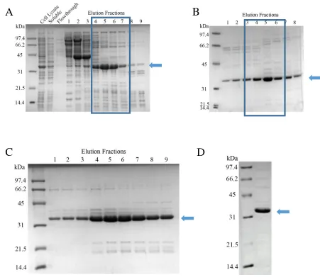
3.2.1 Purification of PCNA
Human PCNA was expressed in E. coli and purified to near homogeneity (Figure
6). Following cell lysis, the sample was clarified using high-speed centrifugation for 15
min at 7000 g. The supernatant was then applied onto prepacked HiTrap Q columns for
anion exchange chromatography (Figure 6A). The protein was eluted from the column
using an increasing gradient of ionic strength. The protein was further purified by heparin
affinity chromatography (Figure 6B). The fractions containing PCNA from anion exchange
chromatography was diluted to reduce the concentration of NaCl before being loaded onto
HiTrap Heparin columns. The protein was again eluted from the column using a gradient
of increasing ionic strength. PCNA was still not sufficiently pure, so the sample was
subjected to mix mode chromatography via ceramic hydroxyapatite (Figure 6C).
Hydroxyapatite chromatography has unique separation properties and can sometimes
separate proteins shown as homogeneous by other chromatographic or electrophoretic
techniques (58). The sample was applied to a 12 mL column and eluted using an increasing
gradient of potassium phosphate. The final low molecular weight contaminants were
removed by size exclusion chromatography (Figure 6D), as PCNA is significantly larger
in solution (as a trimer). Typically, 5 grams of cell paste yielded 25mg of protein.
33
Figure 6. Purification of human proliferating cell nuclear antigen (PCNA) (28.7 kDa). SDS-PAGE analysis after each stage of chromatography. Blue arrows indicate the band of interest. A) Anion exchange chromatography using HiTrap Q columns. Cell lysate, soluble protein, flow-through, and eluted fractions are indicated. Fractions in blue box were pooled. B) Eluted fractions from Heparin affinity chromatography. Fractions 4-6 were pooled. C) Eluted fractions from hydroxyapatite chromatography. D) Peak fraction of PCNA after size exclusion chromatography. PCNA is near homogeneity.
A
B
Despite having a predicted molecular weight of 28.7 kDa, individual PCNA
monomers runs at about 34-35 kDa on a 15% SDS-PAGE gel. Gulbis et al. 1996 had
previously reported that human PCNA runs at a higher apparent molecular weight (47).
PCNA from other organisms such as Schizoscaccharomyces pombe and tomatoes have also
been reported to run higher than their expected molecular weight (59, 60).
3.2.2 Purification of GST-fusion proteins
The GST-fusion proteins (GST-p21, GST-ATAD5_PIP and GST-ATAD5_PIPM)
were expressed in E. coli following the same growth and induction parameters. The vector
(pMCSG10) that encodes the recombinant proteins includes a poly-his tag in the N terminal
of GST, allowing IMAC to be utilized as the first purification step. Figure 7A shows a
representative purification of GST-ATAD5_PIPM.
The proteins were purified to near homogeneity after IMAC and anion exchange
chromatography. All of the proteins were soluble following cell lysis. Clarified lysate was
loaded onto 5 mL prepacked cOmplete His-tag purification columns (Roche). After a wash
with a low concentration of imidazole, the remaining proteins were eluted using a high
concentration of imidazole. The eluted sample was then diluted to reduce the NaCl
concentration and loaded onto HiTrap Q HP columns (GE Healthcare) for anion exchange
chromatography. The protein was then eluted using an increasing gradient of ionic strength.
35
A
37
The GST protein to be used as negative controls for the affinity pull-downs were
purified from the cleavage products of tagged protein with TEV protease. Both the GST
and TEV protease contained the poly-His tag, so GST affinity chromatography was used
to separate them. GST and TEV protease was eluted from the IMAC columns after the
cleaved protein of interest was removed. The sample was directly loaded onto a GSTrap
HP (GE Healthcare) and eluted with a buffer containing 10 mM of reduced glutathione.
Typically, 5 mg of GST was obtained from the digestion of 7 mg of GST-p21.
These proteins were resolved close to their predicted molecular weights during
SDS-PAGE analysis and are of sufficient quality to be used in biochemical assays (Figure
7B). GST-p21 (30.4 kDa), GST-ATAD5_PIP (31.2 kDa), and GST-ATAD5_PIPM (31.3
kDa) ran close to their expected molecular weights. The apparent molecular weight of GST
(28.3 kDa) was slightly higher than expected.
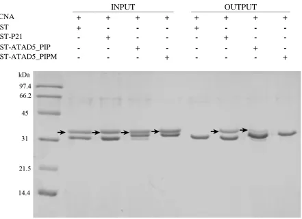
3.2 GST Pull-down assays
Work by a previous student showed that the identified putative PIP box in
ATAD5’s N-terminal was able to bind to PCNA, and that mutation of these residues
abolished the binding. The GST pull-down assay was replicated to confirm the findings. In
brief, GST-bound proteins (bait) were incubated with glutathione agarose resin and
allowed to bind. Unbound protein was then washed off and PCNA (prey) was added to the
resin in excess. After several rounds of washing, only the protein that interacts with the
bait remain. These proteins were eluted from the resin and analyzed using SDS-PAGE on
Figure 8. Pull-down assay showing the interaction between ATAD’s PIP box and PCNA. Excess GST or GST-fused protein was immobilized onto glutathione resin. Unbound protein was then washed off and PCNA (prey) was added to the resin in 2 molar excess. After several rounds of washing, bound protein were eluted from the resin and analyzed by SDS-PAGE on a 15% polyacrylamide gel. Proteins used in each reaction is shown above by a “+”, PCNA is indicated by arrows.
INPUT OUTPUT
PCNA + + + + + + + +
GST + - - - + - - -
GST-P21 - + - - - + - -
GST-ATAD5_PIP - - + - - - + -
39
PCNA was observed to co-elute with the native ATAD5 PIP box along with the
positive control using the PIP box from p21. The intensity of the PCNA band was
significantly lower in the lane containing ATAD5 compared to the lane containing p21.
This suggests that the interaction between the PIP box of ATAD5 is weaker than that of
P21 with PCNA. Mutation of the two aromatic residues (Tyr65 and Phe66) to Ala abolishes
the interaction, as PCNA not present in the eluted proteins (Figure 7).
From the Pull-down experiment, we can conclude that the putative PIP box from
ATAD5 interacts with PCNA. Also, this experiment demonstrates that the two aromatic
residues of this PIP box is important for binding. Therefore, we can infer that the binding
mode of this PIP box to PCNA is similar to that of canonical PIP boxes.
3.3 Yeast two-hybrid assays
To confirm the results of the GST pull-down assays, the interaction between the
N-terminal of ATAD5 containing the PIP box with PCNA was analyzed in a yeast two-hybrid
system. The two-hybrid system takes advantage of a transcriptional activator that has two
separable functions: one to bind to a specific region of DNA, and the second, to increase
the frequency with which transcription is initiated on an adjacent gene (Figure 9A) (61).
The binding domain (BD) and activating domain (AD) of the transcriptional activator Gal4
is encoded separately by the pASI and pACT2 vectors fused to proteins of interest (62).
The ability to synthesize Leu and Trp is conferred by pASI and pACT2 respectlvely. The
yeast stain that is used in this study (PJ69-4A), will express β-galactosidase and be able to
PJ69-4A yeast cells were sequentially transformed with plasmids made from the
pASI vector and then the pACT2 vector. PJ69-4A cells were initially transformed using
the LiAc/single-stranded carrier DNA/PEG method with plasmids made with the pASI
vector and plated on selective media lacking Trp. Colonies were picked for each plasmid
and then subsequently transformed with plasmids made with the pACT2 vector.
Co-transformed cells were selected by their ability to grow on selective media lacking both
Trp and Leu.
Binding of the N-terminal 250 amino acid residues of ATAD5 and PCNA was
assessed by spot plating co-transformed cells on increasingly selective media.
Transcription of the reporter genes responsible for His and Ade synthesis will allow the
cells to survive on media without these amino acid residues. Cells transformed with both
pASI-PCNA and pACT2-A250 were able to grow on plates lacking Trp, Leu, and His
and/or Ade (Figure 9B). Cells transformed with both pASI-PCNA and
pACT2-ATAD_N250M (containing mutations to key PIP box residues), were not able to grow on
the plates lacking His and/or Ade (Figure 9B). From these results, we have confirmed that
the putative PIP box is able to interact with PCNA and mutations of the conserved residues
in the PIP box abolishes binding.
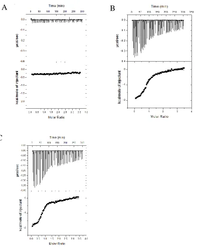
3.4 Isothermal titration calorimetry
With the interaction between ATAD5’s PIP box and PCNA confirmed by both GST
pull-down assays and the yeast two-hybrid system, we wanted to characterize the
interaction. The thermodynamic parameters of the interaction were characterized by ITC
41
Figure 9. Interaction of ATAD5’s N-terminal 250 amino acids with native PCNA in a yeast two-hybrid system. A) Binding of the two proteins fused to Gal4’s binding domain and activating domain will start transcription of t. B) PJ694A cells were successively transformed with the plasmid encoding native human PCNA and then with either the pACT2 vector, encoding only the AD of Gal4, and ATAD5_N250 or pACT2-ATAD5_N250M which encodes the N terminal 250 amino acids of ATAD5 or the mutant. These cells were then spot plated with an initial dilution of 1000x and then serial diluted 4 times on increasing selective media (blue triangles represent dilution).
A
between two components is measured and the thermodynamic terms that define the binding
affinity, enthalpy (∆H) and entropy (∆S), can be determined (63).
The binding affinity of ATAD5’s PIP box with PCNA was determined using a
synthetic peptide. The full-length ATAD5 protein is relatively large, comprising of 1844
amino acid residues, with the first ~1000 residues predicted to be disordered. For this
reason, an 18 amino acid residue peptide was synthesized that contained the PIP box of
ATAD5. The flanking sequence of amino acids on both sides of the PIP box were
conserved. The synthetic peptide containing the PIP box of ATAD5 was diluted in ITC
buffer and thoroughly dialyzed. The pH of the peptide sample after dialysis overnight did
not match that of the buffer, so the time of dialysis was extended to 36 hours total. The
peptide was loaded into the syringe and titrated stepwise into the cell containing PCNA. A
control experiment was conducted in which the peptide was titrated into a cell containing
ITC Buffer only (Figure 10A). The control experiment was subtracted from the
experimental traces and fitted to a one-site binding model (Figure 10B-C). The Kd of the
interaction was determined to be 6.17 0.78 M with an n of 0.93 ± 0.01. The
stoichiometry of the reaction is close to 1, meaning that each peptide binds to one
monomer. The H, S and G of the reaction is -2.18 ± 0.04 kcal, 16.5 cal mol-1 K-1 and
-7.01 kcal/mol respectively.
From the ITC data, we determined that the interaction is in the low micromolar
range, each monomer of PCNA is able to bind to one peptide, and that the interaction occurs
43
Figure 10. Determination of the thermodynamic parameters of the binding between ATAD5’s PIP box and PCNA. Thoroughly dialyzed peptide (800μM) was titrated stepwise into the cell in 2.5μL injections at 25oC. Binding thermograms from titration of a synthetic peptide containing ATAD5’s PIP box into the cell. A) A control experiment was conducted in which the peptide was titrated into a cell containing ITC Buffer only. B) The peptide was titrated into a cell containing 40μM of PCNA. C) Replicate of titration described in (B)