Dynamical patterning modules: a "pattern language"
for development and evolution of multicellular form
STUART A. NEWMAN* and RAMRAY BHAT
Department of Cell Biology and Anatomy, Basic Science Building, New York Medical College, New York, USA
ABSTRACT This article considers the role played by a core set of "dynamical patterning modules" (DPMs) in the origination, development and evolution of complex organisms. These consist of the products of a subset of the genes of what has come to be known as the "developmental-genetic toolkit" in association with physical processes they mobilize. The physical processes are those characteristic of chemically and mechanically excitable mesoscopic systems like cell aggregates: cohesion, viscoelasticity, diffusion, spatiotemporal heterogeneity based on activator-inhibitor interaction, and multistable and oscillatory dynamics. We focus on the emergence of the Metazoa, and show how toolkit gene products and pathways that pre-existed the metazoans acquired novel morphogenetic functions simply by virtue of the change in scale and context inherent to multicellularity. We propose that DPMs, acting singly and in combination with each other, constitute a "pattern language" capable of generating all metazoan body plans and organ forms. This concept implies that the multicellular organisms of the late Precambrian-early Cambrian were phenotypically plastic, fluently exploring morphospace in a fashion decoupled from both function-based selection and genotypic change. The relatively stable developmental trajectories and morphological phenotypes of modern organisms, then, are considered to be products of stabilizing selection. This perspective solves the apparent "molecular homology-analogy para-dox," whereby widely divergent modern animal types utilize the same molecular toolkit during development, but it does so by inverting the neo-Darwinian principle that phenotypic disparity was generated over long periods of time in concert with, and in proportion to genotypic change.
KEY WORDS:
body plan, Cambrian explosion, developmental-genetic toolkit, epithelial-mesenchymal
transformation, lateral inhibition, macroevolution, morphogenesis, morphospace
Introduction
Animal body plans of virtually all the modern Metazoan types emerged relatively suddenly (“compressed in time”; Rokas et al., 2005) during the “Cambrian explosion” about 550-530 million years ago (Ma) (Conway Morris, 2006). The metazoans, in turn, may have had roots (Erwin, 2008) in the Ediacaran period, 575-542 Ma (Shen et al., 2008), derived, in part, from simpler sheet-like, or budded, segmented, tube-like multicellular biota found in deposits of the earlier period (Narbonne, 2004; Droser and Gehling, 2008). The early metazoans, variously, had body cavi-ties, multiple tissue layers and appendages, and were elongated and segmented. Significantly, not only was this emergence of disparate forms rapid, it was all but exhaustive. With the possible exception of the bryozoa, which are not seen until the early
BIOLOGY
www.intjdevbiol.com*Address correspondence to: Dr. Stuart A. Newman. Department of Cell Biology and Anatomy, Basic Science Building,New York Medical College, Valhalla, New York 10595, USA. e-mail: [email protected]
Published online: 25 March 2009.
ISSN: Online 1696-3547, Print 0214-6282 © 2009 UBC Press
Printed in Spain
Ordovician some 40 million years later (though see Passamaneck and Halanych, 2004), exemplars of all the major metazoan bauplans appeared no later than the early Cambrian.
Rapid morphological evolution is compatible with classic neo-Darwinian evolutionary mechanisms of random genetic change followed by natural selection, whereby existing structures are modified in their size or shape (Weiner, 1994; Losos et al., 2006). Nonetheless, the discovery of the phylum-level diversification
(with no evident intermediates) that occurred in the early Cam-brian is not anticipated by this theory (Müller and Newman, 2005). Even more surprising from the viewpoint of the standard evolu-tionary model has been the realization that the Metazoa have used a highly conserved set of gene products, the so-called developmental-genetic toolkit, to generate diverse body and organ forms since their inception more than half a billion years ago (Carroll et al., 2005). If morphological change is supposed to be driven by, and to track, genetic change, why were there not dramatic changes in gene content corresponding to innovation of new organismal architectures (e.g., the dorsal location of nerve cord and heart in chordates vs. the ventral location of these structure in annelids and arthropods; Gerhart, 2000), or of new developmental mechanisms for generating morphologically equivalent structures (e.g., segmentation in beetles and fruit flies; Salazar-Ciudad, 2001b)?
The unexpected gives way to the paradoxical in the recognition that disparate organisms use homologous genes when making structurally dissimilar but functionally similar structures tradition-ally categorized as analogous (e.g., eyes in insects, mollusks and vertebrates; fly and bird wings) (Carroll et al., 2001; Wilkins, 2002). Given that there is nothing eye-like about the twin of eyeless/Pax6 transcription factor (Gehring, 2002), or heart-like about tinman/Nkx-2.5 (Schwartz and Olson, 1999), and consider-ing the immense evolutionary and anatomical divergence be-tween Drosophila and the mouse, why are the same gene regu-latory proteins used, correspondingly, to initiate the developmen-tal pathways that produce structures that have the same function, but which are evolutionarily unrelated (or have been thought so in the past)? The conflict between the expectations of the neo-Darwinian model and these recent findings from the fields of paleontology, comparative anatomy and genomics, and develop-mental biology has been termed the “molecular homology-anal-ogy” paradox (Newman, 2006).
In this paper we suggest a way out of these apparent inconsis-tencies by arguing that pre-metazoans were highly plastic entities and that early morphological diversification was an expression of the primitively loose relationship between phenotype and geno-type (Newman, 2005). Our hypothesis entails organizing the toolkit genes of the presumed pre-metazoan genome into two broad categories. The first of these, which we term the “develop-mental transcription factors” (DTFs), are those gene regulatory molecules which mediate cell type- and region-specific functions: eyeless/Pax6, tinman/Nkx-2.5, Hes, Tbox, Hox, Dlx, Pbx and several others (reviewed in Davidson, 2006 and Newman et al., in press). Our second category of toolkit genes specify products such as cadherins, Notch, Wnt and their ligands, TGF-β/BMP, FGF, Hedgehog, and their receptors, which by mobilizing certain physical processes and effects generic to “soft matter” (de Gennes, 1992) and “excitable media” (Mikhailov, 1990) constitute what we refer to as “dynamical patterning modules” (DPMs) for animal development. The relevant physical processes include adhesion and differential adhesion, induced lateral inhibition of an activated state, dynamical oscillation, cell surface and shape polarization, supracellular gradient formation (reviewed in Forgacs and Newman, 2005; Newman and Bhat, 2008). These physical pro-cesses impart a discernable and at least semi-autonomous role to the functions of the gene products; hence the use of the term "module" (see, for example, von Dassow et al., 2000; Wimsatt and
Schank, 2004).
Our rationale for this binary categorization is the following: the several billion years of unicellular evolution that preceded the origin of multicellularity saw the emergence of numerous signal-ing pathways and transcription factors (Ksignal-ing et al., 2003). With great certainty, cells had acquired the capability to modulate and switch between states in various ways, including by means of multistable transcriptional networks. None of these features, however, are distinguishing for complex multicellular forms like the metazoa. The novelty introduced by multicellularity was the potential for more than one cell type and more than one tissue layer or module to co-exist in the same organism, and for the patterning processes of particular arrangements of such cell types and tissues to be propagated from one generation to the next. Transcription factors, though they are important in determin-ing the states that cells assume, do not, in general, act across cell boundaries (though there may be exceptions; Joliot and Prochiantz, 2004).
When the effects of cell state dynamics, determined transcrip-tionally or otherwise, are communicated between cells and mani-fested in changes of shape or pattern in the multicellular aggre-gate, processes and forces characteristic of condensed, chemi-cally and mechanichemi-cally excitable materials are invariably involved (Newman and Comper, 1990; Forgacs and Newman, 2005). We therefore place toolkit gene products that (along with their down-stream signaling effectors) mediate cell interaction and thereby mobilize these generic physical processes (see below) as DPMs, in a separate category from the DTFs, which mediate the tran-scriptional and gene regulatory aspects of the multicellular pro-cesses.
The DPMs involve members of the adhesion and receptor-mediated signaling classes of toolkit genes asserted by Nichols et al. (2006) to be key elements in the evolution of the metazoans. However, we do not consider signaling and response to the external environment per se, which had many roles in pre-metazoan ancestors, to be decisive in the pre-metazoan-specific burst of morphological diversity. As mentioned above, integral to our definition of DPMs is their role in bringing physical forces and effects, and chemical nonuniformities, to bear on cells within multicellular aggregates and influencing the state of those cells. Concerning the DTFs, as noted by Gehring, “..there is no functional necessity to use a particular transcription factor like Pax6 for particular function e.g., eye morphogenesis, since a transcription factor can regulate any gene, if this gene is endowed with the appropriate regulatory elements in its enhancer or pro-moter” (Gehring, 2002). We therefore suggest that the conserved relationships between particular DTFs and particular region- or organ-specific developmental pathways represent “frozen acci-dents” of the rapid morphological diversification we postulate. The specific roles of the DPMs, in contrast, are tied to the morphoge-netic and patterning effects they mediate, making them (as we will show below), more decisive for the origination of the Metazoa than the DTFs and the cis-regulatory networks by which they indirectly regulate one another. Specifically, networks and path-ways embodying DPMs, whether or not they incorporate tran-scriptional regulation, constitute the driving principles of the evolution of development. This collection of molecular-physical modules in effect constitutes a “pattern language”1 (Alexander et
In this article we have focused on the molecular-genetic and evolutionary aspects of the major DPMs, and their distinction from the developmental transcription factor-cis-regulatory modules that serve as determinants of cell type. Additional details of the physical aspects of these modules are described in Newman and Bhat (2008).
Dynamical patterning modules, developmental
tran-scription factors and “kernels”
Our model for the origination of animal form departs from purely selectionist accounts, since it is based on the idea that a single genotype can be consistent with a variety of multicellular morphologies. While this phenomenon is rare in modern animals (but see West-Eberhard, 2003 and Borges, 2005), it is well known and extensive in protists (Bonner, 1967), fungi (Magee, 1997), bacteria (Shapiro, 1995) and higher plants (Grime et al., 1986).
Our view is consistent with the interpretation that the Burgess Shale-type fauna of the early Cambrian are “crown group” exem-plars of modern phyla (Gould, 1989), but also with the more recent suggestion that they are “stem groups” for the phyla, defined, in Conway Morris’s words, as “the series of extinct organisms that possess some, but not (crucial to note) all, of the defining characters that delineate a phylum” (Conway Morris, 2000). If the latter is the case (see also, Budd and Jensen, 2000), then long periods of natural selection of neo-Darwinian gradualist sort could well have followed before the definitive phyla were in place.
Davidson and his coworkers, in contrast, have proposed that the evolution of body plans leading up to the profusion of forms that appeared in the Burgess Shale-type deposits required no novel processes. They postulate that incremental natural selec-tion was at work for tens of millions of years before the Cambrian explosion, but that the pathways and intermediate forms of that evolutionary episode were lost to the fossil record due to their small size and fragility (Davidson, 2006). By the early Cambrian, in this view, a set of cell type-determining transcription factor networks (“kernels”) had long been in place, and as a result of selective forces they had also become incorporated into numer-ous regulatory hierarchies that deployed them regionally during embryogenesis in reliable phylum-specific modes. The seeming burst of morphotypes during the early Cambrian, according to Davidson and his colleagues, arose not from any unique morpho-genetic events that happened during that period, but rather from the appearance of additional cell populations in the already morphologically advanced organisms that permitted previously evolved patterning mechanisms to operate on a larger scale.
Davidson and colleagues suggest that morphogenesis and spatial patterning of cells is based on earlier-evolved cell type-specific kernels (Erwin and Davidson, 2002). An example of such a kernel is the gene network that controls heart formation, which involves regulation of cardiac muscle differentiation by the homeobox-containing transcription factor tinman in Drosophila, and the homologous protein Nkx-2.5 in mammals (discussed in
Davidson, 2006). But while this particular kernel indeed controls cell differentiation, other transcription factors that were similarly recruited early-on into function-specific developmental modules are not associated with specific cell types. The products of the hairy/enhancer of split (Hes) family, which are involved in seg-mentation in arthropods, annelids and vertebrates (Damen et al., 2005; Song et al., 2004; Giudicelli and Lewis, 2007), and the Pbx1 (Gonzalez-Crespo and Morata, 1996; Selleri et al., 2001) and Dlx families (Panganiban and Rubenstein, 2002; Robledo et al., 2002), which are implicated in marking proximal and distal por-tions, respectively, of insect and vertebrate appendages, function differently. They participate in establishing spatial relationships among tissue domains, as do the products of the well-known Hox class of transcription factors (Wilkins, 2002).
Although products of the DTF class of toolkit genes became linked to DPMs over the course of evolution to form widely used patterning motifs, we do not consider the cell-type differentiation function of transcription factor networks sufficient to explain morphogenesis and pattern formation. Rather, DPMs, by mediat-ing adhesion and tissue multilayermediat-ing, the generation of spatially nonuniform and temporally periodic cell states, and cell polarity (with resulting tissue topological and shape transformations), were required in order to turn pre-metazoan multicellular aggre-gates into metazoan body plans (Newman, 1994; Newman and Müller, 2000; Newman et al. 2006). These physical-genetic pat-terning modules, acting relatively independently and combinato-rially during the early stages of metazoan evolution, provide a plausible basis for the rapid generation of morphological diversity evidenced in the fossil record of the early Cambrian.
Components and properties of core dynamical
pattern-ing modules
In this section we show how a core group of DPMs serve as elements of a pattern language for modern multicellular organ-isms. The DPMs, though emerging simultaneously with multicel-lularity, are nonetheless based on molecules that were present before the split between the architecturally simple poriferans (sponges) (Nielsen, 2008) and placozoans (Miller and Ball, 2005), and the eumetazoans, the evolutionary line that underwent the explosive proliferation of morphologies during the early Cambrian (Nichols et al., 2006). Many of the toolkit genes, therefore, arose first in unicellular organisms, taking on their DPM-associated roles by mobilizing physical processes that only come into play at the “mesoscale”2 in which multicellular aggregates exist.
In the following subsections we describe several of the most important DPMs, focusing on their major molecular components. We describe each DPM’s characteristic initial effects on cells and its distinctiveness in this regard from other DPMs. We also provide examples of how DPMs may combine spatiotemporally to mobilize mesoscale physical phenomena (e.g., biochemical os-cillation, reaction-diffusion patterning instabilities) that are novel in a biological context. Each DPM is given a three-letter
designa-1 This term was introduced by the architectural theorist Christopher Alexander to describe what he considers to be the elements of successful design
in both the natural and artificial realms (Alexander et al., 1977; Alexander, 2002). Except for adopting the term, however, we do not, in this paper, explore the significant relationships between Alexander’s “fundamental properties” and the DPMs.
2 A term referring to the “intermediate scale.” Although it has different referents in different scientific fields, here we use it to refer to condensed
tion that is also used in Fig. 1 and Table 1. With regard to downstream signaling effects of the DPMs, we will focus on only the most proximal components of such pathways. In particular, in order to highlight the causal differences between DPMs and their associated transcription factors, the DTFs, we will defer discus-sion of the latter until a subsequent section.
Cell-cell adhesion and differential adhesion (cadherins, lectins)
Cell adhesion is the defining condition of multicellularity. Free-living cells prior to the origin of metazoa contained proteins on their surfaces that had evolved to serve purposes other than adhesion, such as defense against pathogens and recognition and capture of prey (King et al., 2003). Some of these either evolved further or, possibly due to changed external conditions, were recruited to mediate colonial existence (King et al., 2003) and later, true multicellularity (Nichols et al., 2006). Because protein-protein association is a property that can be modulated by microenvironmental factors, formerly non-adhesive cells could have acquired a tendency to aggregate, as a result of the effects of changes in the ionic content of the aqueous medium on previously evolved cell-surface molecules (Kazmierczak and Kempe, 2004).
Homologs of cadherins, a ubiquitous class of metazoan cell adhesion proteins that depend on Ca2+ for their homophilic
bind-ing function, are present in choanoflagellates (Kbind-ing et al., 2003, 2008; Abedin and King, 2008), a group of organisms that are genetically related to the metazoa (Wainright et al., 1993; King and Carroll, 2001; Snell et al., 2001; Lang et al., 2002) and are
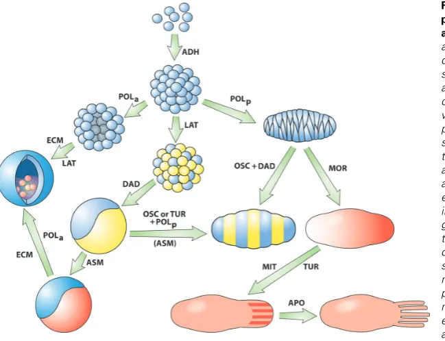
their closest living nonmetazoan relatives (Phillipe et al., 2004). Choanoflagellates are unicellular, or form small colonies, de-pending on laboratory culture conditions. According to our sce-nario, then, cadherins, cell surface molecules previously evolved to serve other functions, would have taken on the new role of mediating cell-cell adhesion when single cells bearing them encountered permissive environments, thus becoming what were perhaps the first DPMs (ADH; see Fig. 1, top, downward arrow and Table 1). (This is an example of an “exaptation” in the
DPM
Characteristic
molecules Physical principle Evo-Devo Role
ADH cadherins adhesion multicellularity
DAD cadherins differential adhesion multilayering
LAT Notch lateral inhibition multiple cell types
POL Wnt anisotropy lumen formation; elongation
OSC Wnt + Notch + Hes synchronous biochemical oscillation
field formation; segmentation
MOR TGF-β/BMP; Hh diffusion pattern formation
ASM FGFs asymmetric interaction induction; epithelial-mesenchymal interaction
TUR MOR + Wnt + Notch chemical waves periodic patterning
ECM collagen; chitin; fibronectin
stiffness; dispersal + cohesion
epithelial elasticity; skeletogenesis; epithelial-mesenchymal transformation
MIT MAPK mass increase (differential) growth
APO Bcl-2 mass decrease (differential) cell loss TABLE 1
PROPERTIES OF DYNAMICAL PATTERNING MODULES (DPMS) DISCUSSED IN THIS ARTICLE
terminology of Gould and Vrba, 1982.) The glycan-binding C-type lectins, which are employed as Ca2+-dependent cell attachment
proteins in multicellular organisms (Kaltner and Gabius 2001), are also produced by choanoflagellates (King et al.,, 2008) and could thus have provided a basis for additional ADH-class DPMs in metazoan ancestors.
If subsets of cells within an aggregate contain sufficiently different amounts of cadherin on their surfaces, the subpopula-tions will sort out into islands of more cohesive cells within lakes composed of their less cohesive neighbors (Steinberg and Takeichi, 1994). Eventually, by random cell movement, the is-lands coalesce and an interface is established across which cells will not intermix (Steinberg, 1998). The physical basis of this sorting-out of cell populations is similar to phase separation of two immiscible liquids, like oil and water (Forgacs and Newman, 2005).
Because a more cohesive tissue (one with stronger bonds between its cells) will always be partly or fully engulfed by a less cohesive one, differential adhesion-driven multilayering acts in a goal-directed fashion (Steinberg, 1998). The “goal” is the thermo-dynamic one of minimization of free energy (Steinberg, 1978; Forgacs and Newman, 2005). Ancient cadherins, then, acting in an environment that permitted them to mobilize the physical force of adhesion, became not only the mediators of colony formation, but of the automatic development of embryo-like structures con-sisting of distinct cell layers or “compartments,” in which no interchange or mixing of cells occurs across the common bound-ary (Crick and Lawrence, 1975; Garcia-Bellido, 1975). This major transition in the history of life can thus have occurred with little genetic change relative to the condition of choanoflagellate-like ancestors.
As components of the DPM designated ADH, cadherins medi-ate the formation of “solid”3 cell clusters, with spherical geometry
(due to minimization of surface tension in a mass of isotropic, mobile cells; Forgacs and Newman, 2005). By bringing differen-tial adhesion into play (DAD, see Fig. 1, center, leftward arrow and Table 1), the DPM in which subpopulations of cells express different levels of a cadherin or lectin can cause the clusters to become multilayered. But in contrast to modern organisms, in which differential adhesion is under precise spatiotemporal regu-lation (e.g., Godt and Tepass, 1998; Gonzales-Reyes and St Johnson, 1998; Damon et al., 2008), the ratio of high-expressing to low-expressing cells would most likely have been unregulated in the earliest such forms, so the resulting organisms would have had many different, poorly defined, morphologies. The following subsection describes how molecules carried over from the unicel-lular ancestors of the metazoa were mobilized in new mecha-nisms to restrict this morphological profligacy. The most straight-forward way this could happen is by a process or pathway that influenced the relative numbers of cells that assume different biosynthetic states.
Cell fate choice and lateral inhibition (the Notch pathway) The Notch-Delta pathway is an ancient signaling system that depends on the interaction of the single-pass integral membrane
protein Notch, which is a receptor for signals that originate outside the cell, and one of a class of other single-pass membrane proteins that act as modulators of Notch activity: Delta, Serrate and Lag2 (the DSL proteins) (Ehebauer et al., 2006). When Notch is activated, an intracellular portion of it is cleaved off and functions in the nucleus to turn a class of transcriptional repressor proteins into transcriptional activators. Notch’s effects are there-fore entirely dependent on which of the dual-action factors are present in a given cell type. The pathway’s role is therefore not to determine the specific fate of a cell, but rather to cause the cell to choose one of two of its potential fates, whatever those may be. The Notch pathway (present in sponges; Nichols et al., 2006, placozoans, Srivastava et al., 2008 and all eumetazoans, but not in choanoflagellates; King et al., 2008), has been extensively characterized during multicellular development, where it oper-ates in a juxtacrine fashion. That is, the Notch receptor on one cell interacts with a DSL-class protein on an adjacent cell, and the two cells, though initially equivalent, come to assume different fates. Although the original cells in such a population will express both Notch and DSL proteins, the population is initially in a metastable state. The receptor-ligand interaction serves to kick it into a stable state where one or a small contiguous group of cells increase their DSL levels and the cells surrounding them are Notch-activated and are thereby prevented from assuming the same fate as the central group. For this reason, the Notch pathway is usually referred to as mediating lateral inhibition (Simpson, 1997).
Recent work, however, suggests that Notch can interact with DSL proteins in a cell-autonomous fashion; that is, the receptor and “ligand” are on the same cell, rather than on adjacent ones (Sakamoto et al., 2002). This may, indeed be the ancestral state of the Notch pathway, with a cell switching between one state and another (consider, for example, sporulation) due to environmen-tally induced association between its Notch and DSL proteins. Indeed, the transcriptional mediators of Notch-dependent cell state switching (but not Notch itself) are present in fungi (Prevorovsky et al., 2007). Protein modules found in Notch receptors are present in a choanoflagellate, though they are not all encoded in the same genes (King et al., 2008). With the ADH-dependent transition to multicellularity there would have been selective pressure to take the few steps necessary to turn a cell autonomous mechanism into a juxtacrine one, and thereby con-vert cell-state switching into lateral inhibition.
The advent of the Notch-related DPM (LAT; see Fig. 1, upper-center, downward arrow and Table 1) (via a change of context and scale, but, in principle, with a minimum of genetic evolution) allowed the relative numbers of cells of different states in an aggregate to be fairly well controlled. Such patterns will be fine-grained, since the spatial scale of juxtacrine signaling is small. If, however, the alternative cell states in question are associated with different levels of cadherin, the subpopulations will sort-out (see previous subsection) and a multilayered aggregate will automatically form.
Apical-basal and planar cell polarity (the Wnt pathway) As described above, the default morphology of a cell
aggre-3 We use the term “solid” here in the topological sense: lacking internal cavities. We also characterize cell aggregates and parcels of tissue as “liquid-like” throughout this paper, but there we refer to the physical state of a material whose subunits (cells in this case) are independently mobile (Forgacs
gate held together by cadherins (or any other uniformly distributed cell-cell adhesive protein) is solid (i.e., without a lumen) and spherical. Wnt, a family of secreted factors that interact with receptors of the Frizzled family, acting through distinct but related pathways, induces cells to become polarized along their apical-basal (A/B) axis (Karner et al., 2006b), or oriented in a plane perpendicular to this axis (planar cell polarity; Mlodzik, 2002; Karner et al., 2006a). Although these are individual cell behaviors, in a multicellular context they permit cell aggregates to overcome the morphological defaults of solidity and sphericality (see below). Although Wnt genes and their cognate secreted proteins are not present in choanoflagellates (King et al., 2008), they and their Frizzled receptors are found in sponges (Nichols et al., 2006) and placozoans (Srivastava et al., 2008). The Wnt pathway has even deeper roots in cellular evolution, however. In the fission yeast Schizosaccharomyces pombe the protein Pmo25p is essential for polar growth; in its absence the actin cytoskeleton becomes depolarized and cells adopt a round morphology (Mendoza et al., 2005). Pmo25p is a homolog of the metazoan MO25 family of proteins, which in nematode, insect and vertebrate cells act as cofactors of the serine-threonine kinase Lkb-1. The latter, by regulating the activity of the cytoplasmic enzyme glycogen syn-thase kinase 3β (GSK3β), acts as a fulcrum of the “canonical” (that is, mediated through the transcriptional co-regulator β-catenin) Wnt pathway (Green, 2004).
The regulation of A/B polarity by Wnt in the metazoa is thus based on an ancient mechanism of polarity control that existed before the fungi and choanoflagellates split off from each other. Furthermore, even in modern metazoa, this pathway, while induc-ible by the secreted Wnt ligand, remains a property of individual cells. Indeed, the entire A/B polarity program is triggered in isolated animal cells if Lkb-1 is activated (Karner et al., 2006a).
Polarization along the A/B axis has a clear role in the regulation of cell division in unicellular organisms like S. pombe (Mendoza et al., 2005), and the same likely held for the single-celled antecedents of the metazoa. In multicellular aggregates, how-ever, it can mediate some entirely unprecedented morphogenetic effects. If cells are polarized in their distribution of an attachment protein, for example, they will not form a solid mass when aggregated. The energetically most favorable configuration would be achieved when the more adhesive portions of the cell mem-branes bind to each other while the less adhesive regions are freed up to enclose an interior lumen (Newman, 1998; Forgacs and Newman, 2005), as seen in the sponges and all eumetazoans. Alternatively, if cells are polarized in different attachment proteins they can form multiple non-mixing layers, such as seen in the three-layered placozoan, Trichoplax (Miller and Ball, 2005).
The characteristic multicellular organisms of the Precambrian had earlier been described as solid-bodied (Seilacher, 1992), but the discovery of small, hollow, cell clusters from the Doushantuo Formation, China (Chen et al., 2004) has qualified this descrip-tion. Although these lumen-containing forms have been termed “embryos” (Chen et al., 2004; Hagadorn et al., 2006), the evi-dence is also consistent with their being definitive adult forms of their time. The Wnt pathway is very ancient, likely originating in the Precambrian (Erwin, 2008). Because A/B polarization accom-panies and appears to be a necessary condition for the formation of interior cavities in cell aggregates, it is plausible that the advent of the DPM enabling A/B polarization helped drive the transition
between the Ediacaran biota and those of the Cambrian explosion (POLa; Fig. 1, upper-center, leftward arrow and Table 1).
The utilization of β-catenin as a transcriptional co-factor in the POLa DPM and certain other Wnt-mediated functions (prolifera-tion, alteration of gene expression), and its use in the ADH and DAD DPMs as a submembrane component of cadherin-based adhesion complexes, provides the basis of regulated switching among Wnt-based DPMs (Nelson and Nusse, 2004). Such alter-native deployment of DPMs is important in a variety of complex morphogenetic changes (Jamora et al., 2003; de Melker et al., 2004).
Planar cell polarity (PCP) is also initiated at Frizzled receptors and is therefore usually described as employing the Wnt pathway (noncanonical in this case, since β-catenin is not involved). Despite the involvement of Frizzled, however, Wnt ligand is not involved in all cases of PCP induction (Veeman et al., 2003). Thus, like A/B polarization, PCP may be based on a cellular mechanism that predates cell-cell communication.
The consequences of PCP in a multicellular context are unintuitive, but highly significant. Elongated cells with anisotropic adhesive properties are predicted on the basis of physical prin-ciples to spontaneously align and intercalate among one another, leading to the tissue mass narrowing in one direction and elongat-ing in the orthogonal direction (Zajac et al., 2003; Keller et al., 2008). These cellular rearrangements and tissue reshaping ef-fects are seen in “convergent extension,” which establishes the elongated body axis during gastrulation in the amphibian embryo, and related phenomena throughout the Metazoa (Keller, 2002) (POLp; Fig. 1, upper-center, rightward arrow and Table 1).
The cell polarization effects associated with the Wnt pathways, then, are based on the mobilization of mechanisms of cytoskeletal rearrangement that appear to have evolved to serve single-cell functions. With the arrival of multicellularity, the existence of these mechanisms enabled those tissue masses in which they were present to acquire internal lumens and elongated shapes, i.e., to move beyond the default solid, spherical morphology of adhesive, mobile cells. They could have done this, we again note, mainly as a result of change in scale and context. Substantial genetic change was not required.
Field formation by synchronized biochemical oscillations (Hes, Notch and Wnt)
A balance of positive and negative feedback interactions in a gene regulatory network can lead to temporal oscillations in concentration of gene products (Goldbeter, 1996; Reinke and Gatfield, 2006). In individual cells, such as the common ancestors of metazoans and choanoflagellates, such oscillations would not have a lasting morphological effect. In a multicellular context, where they can be coordinated across cell boundaries by juxtacrine (e.g., Notch pathway) and short-range paracrine (e.g., Wnt path-way) signaling, however, such oscillations have the potential to drive morphogenetic change.
the Notch pathway mediator Hes1), undergo temporal oscillation with a period similar to the formation of the somites (Pourquié et al., 2003). These oscillations then become synchronized by Notch-mediated juxtacrine signaling (Giudicelli et al., 2007; Kageyama et al., 2007; Riedel-Kruse et al., 2007). In conjunction with an FGF8 morphogen gradient (see below) with its source at one end of the extended embryo, the Hes1- and associated oscillations provide the basis for the generation of somites in vertebrate embryos (Pourquié, 2003).
The recurrent nature of the oscillatory state is an important feature that may utilized in a developmental system, where, as in somitogenesis, temporal periodicity is converted to spatial peri-odicity. But another key property of oscillators is their ability to become synchronized (Garcia-Ojalvo et al., 2004; Masamizu et al., 2006). This highlights a general effect of the OSC DPM that can be developmentally important over durations shorter than the oscillator’s period. The coordination of cell state (e.g., with re-spect to the oscillator’s components) over a broad tissue domain, a phenomenon described in the older embryological literature as a “morphogenetic field” (reviewed in Haraway, 1976; Gilbert, 2006), has long eluded mechanistic explanation but can now be understood as a manifestation of this DPM.
While many gene regulatory networks are capable of sustain-ing oscillatory behavior, Hes-type transcriptional modulators are particularly suitable as elements of the OSC DPM. This is be-cause they are downstream effectors of the Notch pathway, a mediator of juxtacrine cell communication in the multicellular context. Interactions of the Notch pathway with the Wnt pathway (Hofmann et al., 2004; Ishikawa et al., 2004; Hayward et al., 2006) can also mobilize paracrine interactions in the synchronization of Hes oscillations. Hes-type transcription factors are ancient mem-bers of the developmental-genetic toolkit, with two representa-tives in the placozoan genome (Srivastava et al. 2008).
Single- and dual-tissue morphogen gradients (the TGF-βββββ/ BMP superfamily, Hedgehog and FGFs)
Unicellular eukaryotes such as protozoan ciliates have the ability to change their physiological state in response to mol-ecules secreted into the microenvironment by other such cells (Luporini et al., 2006). It is a reasonable assumption that this capacity had already evolved in the unicellular progenitors of the metazoa. In the multicellular context, a secreted molecule for which these ancient cells had evolved concentration-dependent responses could serve as a patterning molecule or “morphogen” (MOR; Fig. 1, upper-right, right-downward arrow and Table 1). To perform this function, however, it would need to have the capacity to spread a significant distance across a cluster of cells using their lipid membranes or the extracellular space as a medium for diffusion. The locally acting Wnts have a limited capacity to act as morphogens (Sick et al., 2006); in general they stabilize, rather than specify, cell fate (Martinez-Arias, 2003).
The ability of one or a small group of cells to influence other cells via morphogens enables the generation of heterogeneous patterns on a spatial scale of 100 μm - 1 mm over tens of hours, consistent with the time-distance-concentration relationships in-herent in macromolecular diffusion (Crick, 1970). Morphogens fall into two main classes. Some act on cells similar to those that produce them, causing them to assume one of several new states depending on the concentration of morphogen to which they are
exposed. Others act on cells that are already different from the producers (perhaps residing in a separate layer), causing them to become different from their unexposed neighbors. Different mor-phogens also move through different media. Those of the TGF-β/ BMP and FGF classes diffuse in the aqueous interstices and extracellular matrix at variable rates, dependent on their capacity to bind to specific extracellular molecules, which may themselves be distributed nonuniformly (Ohkawara et al., 2002). Molecules of the Hedgehog class of morphogens are alternately tethered to cell membranes by covalently attached lipid moieties and diffusible through the aqueous interstitial phase beyond the membrane (Goetz et al., 2006). Because the lipid component of the Hedge-hog morpHedge-hogens limits their spread (Guerrero and Chiang, 2007), their diffusion rate is probably intermediate between the TGF-β/ BMP class and the very short-range Wnts.
While each of these morphogens activates signaling pathways that apparently preexisted the metazoa, the morphogen mol-ecules themselves arose at various times during the metazoan radiation. For example, while demosponges, the most primitive metazoan group, have a Hedgehog-type morphogen, they lack TGF-β/BMP and FGFs (though they have a receptor homolog for the latter class; Nichols et al., 2006). Morphogens of all three classes are present in cnidarians (Rentzsch et al., 2006; 2008; Matus et al., 2008) and echinoderms (Lapraz et al., 2006; Walton et al., 2006), while the placozoan Trichoplax appears to only contain the TGF-β/BMP pathway (Srivastava et al., 2008).
FGFs in arthropods and chordates have multiplied by gene duplication and their receptors are alternatively spliced in such a fashion that an FGF produced by one tissue layer typically only affect cells in a different layer, and not those cells that produce it (Huang and Stern, 2005). This type of tissue interaction consti-tutes a novel and fruitful developmental principle not seen in other animal groups, nor, generally, with respect to other morphogen systems. In particular, it enables asymmetric interaction between different tissue layers, exemplified by induction and epithelial-mesenchymal interaction (ASM; Fig. 1, left-center, left-downward arrow and Table 1).
Morphogen function is inextricably tied to the physical principle of diffusion, a mechanism that signifies a different biological phenomenon in multicellular and unicellular contexts. By the simple effect of setting up molecular gradients across a cluster of initially equivalent but responsive cells, morphogen-based DPMs generated organismal forms that ultimately contained heteroge-neously distributed cell types. Whether or not any given form was functionally compatible with survival was a matter subject to natural selection. But as with the DPMs described earlier, the potential to generate a panoply of morphologies is tied to material properties of the system rather than to incremental selective advantage.
Spot, stripe and boundary patterns arising from local autoactivation-lateral inhibition
positively autoregulatory morphogen that became linked, how-ever, in a composite DPM, to a mechanism of lateral inhibition (like the LAT DPM associated with Notch signaling), will induce a zone around any peak of its activity within which no local activa-tion can occur (Gierer and Meinhardt, 1972; Meinhardt and Gierer, 2000; Meinhardt, 2008). New peaks of activation would only form at distances sufficiently far from other peaks so that the inhibitor’s effects will have attenuated. Such systems, involving short-range local activation and long-range lateral inhibition (simi-lar to the chemical pattern forming systems described by Turing, 1952), or formally equivalent local autoactivation-lateral inhibition (LALI) systems (Nijhout 2003; Newman and Bhat, 2007), can produce regularly spaced spots or stripes of morphogen concen-tration (TUR; Fig. 1, far-right-center, left downward arrow and Table 1). These, in turn, can induce primordia of skeletal elements (Newman and Frisch, 1979; Hentschel et al., 2004) and other serially repeated structures such as teeth (Salazar-Ciudad and Jernvall, 2002), feather germs (Jiang et al., 2004) and hair follicles (Sick et al., 2006). Tissue boundary formation (von Dassow et al., 2000) and axial polarization (De Robertis, 2006) regulated by such mechanisms are more robust than those that depend solely on differential adhesion or diffusion gradients (Ingolia, 2004).
Epithelial elasticity, epithelial-mesenchymal transformation and global organization of cell polarity (extracellular matrix) Up to this point we have described “epithelioid” cell clusters in which cells are directly attached to each other. The viscosity of such clusters depends on the ease with which cells slip past one another while maintaining their attachments. (Cell-cell bonds, even if strong, can have short lifetimes). Their elasticity is prima-rily a function of the cytoskeleton, and their cohesivity is deter-mined by the force required to separate the cells. The other major category of cell aggregate or tissue in multicellular metazoan forms is termed “mesenchyme.” In mesenchymal aggregates the constituent cells secrete a complex macromolecular microenvi-ronment, the extracellular matrix (ECM) (Comper, 1996), and it is the properties of this material that determine the aggregate’s viscoelasticity and cohesivity, collectively, its rheological proper-ties. ECMs are often quite elaborate on the molecular scale and mesenchymes thus have different morphogenetic capabilities from the epithelioid cell aggregates subject to the DPMs dis-cussed above.
The most ancient metazoans, the Porifera or marine sponges, produce skeletal structures whose morphology is, in part, a function of environmental factors (Uriz et al., 2003). Most sponge cells (sclerocytes) reside within an ECM called the “mesohyl,” which constitutes the bulk of the organism’s body. These cells bind to the ECM via integrin transmembrane proteins (Wimmer et al., 1999), as in eumetazoans. Sponges also contain epithelial cells (Schröder et al., 2004) and homologs of type IV-like collagen (Boute et al., 1996), which in eumetazoans forms a major compo-nent of the sheet-like supporting structure for epithelia, the basal lamina. Despite the active remodeling of the internal anatomy of sponges by the continuous movement of their cells (Bond, 1992), and the presence of most of the molecular ingredients of the major DPMs (see above), poriferans branched off from the metazoan lineage early on, as a morphological dead-end (Nichols et al., 2006). One reason for this may have been their great reliance on ECM, a stiff, relatively non-dynamic medium at the mesoscopic
scale, as their major morphogenetic component. While sponges produce both metalloproteinases (Arreguin et al., 1995) and metalloproteinase inhibitors (Fujita et al., 2003), two classes of molecules used by eumetazoans to break down and remodel their ECMs, it is possible that these functions never became integrated in these groups of organisms into a composite DPM that would have facilitated the mobilization of other DPMs.
In the eumetazoa ECM is employed in a more limited fashion, one that permits the other DPMs to have freer play. Cnidarians have a thin sheet-like mesoglea consisting of separate regions of basement membrane-like and interstitial matrix-like properties (Zhang et al., 2007), whereas true interstitial ECM is found only in triploblasts (Huxley-Jones et al., 2006). A basement membrane permits an epithelium to behave as an elastic sheet in the direction perpendicular to the plane, though it may retain in-plane liquid-like properties (Mittenthal and Mazo, 1983; Newman, 1998). Elastic sheet epithelia exhibit a range of folding, buckling and wrinkling effects that are not seen in liquid-like epithelioid tissues, and which provide the basis for some modes of gastrulation and for formation of appendages (Gierer, 1977; Forgacs and Newman, 2005).
The mesodermal layer of many triploblasts and some cnidarians (Fritzenwanker et al., 2004; Seipel and Schmid, 2006) is com-posed of cells that have undergone epithelial-mesenchymal trans-formation (EMT) (Hay, 2005). This change in the physical state of tissues is enabled by the ECM, which permits cells that are not directly attached to one another to remain part of an integral tissue. Whereas skeletogenesis and elastic sheet behavior are both based on the stiffness of ECM, EMT employs its space-filling properties.
Cell polarity, which we have seen above is mediated by the Wnt signaling pathway, can be oriented in preferred directions by interaction with previously deposited ECM (Thery et al., 2006). In analogy to the effects of diffusible morphogens in organizing spatial patterns of cell state across a cluster of cells (see above), ECM cues can help determine global patterns of cell polarity in multicellular aggregates. Although ECM molecules and integrins pre-existed the origins of the metazoa – homologs are present in the non-colonial choanoflagellate M. brevicollis (King et al., 2008) – the emergence of multicellularity recruited these molecules into “internal substrata,” constituting a distinct DPM (ECM; Fig. 1, bottom-left, left upward arrow and Table 1), with new capacities for generating pattern motifs.
Developmental function-dedicated transcription
fac-tors as “frozen accidents”
and responding centers (Ciudad et al., 2003; Salazar-Ciudad, 2006).
Certain of the transcription factors included among the “toolkit” gene products (the developmental transcription factors, or DTFs, in our terminology), were tied to functions in single-celled ances-tors that were later utilized in differentiated cells of metazoans (cell contractility, light sensitivity). But in other cases (participation in segmentation, anteroposterior or proximodistal tissue identity) they were unlikely to have corresponding functional roots in the unicellular world. Given the scenario we have described above, we can speculate that in the first metazoa, fortuitous associations of one or more DPMs with the DTFs existing at the time produced “generalized” eyes, appendages, heart-like contractile tubes and segments. If, as seems likely, they were loosely interacting, subject to external conditions, and not yet organized into the hierarchical regulatory schemes that came with subsequent evo-lution (Salazar-Ciudad et al., 2001a,b), these DPM-DTF com-plexes would not have programmed unique, definitive body plans and organs, but rather versions of these morphological motifs that were plastic within populations and even individual proto-organ-isms. In this view, radically different types of eyes (Gehring, 2002) and appendages, and even wholesale inversions of the body axis (Gerhart, 2000), were possible for organisms of fixed genotype, in different environments and across generations.
With subsequent selection for reliability of developmental outcome (Waddington, 1942; Schmalhausen, 1949) the “proto”-bodies and -organs resulting from particular DPM-DTF associa-tions would have become uniquely associated with specific pro-grams of gene expression having homologous molecular bases and analogous morphological outcomes (Newman et al., 2006). Examples of developmental outcomes based on such “homolo-gous-analogous” DTF-DPM associations across metazoan phyla include skeletogenesis, appendage formation, eye formation, and heart development.
Conclusion: a pattern language for development and
morphological evolution
We have proposed that the developmental mechanisms of biologically modern animals can be understood in terms of a pattern language of metazoan form. The elements of this lan-guage are transformations away from the physical default of an aggregate of undifferentiated cells: topologically solid, geometri-cally spherical, and spatially uniform. The major transformations include (i) formation of stable mixtures of cells in more than one biochemical state, (ii) stable formation of distinct cell layers, (iii) formation of internal cavities, (iv) elongation of cell masses, (v) generation of nonuniform patterns of cell type, and (vi) dispersal of subpopulations of cells without disintegration of the organism. We have further proposed that each pattern-language element is tied to a set of gene activities of the so-called developmental-genetic toolkit many of which pre-evolved the metazoa in single-celled ancestors. The elements of the pattern language, however, are not genes, gene products, or even merely gene networks. They are, rather, what we term dynamical patterning modules or DPMs, in which a complex of the toolkit gene products mobilizes a mesoscale physical process. Thus, cell adhesion is mobilized by cadherins, lateral inhibition enabling maintenance of stable cell mixtures by the Notch pathway, tissue multilayering by differential
expression of cadherins, cell polarization, leading to cavity forma-tion and elongaforma-tion, induced by the Wnt pathway, coordinaforma-tion of cell state by the Notch- and Wnt-dependent synchronization of Hes oscillations, and so forth.
The collection of DPMs we have described here is relatively comprehensive, but not exhaustive. Increase and decrease in cell number, for example, have only a quantitative impact on popula-tions of free-living protists. In a multicellular context, however, these processes constitute reciprocal elements of a patterning system in which organismal shape and form are altered, but not cell state (Salazar-Ciudad et al., 2003; Salazar-Ciudad, 2006). For this reason the mitogen-activated protein kinase (MAPK) pathway, one of whose functions is to mediate proliferation in response to external signals (Krens et al., 2006), and the apop-totic pathway, both of which have roots in the premetazoan unicellular world (Widmann et al., 1999; Blackstone and Green, 1999), can be considered DPMs which modulate the physical mass of a multicellular aggregate (MIT, Fig. 1, center-right, left downward arrow, and APO; Fig. 1, bottom-center, rightward arrow and Table 1).
An additional aspect of our hypothesis is that the earliest metazoan developmental mechanisms did not arise primarily by incremental natural selection. We suggest instead that the change in context and spatial scale inherent to the multicellular state led to the relatively abrupt appearance of such mechanisms. Thus, for example, Notch signaling, which plausibly evolved in unicellu-lar forms to elicit transitions in cell state in response to environ-mental cues, would come to mediate the coexistence of alterna-tive cell states in a single colony, where cells serve as each other’s microenvironmental determinants.
Finally, autoregulatory networks of transcription factors and cis-regulatory modules, a major mechanism in the specification of cell fate in modern-day developmental systems (Davidson, 2006), are nonetheless not central to our concept of a pattern language for multicellular form. To paraphrase the statement by Gehring (2002) quoted in the introduction to this paper, transcription factors are, in principle, interchangeable in their roles and are not intrinsically connected to the functions of the genes they happen to regulate. This contrasts with the gene products that mediate the DPMs, which perform not one common gene expression-associ-ated function (transcription), but qualitatively different, physically-associated functions (adhesion, diffusion, geometric polarization, etc.).
(e.g., Ruiz-Trillo et al., 2007) cannot be entirely successful. In general, the recruitment of toolkit gene products and path-ways into basic and composite DPMs must lead to very different outcomes depending on the topology (i.e., connectivity) and the relative strengths of interaction among the components (Salazar-Ciudad, 2006). In extreme cases, only certain DPMs might have become active in certain lineages and other DPMs largely fore-closed. We may speculate, for example, that with multicellularity in place, the Wnt-mediated DPM in the ancestors of sponges, via its induction of apical-basal polarity, enabled the generation of multicellular forms with simple and branched interior cavities, but that an overactive integrin-ECM-mediated DPM (or underactive metalloproteinases), may have rendered the tissue microenviron-ment nonpermissive for employmicroenviron-ment of other DPMs.
In the ancestors of the eumetazoa, in contrast, the action of TGF-β/BMP and hedgehog-mediated DPMs readily produced tissue masses nonuniform in cell state, and Notch-mediated lateral inhibition must have stabilized these patterns. One result of this would have been cell aggregates compartmentalized into distinct layers. Then, heterotypic tissue interactions, mediated by FGFs and their receptors, would have promoted the emergence of sophisticated diploblastic forms, such as the Cnidaria. Next, the functional linking of simple DPMs, such as the cadherin and Wnt pathways (via their common effector, β-catenin), and the Notch and Wnt pathways (via their common mediator, GSK3β), into composite DPMs, opened the way for unprecedented dynamical processes acting on cell masses: global coordination of cell states via synchronized oscillations and reaction-diffusion patterning, to name two important ones. Finally, secreted ECMs, assembled on the mesoscopic scale of multicellular aggregates, became DPMs that mobilized mechanical effects, introducing elastic properties and solid skeletons to tissue masses and sheets. Once estab-lished, the DPMs remained highly conserved across the diploblasts and triploblasts, protostomes and deuterostomes, invertebrates and vertebrates.
Development of modern organisms exhibits extensive plastic-ity, permitting alternative outcomes depending on different exter-nalities (West-Eberhard, 2003). By our hypothesis, development in metazoan ancestors would have been even more plastic, since prior to the evolution of canalizing mechanisms the physical processes mobilized by the various DPMs would have been highly sensitive to environmental conditions (see also Newman, 1994; Newman and Müller, 2000; Newman et al., in press). This bears on recent attempts to relate mechanisms of axis formation and arrangement of molecular signaling centers in the embryos of metazoan groups to presumed ancestral relationships among those groups (e.g., chordates, tunicates, hemichordates, cephalochordates; Passamaneck and Di Gregorio, 2005; Lowe et al., 2006; Yu et al., 2007). Because the DPMs could have been deployed combinatorially and non-hierarchically in ancient form the taxonomic relationships among these groups may not be direct, nor would they necessarily conform either to anatomical affinities or a straightforward developmental logic.
The deployment of DPMs, in modern metazoa, but even in their primitive ancestors, cannot generate an unlimited array of forms. Despite their potential (presumably greater in early metazoan evolution than at present) to transform different anatomies one into the other with little or no genetic change, DPMs are con-strained to mold pre-metazoan cell masses into only those
mor-phologies characteristic of chemically and mechanically excitable mesoscopic materials, among these hollow, multilayered, elon-gated, segmented forms. If the “tape of life” were to be replayed (Gould, 1989) things would probably turn out not too differently at the level of bauplan.
Comparative anatomists have long recognized that animal bodies share a common morphological phrase book. More re-cently, molecular evolutionists have discovered that the metazoa share a common developmental-genetic vocabulary. Both of these findings, as we have shown, stem from the existence of a pattern language for animal development. The grammar of this language emerged abruptly more than 500 million years ago when a group of proteins and pathways of the unicellular world, by coming to operate on the mesoscale, mobilized the physical laws pertaining to soft-matter and excitable media in the construction of multicellular organisms.
Acknowledgments
We acknowledge the U.S. National Science Foundation for support.
References
ABEDIN, M. and KING, N. (2008). The premetazoan ancestry of cadherins. Science
319: 946-8.
ALEXANDER, C. (2002). The nature of order: An essay on the art of building and the nature of the universe. Center for Environmental Structure, Berkeley, CA.
ALEXANDER, C., ISHIKAWA, S. and SILVERSTEIN, M. (1977). A pattern lan-guage: Towns, buildings, construction. Oxford University Press, New York.
AMEISEN, J.C. (2002). On the origin, evolution, and nature of programmed cell death: A timeline of four billion years. Cell Death Differ 9: 367-393.
ARREGUIN, R., ARREGUIN, B., HERNANDEZ-ARANA, A. and RODRIGUEZ-ROMERO, A. (1995). Metal content and conformation of the metalloprotease from the marine sponge Spheciospongia vesparia. Biochem Mol Biol Int 36:
827-833.
BLACKSTONE, N.W. and GREEN, D.R. (1999). The evolution of a mechanism of cell suicide. Bioessays 21: 84-88.
BONNER, J.T. (1967). The cellular slime molds. Princeton University Press,
Princeton.
BORGES, R.M. (2005). Do plants and animals differ in phenotypic plasticity? J Biosci 30: 41-50.
BOUTE, N., EXPOSITO, J.Y., BOURY-ESNAULT, N., VACELET, J., NORO, N., MIYAZAKI, K., YOSHIZATO, K. and GARRONE, R. (1996). Type IV collagen in sponges, the missing link in basement membrane ubiquity. Biol Cell 88: 37-44.
CARROLL, S.B., GRENIER, J.K. and WEATHERBEE, S.D. (2005). From DNA to diversity: Molecular genetics and the evolution of animal design. Blackwell Pub.,
Malden, MA.
CHEN, J.Y., BOTTJER, D.J., OLIVERI, P., DORNBOS, S.Q., GAO, F., RUFFINS, S., CHI, H., LI, C.W. and DAVIDSON, E.H. (2004). Small bilaterian fossils from 40 to 55 million years before the cambrian. Science 305: 218-222.
COMPER, W.D. (Ed.) (1996). Extracellular matrix, vol. I. Tissue Function; II. Molecular Components and Interactions. Amsterdam: Harwood Academic Publishers.
CONWAY MORRIS, S. (2000). Nipping the Cambrian "explosion" in the bud?
Bioessays 22: 1053-1056.
CONWAY MORRIS, S. (2006). Darwin’s dilemma: The realities of the Cambrian ‘explosion’. Philos Trans R Soc Lond B Biol Sci 361: 1069-1083.
CRICK, F.H.C. (1970). Diffusion in embryogenesis. Nature. 225: 420-422.
CRICK, F.H.C. and LAWRENCE, P.A. (1975). Compartments and polyclones in insect development. Science. 189: 340-347.
DAVIDSON, E.H. (2006). The regulatory genome: Gene regulatory networks in development and evolution. Elsevier/Academic Press, Amsterdam; London.
DE GENNES, P.G. (1992). Soft matter. Science 256: 495-497.
DEQUEANT, M.L., GLYNN, E., GAUDENZ, K., WAHL, M., CHEN, J., MUSHEGIAN, A. and POURQUIÉ, O. (2006). A complex oscillating network of signaling genes underlies the mouse segmentation clock. Science 314: 1595-1598.
DE MELKER, A.A., DESBAN, N. and DUBAND, J.L. (2004). Cellular localization and signaling activity of beta-catenin in migrating neural crest cells. Dev Dyn
230: 708-726.
DE ROBERTIS, E.M. (2006). Spemann’s organizer and self-regulation in amphib-ian embryos. Nat Rev Mol Cell Biol 7: 296-302.
DROSER, M.L. and GEHLING, J.G. (2008). Synchronous aggregate growth in an abundant new Ediacaran tubular organism. Science 319: 1660-1662.
EHEBAUER, M., HAYWARD, P. and ARIAS, A.M. (2006). Notch, a universal arbiter of cell fate decisions. Science 314: 1414-5.
ERWIN, D.H. (2008). Wonderful Ediacarans, wonderful cnidarians? Evol Dev 10:
263-264.
ERWIN, D.H. and DAVIDSON, E.H. (2002). The last common bilaterian ancestor.
Development 129: 3021-3032.
FORGACS, G., FOTY, R.A., SHAFRIR, Y. and STEINBERG, M.S. (1998). Vis-coelastic properties of living embryonic tissues: A quantitative study. Biophys J
74: 2227-2234.
FORGACS, G. and NEWMAN, S.A. (2005). Biological physics of the developing embryo. Cambridge Univ. Press, Cambridge.
FRITZENWANKER, J.H., SAINA, M. and TECHNAU, U. (2004). Analysis of forkhead and snail expression reveals epithelial-mesenchymal transitions dur-ing embryonic and larval development of Nematostella vectensis. Dev Biol 275:
389-402.
FUJITA, M., NAKAO, Y., MATSUNAGA, S., SEIKI, M., ITOH, Y., YAMASHITA, J., VAN SOEST, R.W. and FUSETANI, N. (2003). Ageladine A: An antiangiogenic matrixmetalloproteinase inhibitor from the marine sponge Agelas nakamurai. J Am Chem Soc 125: 15700-15701.
GEHRING, W.J. (2002). The genetic control of eye development and its implica-tions for the evolution of the various eye-types. Int J Dev Biol 46: 65-73.
GERHART, J. (2000). Inversion of the chordate body axis: Are there alternatives?
Proc Natl Acad Sci USA 97: 4445-4448.
GIERER, A. (1977). Physical aspects of tissue evagination and biological form.
Quarterly Reviews of Biophysics 10: 529-593.
GIERER, A. and MEINHARDT, H. (1972). A theory of biological pattern formation.
Kybernetik 12: 30-39.
GILBERT, S.F. (2006). Developmental biology, 8th Ed. Sinauer Associates, Sunderland, Mass.
GIUDICELLI, F., OZBUDAK, E.M., WRIGHT, G.J. and LEWIS, J. (2007). Setting the tempo in development: An investigation of the zebrafish somite clock mecha-nism. PLoS Biol 5: e150: 1309-1323.
GODT, D. and TEPASS, U. (1998). Drosophila oocyte localization is mediated by
differential cadherin-based adhesion. Nature 395: 387-391.
GOETZ, J.A., SINGH, S., SUBER, L.M., KULL, F.J. and ROBBINS, D.J. (2006). A highly conserved amino-terminal region of Sonic hedgehog is required for the formation of its freely diffusible multimeric form. J Biol Chem 281: 4087-4093.
GOLDBETER, A. (1996). Biochemical oscillations and cellular rhythms: The mo-lecular bases of periodic and chaotic behaviour. Cambridge University Press,
Cambridge.
GONZALEZ-CRESPO, S. and MORATA, G. (1996). Genetic evidence for the subdivision of the arthropod limb into coxopodite and telopodite. Development
122: 3921-3928.
GONZALEZ-REYES, A. and ST JOHNSTON, D. (1998). The Drosophila AP axis is
polarised by the cadherin-mediated positioning of the oocyte. Development
125: 3635-3644.
GOULD, S.J. (1989). Wonderful life. W.W. Norton, New York.
GOULD, S.J. and VRBA, E. (1982). Exaptation; a missing term in the science of form. Paleobiology 8: 4-15.
GREEN, J.B. (2004). Lkb1 and GSK3-beta: Kinases at the center and poles of the action. Cell Cycle 3: 12-14.
GRIME, J.P., CRICK, J.C. and RINCON, J.E. (1986). The ecological significance of plasticity. Symp Soc Exp Biol 40: 5-29.
GUERRERO, I. and CHIANG, C. (2007). A conserved mechanism of hedgehog gradient formation by lipid modifications. Trends Cell Biol 17: 1-5.
HAGADORN, J.W., XIAO, S., DONOGHUE, P.C., BENGTSON, S., GOSTLING, N.J., PAWLOWSKA, M., RAFF, E.C., RAFF, R.A., TURNER, F.R., CHONGYU,
Y. et al. (2006). Cellular and subcellular structure of neoproterozoic animal
embryos. Science 314: 291-294.
HARAWAY, D.J. (1976). Crystals, fabrics and fields; metaphors that shape em-bryos. Yale University Press, New Haven.
HAY, E.D. (2005). The mesenchymal cell, its role in the embryo, and the remarkable signaling mechanisms that create it. Dev Dyn 233: 706-720.
HAYWARD, P., BALAYO, T. and MARTINEZ ARIAS, A. (2006). Notch synergizes with axin to regulate the activity of armadillo in Drosophila. Dev Dyn 235:
2656-2666.
HENTSCHEL, H.G., GLIMM, T., GLAZIER, J.A. and NEWMAN, S.A. (2004). Dynamical mechanisms for skeletal pattern formation in the vertebrate limb.
Proc R Soc Lond B Biol Sci 271: 1713-1722.
HOFMANN, M., SCHUSTER-GOSSLER, K., WATABE-RUDOLPH, M., AULEHLA, A., HERRMANN, B.G. and GOSSLER, A. (2004). Wnt signaling, in synergy with T/TBX6, controls Notch signaling by regulating Dll1 expression in the presomitic mesoderm of mouse embryos. Genes Dev 18: 2712-2717.
HUANG, P. and STERN, M.J. (2005). FGF signaling in flies and worms: More and more relevant to vertebrate biology. Cytokine Growth Factor Rev 16: 151-158.
HUXLEY-JONES, J., ROBERTSON, D.L. and BOOT-HANDFORD, R.P. (2007). On the origins of the extracellular matrix in vertebrates. Matrix Biol 26: 2-11.
INGOLIA, N.T. (2004). Topology and robustness in the Drosophila segment polarity
network. PLoS Biol 2: 805-15.
ISHIKAWA, A., KITAJIMA, S., TAKAHASHI, Y., KOKUBO, H., KANNO, J., INOUE, T. and SAGA, Y. (2004). Mouse Nkd1, a Wnt antagonist, exhibits oscillatory gene expression in the PSM under the control of Notch signaling. Mech Dev 121:
1443-1453.
JAMORA, C., DASGUPTA, R., KOCIENIEWSKI, P., FUCHS, E. (2003). Links between signal transduction, transcription and adhesion in epithelial bud development. Nature 422: 317-322.
JIANG, T.X., WIDELITZ, R.B., SHEN, W.M., WILL, P., WU, D.Y., LIN, C.M., JUNG, H.S. and CHUONG, C.-M. (2004). Integument pattern formation involves genetic and epigenetic controls: Feather arrays simulated by digital hormone models. Int J Dev Biol 48: 117-135.
JOLIOT, A. and PROCHIANTZ, A. (2004). Transduction peptides: From technology to physiology. Nat Cell Biol 6: 189-196.
KALTNER, H. and GABIUS, H.J. (2001). Animal lectins: From initial description to elaborated structural and functional classification. Adv Exp Med Biol 491: 79-94.
KARNER, C., WHARTON, K.A., JR. and CARROLL, T.J. (2006a). Planar cell polarity and vertebrate organogenesis. Semin Cell Dev Biol 17: 194-203.
KARNER, C., WHARTON, K.A. and CARROLL, T.J. (2006b). Apical-basal polarity, Wnt signaling and vertebrate organogenesis. Semin Cell Dev Biol 17: 214-222.
KAZMIERCZAK, J. and KEMPE, S. (2004). Calcium build-up in the Precambrian sea: A major promoter in the evolution of eukaryotic life. In Origins, (ed.
SECKBACH, J.). Kluwer, Dordrecht, pp.329-345.
KELLER, R. (2002). Shaping the vertebrate body plan by polarized embryonic cell movements. Science 298: 1950-1954.
KELLER, R., SHOOK, D. and SKOGLUND, P. (2008). The forces that shape embryos: Physical aspects of convergent extension by cell intercalation. Phys Biol 5: 15007.
KING, N. and CARROLL, S.B. (2001). A receptor tyrosine kinase from choanoflagellates: Molecular insights into early animal evolution. Proc Natl Acad Sci USA 98: 15032-15037.
KING, N., HITTINGER, C.T. and CARROLL, S.B. (2003). Evolution of key cell signaling and adhesion protein families predates animal origins. Science 301:
361-363.
KING, N., WESTBROOK, M.J., YOUNG, S.L., KUO, A., ABEDIN, M., CHAPMAN, J., FAIRCLOUGH, S., HELLSTEN, U., ISOGAI, Y., LETUNIC, I. et al. (2008).