O R I G I N A L R E S E A R C H
Calcium Silicate-Based Cements as Root Canal
Medicament
This article was published in the following Dove Press journal: Clinical, Cosmetic and Investigational Dentistry
Okba Mahmoud 1,2
Walid Ali Al-Meeri2
Mohideen Salihu Farook2
Nashwan Abdullah Al-Afifi2
1Department of Restorative Dentistry,
College of Dentistry, Ajman University, Ajman, United Arab Emirates; 2Department of Restorative Dentistry,
Faculty of Dentistry, University of Malaya, Kuala Lumpur 50603, Malaysia
Purpose: This study aims to retard the setting reaction of CSC by mixing it with 2% chlorhexidine gel (CHX) which will be used as an intracanal medicament, and to evaluate the removal of the experimental medicaments from the root canal.
Materials and Methods: White Portland cement, white ProRoot MTA and Biodentine were mixed with 2% CHX. The setting time,flowability andfilm thickness of the CSC/CHX mixture (experimental medicaments) were assessed and measured following the standards of ISO specification. Calcium ion release was measured using ICP-OES, while pH was tested using a pH meter. Moreover, twenty single-rooted teeth were filled with the experimental medicaments for seven days, then the medicaments were removed and the samples analyzed using SEM. Calcium hydroxide paste was used as a control.
Results:The setting time of the experimental medicaments was inhibited until 84 days. The calcium ion release of the experimental medicaments was significantly higher compared to the control over the period of 14 days (P<0.001). The mean pH value was above 11.45 for all tested materials over a period of 14 days, with no significant difference between them (P<0.05). There was no significant difference infilm thickness of the experimental medica-ments compared to the control (P> 0.05). However, the flowability of the experimental medicaments was significantly higher than the control (P<0.05). SEM showed no significant differences in the removal of the intracanal medicaments between all the tested groups.
Conclusion:The addition of 2% CHX to CSCs retarded or inhibited its setting reaction over a period of 84 days. The calcium ion release andflowability of these experimental ments was found to be better than calcium hydroxide. Removal of the intracanal medica-ments from the root canal was successfully achieved in all groups. Therefore, these experimental medicaments have the potential to be used as an enhanced root canal medicament.
Keywords:calcium silicate-based cements, chlorhexidine gel, intracanal medicament
Introduction
Mineral trioxide aggregate (MTA) is a calcium silicate-based cement (CSC) which launched into the dental field in the mid-1990s and it has gained much popularity since.1,2MTA has been a material of choice for several endodontic procedures. It has been used to repair root defects such as perforations and internal and external resorptions.3Furthermore, it has been successfully used as a root-endfilling material during periapical surgeries and as an apical barrier for the open apex of the canals. More recently, it has been used as a root canal sealer and for treatment of dentine hypersensitivity.4–6Additionally, MTA has been proposed to be used to stimulate a dentinogenic process in human pulp capping7 and obturation of retained primary Correspondence: Okba Mahmoud
College of Dentistry, Ajman University, Ajman, United Arab Emirates Tel +971 67 056046 Fax + 971 67 438888 Email [email protected]
Clinical, Cosmetic and Investigational Dentistry
Dove
press
open access to scientific and medical research
Open Access Full Text Article
Clinical, Cosmetic and Investigational Dentistry downloaded from https://www.dovepress.com/ by 118.70.13.36 on 24-Aug-2020
teeth.8Furthermore, CSC has an antibacterial activity due to the release of hydroxide ions (OH−) by calcium silicate that triggers a rise in pH values.9,10
In root canal treatment, the infected pulp tissues (a source of infection) are removed to eliminate or decrease pain through chemo-mechanical cleaning.11 It has been shown that following chemo-mechanical preparation of the root canal system, bacteria can still be detected in the empty canals and that the bacteria may grow rapidly in between the dental appointments.12Shaping is usually not enough to complete the removal of bacteria; therefore, the combination of irrigation, instrumentation and intercanal medication is recommended.13The antibacterial agent that is placed inside the canal, acting as an interappointment dressing to eliminate the residual bacteria and prevent further infection, is called intracanal medicament. It is used after shaping and cleaning the root canal to reduce periapical tissue inflammation and to achieve a root canal free of bacteria after instrumentation.14 Several intracanal medicaments such as calcium hydroxide, phenolics, aldehydes, halides, steroids, antibiotics and chlor-hexidine are used in endodontics.15,16
Among all the intracanal medicaments, calcium hydro-xide is considered the gold standard.17 It is more often used as an interappointment dressing during root canal treatment because of its potential antibacterial actions in primarily infected root canals.18 However, it may fail to destroy certain facultative bacteria such as Enterococcus faecalis andCandida albicans found in secondary endo-dontic infections.19 Furthermore, its distribution through the whole canal is usually troublesome20,21 and its com-plete removal is problematic.22
Although CSC has been used in different dental proce-dures as mentioned previously, it has not been used as intra-canal medicament due to its setting reaction. To use CSC as a potential intracanal medicament for endodontic therapy, the setting reaction of the cement must be retarded or inhibited and antibacterial activity needs to be enhanced. Some investigators had replaced its mixing liquid with an antibacterial agent to enhance its antimicrobial properties. It should be noted that chlorhexidine has been used as endodontic irrigant and intra-canal medicament during root intra-canal treatment.23 It has an antibacterial effect againstE. faecaliswhich affects the endo-dontic success.24 However, chlorhexidine does not act as a physical barrier against microbial recolonization and is inef-fective in detoxifying bacterial endotoxins. Furthermore, it is not radiopaque if it is used as an intracanal medicament.25It is well established that the combination of 2% chlorhexidine liquid with MTA powder improves the antibacterial effect of
white and grey MTA againstE. Faecalis.26Another in vitro study concluded that a concentration of 2% chlorhexidine mixed with MTA powder had a larger zone of inhibition againstE. faecaliscompared to lesser concentrations.27
The setting reaction of CSC can be interfered by inhi-biting the process of hydration.28This can be achieved by substituting its mixing liquid with gel-based agents. A study performed by Kogan et al showed that the addition of chlorhexidine gel to MTA prevented the material from setting for four hours.29
The purpose of this study is to retard the setting time and improve the handling characteristics of the CSC by mixing it with 2% chlorhexidine (CHX) gel and to determine the phy-sical properties of this experimental medicament which is to be used as an intracanal medicament. Additionally, it aims to evaluate the removal of different experimental root canal medicaments and calcium hydroxide from the root canal using radiographs and scanning electron microscoe (SEM).
Materials and Methods
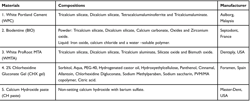
Table 1 shows the composition and manufacture of each material used in the present study. Experimental medica-ments used in this study are as follows:
● White Portland Cement (WPC) mixed with 2% chlor-hexidine gel (CHX)
● White ProRoot MTA (WMTA) mixed with 2% chlor-hexidine gel (CHX)
● Biodentine (BIO) mixed with 2% chlorhexidine gel (CHX)
● Calcium Hydroxide paste (CH paste)
The Physical Properties of the Intracanal
Medicaments
Setting Time
Disc-shaped samples were prepared to assess the setting time of three CSC (n=5) namely: White Portland Cement (WPC), White ProRoot MTA (WMTA), and Biodentine (BIO) after mixing with 2% chlorhexidine gel (CHX). A custom made Perspex mold (Zectron Sdn Bhd, Kuala Lumpur, Malaysia) measuring 2mm in height and 10mm in diameter was used to prepare the disc-shaped samples. Experimental medicaments (a mixture of CSC and 2% CHX) was prepared by mixing the cement with the gel in the ratio of 2:1 (0.3g powder: 0.15g gel). The ratio of 2:1 was selected based on our preliminary study to have a
Clinical, Cosmetic and Investigational Dentistry downloaded from https://www.dovepress.com/ by 118.70.13.36 on 24-Aug-2020
medicament with workable consistency. Air entrapments inside the mixture were avoided by using mechanical vibration for 10 seconds to evenly distribute the mixture. Samples were then kept inside an incubator at 37°C, 100% humidity for 24 hrs until the test began.
The setting time was analyzed using the Vicat apparatus with a modified Gilmore needle based on the American Society for Testing and Materials C266-08 specifications and according to the ISO Standard 6876/2001 method.30During the test, the samples were subjected to a load of 456g. Thefinal setting time for the specimens was taken as the time from mixing until the needle failed to make an observable indenta-tion on the material. Samples were assessed for the following time periods: 1, 2, 3, 4, 5, 6, 7, 14, 28, 60 and 84 days.
pH and Calcium Ion Release Analysis
The pH and calcium ion tests were performed by filling polyethylene tubes 10 mm long and 1 mm in diameter with each tested material: WPC; WMTA; BIO that mixed with 2% CHX gel in the ratio of 2:1 (0.3g powder: 0.15g gel); and calcium CH paste which was used as a control group in this test. The CH paste group was mixed and prepared according to the manufacturer’s recommendations. All samples were then stored in an incubator at 37°C, 100% humidity for 24 hrs until the analysis began.
For pH analysis of the tested materials (n=8), a digital pH meter (pH 700, Eutech, Thermo Fisher Scientific Inc, Waltham, USA) was used. The device was calibrated before each reading with buffer solutions at pH 4, 7, and 10. Fresh distilled water (5 mL) was added into the vial of each sample and allowed to equilibrate for 24 hrs in the
incubator before testing. Subsequently, the pH of the water surrounding the specimens was measured. After recording the readings, the sample was stored in the incubator until the next test. The replenishing of the distilled water into the vials was done one day prior to each of the following testing periods: 1, 7, 14, 21, and 28 days.
For calcium ion release of the tested materials (n=8), inductively coupled plasma-optical emission spectrometry (ICP-OES) (OPTIMA® 7000 DV, PerkinElmer, Inc., Waltham, USA) was used. The amount of 0.1g was mea-sured from the stored samples and used for acid digestion and analysis. The acids used for the digestions were 67% concentrated nitric acid (HNO3), 30% hydrogen peroxide
(H2O2) and 37% concentrated hydrochloric acid (HCl).
The digested sample was eventually analyzed for the fol-lowing time periods: 1, 7, 14, 21, and 28 days. The method used to analyze the calcium ion release was based on the American Public Health Association 3120 (APHA) stan-dard. The equipment was standardized using calibration standards and was se to blank before the readings.
Flowability and Film Thickness Analysis
Fresh samples of the same materials used in this study were mixed in the same manner to test theflowability and
film thickness and then subjected to analysis. The control group (CH paste) was mixed and prepared according to the manufacturer’s recommendations.
Flowability was analyzed based on ISO specification 6876/2001.30Two opticallyflat glass plates with a mass of 20 ±2 g were used. An amount of 0.5 mL for each sample (n=5) was placed on the center of a glass plate using a 1mL Table 1Different Types of CSC and CHX Gel with the Control Used in This Study
Materials Compositions Manufacturer
1. White Portland Cement (WPC)
Tricalcium silicate, Dicalcium silicate, Tetracalciumaluminoferrite and Tricalciumaluminate. Aalborg, Malaysia
2. Biodentine (BIO) Powder: Tricalcium silicate, Dicalcium silicate, Calcium carbonate, Oxides and Zirconium oxide.
Liquid: Iron oxide, calcium chloride and a water–soluble polymer.
Septodont, France
3. White ProRoot MTA (WMTA)
Tricalcium silicate, Dicalcium silicate, Tricalcium aluminate, Silicate oxide and Bismuth oxide. Dentsply, USA
4. 2% Chlorhexidine Gluconate Gel (CHX gel)
Sorbitol, Aqua, PEG-40, Hydrogenated castor oil, Hydroxyethylcellulose, Panthenol, Cinnamal, Allantoin, Chlorhexidine Digluconate, Sodium Methylparaben, Sodium saccharin, PVM/MA copolymer, Citric acid.
Foramen, Spain
5. Calcium Hydroxide paste (CH paste)
Non-setting calcium hydroxide with barium sulfate. Master-Dent,
USA
Clinical, Cosmetic and Investigational Dentistry downloaded from https://www.dovepress.com/ by 118.70.13.36 on 24-Aug-2020
syringe. At 180 ±5 seconds after mixing, another plate was placed carefully on top of the mixed sample, followed by a load of 100g that was carefully applied onto the top surface of the plate. Ten minutes after mixing, the load was removed, and the major and minor diameters of the compressed discs were measured using a digital caliper with a resolution of ±0.01 µm (Absolute Digimatic 500–197, Mitutoyo Corp, Kawasaki, Japan). The mean of three such determinations for each sample was taken as theflow of the material.
Film thickness (n=5) was analyzed using two 5 mm thick opticallyflat glass plates with a contact surface area of 200 ±25 mm2that were positioned together, and their combined thickness was taken with a digital caliper (Absolute Digimatic 500–197, Mitutoyo Corp, Kawasaki, Japan) to an accuracy of ±0.01 µm. Freshly mixed samples were placed between the glass plates after 2 mins after the start of mixing, and a load of 150N was applied on the top surface of the glass plate for 10 mins according to the method described in ISO Standard 6876/2001.30The difference in thickness of the two glass plates with and without the sample was recorded as the
film thickness of the material.
Removal the Intracanal Medicaments
from the Root Canal
Teeth Selection
Twenty single-rooted human teeth with straight canals were used for this study. The teeth were examined visually for no visible root fractures, caries or cracks. The teeth were radio-graphed to confirm that they had a single canal without any internal calcifications, irregularities, and other anomalies and a completely formed root apex. The root surfaces were scaled with an ultrasonic instrument to remove any calculus or soft tissue from the surface, washed with distilled water and then stored in normal saline for two days.
The crown of the teeth was removed to obtain a stan-dardized root length of 15 mm by using a diamond disc. The teeth were randomly divided into four experimental groups (n=5).
Root Canal Instrumentation
All root canals of the teeth were negotiated using K-files size 10 (Dentsply Maillefer) and a glide path was created using K
files size 20. The working length was confirmed at 1 mm shorter than the apical foramen. Mechanical instrumentation of the root canal was performed using ProTaper Next rotary
files (Dentsply Maillefer, Ballaigues, Switzerland) to size X3 by following the sequence prescribed by the manufacturer. The canals were irrigated with 5.2% NaOCl (Onemed,
Cairan Dental, Kuala Lumpur, Malaysia) and 17% EDTA paste (Vista Dental Products, South Street Racine, WI, USA) was used between eachfile during instrumentation. Patency of the root canals was verified by passing a Kfile size 10. Radiographs were taken for each tooth after completing the shaping and cleaning of the root canal.
Following irrigation with ultrasonicfile size 25 (Irrisafe File, Satelec Acteon, Mendota Heights, MN, USA), the root canals were dried with X3 paper points. Experimental med-icaments and the control were mixed using the same protocol used for evaluating the physical properties in this study. The root canals werefilled with the medicaments using lentulo spiral size 25 (Dentsply Maillefer, Ballaigues, Switzerland) at a speed of 600 rpm. Radiographs were taken to confirm the complete filling and distribution of the medicament in the root canals. The access cavities were sealed with cotton pellets and temporaryfilling material (3M ESPE, CavitTM W, Germany). Teeth were incubated for 7 days in an incu-bator at 37°C temperature, 100% humidity (Memmert, Schwabach Germany).
Intracanal Medicament Removal
The intracanal medicaments in each group were removed using a combination of manual k-file size 15 and ultra-sonicfile size 25, together with irrigation solutions 5.25% NaOCl and finally irrigated by 17% EDTA solution. Radiographs were taken to ensure the complete removal of the intracanal medicaments from the root canal.
Root Splitting
The roots were grooved on the buccal and lingual aspects using a water-cooled cutflex diamond disc (DFS, Germany). The grooves were prepared short of the apical foramen by 0.5 mm, along the long axis of the root without touching the canal walls. All roots were split in the buccolingual direction using a chisel and mallet into two halves. Both halves of the root were marked at 12 mm (coronal third), 8 mm (middle third) and 4 mm (apical third) from the root apex for SEM evaluation. The method for splitting the teeth was adapted from previous studies.31
SEM Evaluation
Each Sample was dehydrated to reduce image noise thus facilitating the assessment of samples and minimizing arti-facts. Therefore, all samples were placed in a desiccator for 24 hrs before SEM evaluation. Each sample was mounted on an aluminum stub. Then, images were viewed at 1000 times magnification and photographed at three different levels (apical, middle and coronal thirds, respectively).
Clinical, Cosmetic and Investigational Dentistry downloaded from https://www.dovepress.com/ by 118.70.13.36 on 24-Aug-2020
Scoring and Calibration
A scoring procedure was defined to assess the quantity of residue on the canal walls. Two experienced endodontists (who were unaware of the experimental groups to which each section belonged) conducted the analysis using the following 5-grade scale:31
Score 1: 80%–100% removal of medicament (total cleanliness)
Score 2: 60%–80% removal of medicament (great cleanliness)
Score 3: 40%–60% removal of medicament (partial cleanliness)
Score 4: 20%–40% removal of medicament (light cleanliness)
Score 5: 0%–20% removal of medicament (no cleanliness)
Statistical Analysis
All data was statistically analyzed by using SPSS 20.0. One-way analysis of variance (ANOVA) with Tukey’s post hoc test was used to analyze all the results for the tested materials for all the experiments. The significant level was set at p<0.05 or p<0.001.
For evaluation of intracanal medicament removal, scores for each sample were calculated, tabulated and analyzed with SPSS. Cohen’s kappa coefficient was used to measure inter-rater reliability and indicated substantial agreement between two examiners. The Kruskal–Wallis test was used to determine differences between all groups. The significant level was set at p<0.05.
Results
The Physical Properties of the Intracanal
Medicaments
Setting Time
The setting time of experimental medicaments was consis-tent between groups. The three experimental medicaments
that mixed with 2% CHX gel did not set until the end of the testing period (1, 2, 3, 4, 5, 6, 7, 14, 28, 60, and 84 days).
pH Analysis
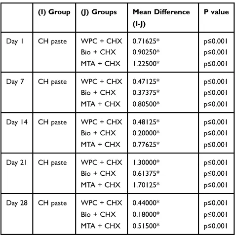
Throughout the experimental period, the results of pH mea-surements were alkaline. The mean pH values for all the experimental medicaments varied between 10.55–12.18 (Table 2). Analysis of variance (one-way ANOVA) showed a significant difference at all times of the study (p≤0.001) as shown inTable 3. However, BIO/CHX gel mixture showed the highest pH values among other experimental medica-ments used in this study except for day 1. CH paste was significantly higher than other experimental medicaments used in this study during the testing periods (p≤0.001).
Calcium Ion Release
Analysis of variance (one-way ANOVA) showed a signif-icant difference at all times of the study (p≤0.001) as
Table 2Mean and Standard Deviation of pH Value for the Experimental Medicaments and CH Paste Used in This Study Over 28 Days Period
Periods WPC + 2% CHX Gel BIO + 2% CHX Gel WMTA + 2% CHX Gel CH Paste P value
Day 1 11.84±(0.027) 11.66±(0.028) 11.34±(0.029) 12.56±(0.029) p≤0.001
Day 7 12.09±(0.025) 12.18±(0.028) 11.75±(0.035) 12.56±(0.027) p≤0.001
Day 14 11.74±(0.026) 12.02±(0.027) 11.45±(0.029) 12.22±(0.039) p≤0.001
Day 21 10.95±(0.029) 11.64±(0.028) 10.55±(0.037) 12.25±(0.029) p≤0.001
Day 28 11.71±(0.027) 11.97±(0.037) 11.56±(0.202) 12.16±(0.035) p≤0.001
Note:p value (p≤0.001) significant value.
Table 3Tukey HSD Post Hoc Test for pH Value
(I) Group (J) Groups Mean Difference (I-J)
P value
Day 1 CH paste WPC + CHX 0.71625* p≤0.001
Bio + CHX 0.90250* p≤0.001
MTA + CHX 1.22500* p≤0.001
Day 7 CH paste WPC + CHX 0.47125* p≤0.001
Bio + CHX 0.37375* p≤0.001
MTA + CHX 0.80500* p≤0.001
Day 14 CH paste WPC + CHX 0.48125* p≤0.001
Bio + CHX 0.20000* p≤0.001
MTA + CHX 0.77625* p≤0.001
Day 21 CH paste WPC + CHX 1.30000* p≤0.001
Bio + CHX 0.61375* p≤0.001
MTA + CHX 1.70125* p≤0.001
Day 28 CH paste WPC + CHX 0.44000* p≤0.001
Bio + CHX 0.18000* p≤0.001
MTA + CHX 0.51500* p≤0.001
Note:*p value (p≤0.001) significant value.
Clinical, Cosmetic and Investigational Dentistry downloaded from https://www.dovepress.com/ by 118.70.13.36 on 24-Aug-2020
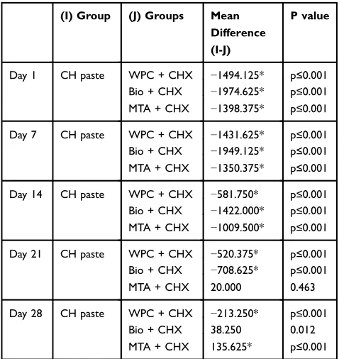
shown in Table 4. Tukey HSD Post hoc test (Table 5) showed that all the experimental medicaments used in this study released significantly higher calcium ions (mean values, mg/l) compared to CH paste until 14 days (p≤0.001). Among the experimental medicaments, BIO/ CHX mixture showed the highest calcium ions release at day 21 compared to CH paste (p≤0.001). However, WPC/ CHX mixture showed significantly higher calcium ions release compared to CH paste at day 28 (p≤0.001).
Flowability
Analysis of variance (one-way ANOVA) showed a signif-icant difference between the groups p≤0.05, as shown in
Table 6. Tukey HSD Post hoc testing showed a higher significant difference in theflowability between the experi-mental medicaments and CH paste (Table 7). All the
experimental medicaments except CH paste showed the
flow to be greater than 17 mm, which complied with the ISO standards.30,32 It was demonstrated that the MTA/ CHX mixture had significantly higher flowability than others.
Film Thickness
The results of the film thickness are shown inTable 6. In the present study, no statistically significant difference was presented in film thickness for the experimental medica-ments compared to CH paste (p=0.844). Film thickness for all the experimental medicaments was 0.10 µm, while the Table 4Mean and Standard Deviation of Calcium Ion Release (Mg/l) for the Experimental Medicaments and CH Paste Used in This Study Over 28 Days Period
Periods WPC + 2% CHX gel (mg/l) BIO + 2% CHX Gel (mg/l) WMTA + 2% CHX Gel (mg/l) CH Paste (mg/l) P value
Day 1 1979.75±(06.32) 2460.25±(33.96) 1884.00±(52.48) 485.63±(03.15) p≤0.001
Day 7 1926.25±(27.11) 2443.75±(35.38) 1845.00±(64.18) 494.63±(02.61) p≤0.001
Day 14 1058.38±(25.90) 1898.62±(43.68) 1486.12±(33.430) 476.62±(02.82) p≤0.001
Day 21 1050.00±(26.75) 1238.25±(07.166) 509.62±(46.356) 529.62±(03.37) p≤0.001
Day 28 745.88±(28.68) 494.38±(21.567) 397.00±(28.005) 532.62±(03.37) p≤0.001
Note:p value (p≤0.001) significant value.
Table 5Tukey HSD Post Hoc Test for Calcium Ion Release
(I) Group (J) Groups Mean Difference (I-J)
P value
Day 1 CH paste WPC + CHX −1494.125* p≤0.001
Bio + CHX −1974.625* p≤0.001
MTA + CHX −1398.375* p≤0.001
Day 7 CH paste WPC + CHX −1431.625* p≤0.001
Bio + CHX −1949.125* p≤0.001
MTA + CHX −1350.375* p≤0.001
Day 14 CH paste WPC + CHX −581.750* p≤0.001
Bio + CHX −1422.000* p≤0.001
MTA + CHX −1009.500* p≤0.001
Day 21 CH paste WPC + CHX −520.375* p≤0.001
Bio + CHX −708.625* p≤0.001
MTA + CHX 20.000 0.463
Day 28 CH paste WPC + CHX −213.250* p≤0.001
Bio + CHX 38.250 0.012
MTA + CHX 135.625* p≤0.001
Note:*p value (p≤0.001) significant value.
Table 6Mean and Standard Deviation of Flowability (Mm) and Film Thickness (μm) for the Experimental Medicaments and CH Paste Used in This Study
Materials Flowability (mm)
P value-One-Way ANOVA test
Film Thickness (μm)
P value-One-Way ANOVA test
WPC + 2%
CHX gel
18.27±(1.79) p≤0.05 0.10±(0.016) 0.844
BIO + 2%
CHX gel
17.32±(1.80) 0.10±(0.031)
MTA + 2%
CHX gel
22.80±(1.00) 0.10±(0.019)
CH paste 14.15±(0.52) 0.11±(0.016)
Note:p value (p<0.05) significant value.
Table 7Tukey HSD Post Hoc Test for Flowability
(I) Groups
(J) Groups Mean Difference (I-J)
P value
Flowability CH paste WPC + CHX −4.11800* 0.001
Bio + CHX −3.17000* 0.011
MTA + CHX −8.65400* p≤0.05
Note:*The mean difference is significant at the 0.05 level.
Clinical, Cosmetic and Investigational Dentistry downloaded from https://www.dovepress.com/ by 118.70.13.36 on 24-Aug-2020
film thickness of CH paste was 0.11 µm, which is in agreement with the ISO standards.30,32
Removal the Intracanal Medicaments
from the Root Canal
The conventional radiograph showed that the intercanal medicaments were successfully removed from the root canal (Figure 1).
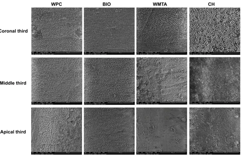
SEM for all the samples tested in this study showed a small amount of debris left in the root canal walls. Residue of intracanal medicaments was found in all experimental groups regardless of the material used in this study. Representative SEM micrographs are shown in Figure 2.
The Cohen’s kappa coefficient of inter-rater reliabil-ity between the two examiners was 0.78 and there was high reproducibility with a significance level of P=0.001, while the intra-rater reliability scores were 94% and 96%.
The Kruskal–Wallis test showed that there was no significant difference between the four groups in terms of intracanal medicaments removal (P=0.087), as shown in
Table 8 and Figure 2. Furthermore, the results revealed that there was no significant difference between the cor-onal third compared to the middle and apical thirds in all samples respectively. However, the mean of WPC (18.17) was less than CH (21.92), BIO (26.17), and WMTA (31.75). Thus, the removal of WPC from the root canal walls was superior compared to other intracanal medica-ments used in this study.
Discussion
Mineral Trioxide Aggregate has been widely used in endo-dontics due to its good physicochemical properties and excel-lent biocompatibility.33Although MTA is preferred in many dental treatments due to its numerous advantages,34it has not been used as root canal medicament. This is because the removal of MTA from the root canal after being fully set and cured is extremely difficult.33 This in vitro study explored the possibility of using calcium silicate-based cement (CSC) as an intracanal medicament by retarding its setting time. The experimental medicaments were a mixture of CSCs and 2% CHX. Furthermore, the physical properties such as setting time, pH, calcium ion release of the water surrounding the specimens, flowability and film thickness were analyzed. The application of intracanal medicament is a routine procedure throughout root canal treatment. Breaking the coronal seal could happen because the tempor-ary restorations may dislodge or crack during treatment that may lead to contamination of the root canal. Therefore, using intracanal medicament is necessary to protect the root canal system during root canal treatment.35,36
Several additives have been combined with MTA to enhance the working properties of the cement. It has been demonstrated that NaOCl gel may reduce the setting time and improve the handling characteristics of the mixture. It may also provide antimicrobial action. Additionally, chlorhexidine glu-conate gel may retard and impair the setting time of the cement.29Researchers were using CHX in different concentra-tions and forms such as liquid, gel and powder with MTA to improve its antimicrobial properties.27,29,37 In a study
B
A
Figure 1(A) Intracanal medicament placed into the canal after instrumentation and (B) after removal of intracanal medicament.
Clinical, Cosmetic and Investigational Dentistry downloaded from https://www.dovepress.com/ by 118.70.13.36 on 24-Aug-2020
performed by Mittag et al27 the antimicrobial properties of MTA were increased when mixed with CHX in a dose-depen-dent manner, whereas MTA with water had little antibacterial effect. Additionally, in an ex vivo study by Ramezani et al38 mixing MTA with 0.12% CHX reduced the amount of bacterial leakage. The current study is the first which investigates the potential use of CSC mixing with 2% CHX gel as an intracanal medicament that could be used during inter appointment of root canal treatment. Delayed setting times is considered a problem and may limit the use of mineral trioxide aggregate (MTA) in endodontic procedures.29However, in this study, this limitation can be transformed into an advantage with the associated anti-bacterial efficacy of MTA, where the inhibition of the setting
time will add a new application for CSC and allow this material to be used as an intracanal medicament. Recently, MTA has become a common substitute for Ca(OH)2, and the outcomes of
the available clinical studies comparing MTA with Ca(OH)2
indicate its superiority.39
The setting time of MTA has been investigated exten-sively in previous studies. It has been reported that the
final setting time of MTA is approximately 2 hrs and 45 mins.2,40 Another study reported 2 hrs and 55 mins for Grey MTA and 2 hrs and 20 mins for White MTA.41In this study, the setting time of the experimental medicament (CSC/CHX mixture) was analyzed using a Gilmore needle and the procedure was based on the previous study which had determined the setting time of CSC.42 This study analyzed the setting time of experimental medicament for a period of 84 days. The results showed that 2% CHX gel prohibited the setting of all three cements until the end of the experimental period. Thisfinding is in accordance with the study done by Kogan et al29 however, they evaluated the setting time only for 4 hrs. Among the different experi-mental medicaments used in the study, Biodentine has faster setting time compared to others.43 However, the addition of 2% CHX gel to CSC altered the ideal setting
WPC BIO WMTA CH
Coronal third
Middle third
Apical third
Figure 2High power SEM (1000×) micrographs of a longitudinal section of the root canal retrieved from intracanal medicaments (WPC, BIO, WMTA and CH paste).
Table 8Difference in Intercanal Medicaments Removal Between the Different Groups Used in This Study Using SEM
Materials N Mean df Siga
Scores Measure WPC + CHX 12 18.17 3 0.087
BIO + CHX 12 26.17
WMTA + CHX 12 31.75
CH paste 12 21.92
Total 48
Note:aNKruskal–Wallis Test.
Clinical, Cosmetic and Investigational Dentistry downloaded from https://www.dovepress.com/ by 118.70.13.36 on 24-Aug-2020
time of the cement. Further research is needed to investi-gate the exact chemical reaction occurring in the experi-mental medicament.
Calcium ion release and an alkaline pH would stimulate tissue repair and provide an antimicrobial environment. It was also suggested that the material should have a pH of more than 7.0, along with calcium ion release to stimulate mineralization.44In this study, the release of calcium ions was assessed to check that CHX gel use does not interfere with the dissociation of calcium ions from experimental medicaments and also that it does not decrease pH values. Misra et al35studied the release of calcium ions and the pH of calcium hydroxide paste when mixed with different vehicles, and found that the calcium hydroxide with glycerine showed a highest calcium ion release followed by calcium hydroxide with chlorhexidine. The methodology used in this study to analyze the calcium ion release was similar to work done in the previous studies.45,46 All the materials analyzed presented the suitable release of calcium in the ICP-OES analysis. The experimental medica-ments used in this study demonstrated a reduction in calcium ions over time and presented higher values at the initial period. The three experimental medicaments mixed with 2% CHX gel had an overall reduction in calcium ion release after 21 days, except for the WMTA/CHX gel mixture which showed lower calcium ion release than CH paste at 21 days, while CH paste was likely to be stable during all periods. Thisfinding is similar to the results of other studies using the same analyzing equip-ment without using chlorhexidine.47,48Among the group of experimental medicaments, BIO/CHX gel mixture showed the highest release of calcium ions over three weeks. The results of this study would suggest that the addition of 2% CHX gel to CSC has no inhibitory action on calcium ion release, thus corresponding with the results of previous studies.37 The released calcium ions maximize alkalinizing activity and con-sequently increase the bactericidal effect of MTA.49It is inter-esting to note that the effective release of calcium ions for three weeks by all experimental medicaments tested in this study would be sufficient to allow their use as an intracanal medica-ment. This is based on the study by Sjögren et al50that demon-strated a one-week application of calcium hydroxide medicament is sufficient to reduce the bacterial load in the canal which was evident by negative culture.
In this study, CH paste showed the highest pH values compared to the experimental medicaments. Its pH was consistent until four weeks within a range of about 12.1– 12.5. This is similar to the values mentioned in the literature.51Among the different experimental medicaments used in this study, BIO/CHX gel mixture showed the
highest alkalizing activity until the end of the experiment. The current study demonstrated that all experimental med-icaments had pH values of more than 10.5 until the end of the study. On the other hand, the WPC/CHX gel mixture and BIO/CHX gel mixture had a pH value of more than 11.5 until two weeks. It can be concluded that the pH values of these experimental medicaments may not be affected by the addition of CHX gel and could have bacteriostatic and bactericidal properties against Enterococcus faecalis. It is theorized that the high pH is responsible for the material’s antimicrobial action and biological activity. This high pH is achieved because of the constant calcium release from MTA and Ca(OH)2formation.49
Ideal intracanal medicaments should be easily applic-able within the canal for proper contact within the canal walls and be able to be retrieved from the canal with minimal effort.52Therefore,flowability andfilm thickness were assessed to achieve these objectives. This study showed that the flowability of the experimental medica-ments was statistically significant compared to CH paste. The WMTA/CHX gel mixture had better flowability among the experimental medicaments used in this study. This could be due to differences in the particle size and shape of CSC. A few investigators have reported that MTA Angelus has finer and more homogeneous particle size compared to WPC and other types of MTA.53,54 One study reported that some particles of MTA have a diameter of less than 1.5 micrometers, which is smaller than the diameter of some dentinal tubules.28 These studies were supporting thefindings of this current study; however, the
film thickness of the CH paste was similar to the experi-mental medicaments.
It has been emphasized that intracanal medication should be removed completely from the root canal because its presence on the dentinal wall can interfere negatively with the root canal obturation.55 Thus, complete removal of the intracanal medication (calcium hydroxide) from the root canal before root canal obturation is necessary.56The effect of ultrasonic agitation of the irrigants has been evaluated and presented its efficacy in improving cleanli-ness and removing some calcium hydroxide from straight root canals.57 However, complete removal of the calcium hydroxide from the root canal system could not be achieved.55–57
Ultrasonic and rotary instrumentation without irrigates have been shown to be ineffective in the removal of set MTA from the root canal.58 Similarly, in our study, the result of SEM showed that complete removal of intracanal
Clinical, Cosmetic and Investigational Dentistry downloaded from https://www.dovepress.com/ by 118.70.13.36 on 24-Aug-2020
medications from the root canal walls could not be achieved. This could be attributed to the use of chlorhex-idine which has the ability to decrease the surface micro-hardness of a partially set MTA,59 and it is water-soluble and can be removed from the root canal by irrigation with distilled water.60
Saghiri et al61suggested that by decreasing the micro-hardness of MTA, the MTA can be removed more easily than hard and set cement. He demonstrated that a 37% hydrochlo-ric acid (HCl) was an effective solution for the removal of MTA from the canal wall due to its ability to reduce micro-hardness of MTA. Therefore, the removal of CSC/CHX mixture from the root canal was easier than MTA because of the delay in the setting reaction of the cement. Regarding the calcium hydroxide paste, the vehicle used in its prepara-tion plays an important role where the oil-based calcium hydroxide is more difficult to remove than powder form calcium hydroxide mixed with distilled water.62
Conclusions
The addition of 2% CHX gel to CSC retarded or inhibited the setting reaction and significantly improved the calcium ion release and flowability of the experimental medica-ments. The pH and film thickness of the experimental medicament were in acceptable range compared to CH paste. Among different experimental medicaments, BIO/ CHX gel mixture showed superior physical properties compared to the others. Removal of the intracanal medica-ments from the root canal was successfully achieved in all groups tested in this study. Based on the initial finding, these experimental medicaments have the potential to be used as an enhanced root canal medicament. Further research on this formulation (particularly biocompatibility and antibacterial activity) is needed.
Acknowledgment
This study wasfinancially supported by the UMRG Grant (RG530-13HTM) and the PPP Grant (PG059-2014A) at the University of Malaya, Malaysia.
Author Contributions
All authors contributed to data analysis, drafting or revising the article, gavefinal approval of the version to be published, and agree to be accountable for all aspects of the work.
Disclosure
Financial support: A postgraduate research grant (PPP) from the Faculty of Dentistry, University of Malay (grant no. PS
169/2010B) was received by Okba Mahmood, Walid Ali Al-Meeri and Mohideen Salihu Farook. The authors report a patent Calcium Silicate-Based Cement (CSC) as Root Canal Medicament pending to PI2014703468. The authors report no other conflicts of interest in this work.
References
1. Torabinejad M, Hong CU, Lee SJ, Monsef M, Ford T. Investigation of mineral trioxide aggregate for root-endfilling in dogs. J Endod.
1995;21(12):603–608. doi:10.1016/S0099-2399(06)81112-X 2. Torabinejad M, Hong C, McDonald F, Ford T. Physical and chemical
properties of a new root-end filling material. J Endod. 1995;21 (7):349–353. doi:10.1016/S0099-2399(06)80967-2
3. Dawood AE, Parashos P, Wong RH, Reynolds EC, Manton DJ. Calcium silicate-based cements: composition, properties, and clinical applications.J Investig Clin Dent.2015.
4. Torabinejad M, Parirokh M, Dummer PMH. Mineral trioxide aggregate and other bioactive endodontic cements: an updated overview -part II: other clinical applications and complications. Int Endod J.
2018;51(3):284–317.
5. Berkathullah M, Farook MS, Mahmoud O. The effectiveness of remineralizing agents on dentinal permeability. Biomed Res Int.
2018;2018:4072815. doi:10.1155/2018/4072815
6. Jafari F, Jafari S. Composition and physicochemical properties of calcium silicate based sealers: a review article. J Clin Exp Dent.
2017;9(10):e1249–e1255. doi:10.4317/jced.54103
7. Min K-S, Park H-J, Lee S-K, et al. Effect of mineral trioxide aggre-gate on dentin bridge formation and expression of dentin sialoprotein and heme oxygenase-1 in human dental pulp. J Endod 2008;34 (6):666–670.
8. O’sullivan SM, Hartwell G. Obturation of a retained primary mandibular second molar using mineral trioxide aggregate: a case report.J Endod.
2001;27(11):703–705. doi:10.1097/00004770-200111000-00013 9. Chen Y-W, Hsu -T-T, Wang K, Shie M-Y. Preparation of the fast
setting and degrading Ca–Si–Mg cement with both odontogenesis and angiogenesis differentiation of human periodontal ligament cells.Mater Sci Eng.2016;60:374–383.
10. Shie M-Y, Chiang W-H, Chen I-WP, Liu W-Y, Chen Y-WJMS, C E. Synergistic acceleration in the osteogenic and angiogenic differentia-tion of human mesenchymal stem cells by calcium silicate–graphene composites. Mater Sci Eng C. 2017;73:726–735. doi:10.1016/j. msec.2016.12.071
11. Dall AQ, Jouhar R, Khoso NA. Comparison of inter-appointment pain between ledermix and no intracanal medicament in acute apical periodontitis.JLUMHS.2011;10(03):106.
12. Morrier JJ, Benay G, Hartmann C, Barsotti O. Antimicrobial activity of Ca(OH)2 dental cements: an in vitro study. J Endod. 2003;29 (1):51–54. doi:10.1097/00004770-200301000-00014
13. Torabinejad M, Handysides R, Khademi AA, Bakland LK. Clinical implications of the smear layer in endodontics: a review.Oral Surg Oral Med Oral Pathol Oral Radiol Endod. 2002;94(6):658–666. doi:10.1067/moe.2002.128962
14. Chong B, Ford TP. The role of intracanal medication in root canal treatment. Int Endod J. 1992;25(2):97–106. doi:10.1111/j.1365-2591.1992.tb00743.x
15. Barbosa CA, Goncalves RB, Siqueira JF, De Uzeda M. Evaluation of the antibacterial activities of calcium hydroxide, chlorhexidine, and camphorated paramonochlorophenol as intracanal medicament. A clinical and laboratory study. J Endod. 1997;23(5):297–300. doi:10.1016/S0099-2399(97)80409-8
16. Gomes B, Souza S, Ferraz C, et al. Effectiveness of 2% chlorhexidine gel and calcium hydroxide against Enterococcus faecalis in bovine root dentine in vitro.Int Endod J.2003;36(4):267–275.
Clinical, Cosmetic and Investigational Dentistry downloaded from https://www.dovepress.com/ by 118.70.13.36 on 24-Aug-2020
17. Zehnder M, Söderling E, Salonen J, Waltimo T. Preliminary evaluation of bioactive glass S53P4 as an endodontic medication in vitro.J Endod.
2004;30(4):220–224. doi:10.1097/00004770-200404000-00009 18. Byström A, Claesson R, Sundqvist G. The antibacterial effect of
camphorated paramonochlorophenol, camphorated phenol and cal-cium hydroxide in the treatment of infected root canals. Dental Traumatol.1985;1(5):170–175. doi:10.1111/edt.1985.1.issue-5 19. Evans M, Davies J, Sundqvist G, Figdor D. Mechanisms involved in the
resistance of Enterococcus faecalis to calcium hydroxide.Int Endod J.
2002;35(3):221–228. doi:10.1046/j.1365-2591.2002.00504.x 20. Lohbauer U, Gambarini G, Ebert J, Dasch W, Petschelt A. Calcium
release and pH-characteristics of calcium hydroxide plus points.Int Endod J.2005;38(10):683–689. doi:10.1111/j.1365-2591.2005.00972.x 21. Shetty S, Manjunath M, Tejaswi S. An in-vitro evaluation of the pH change through root dentin using different calcium hydroxide pre-parations as an intracanal medicament.J Clin Diagn Res.2014;8(10): ZC13. doi:10.7860/JCDR/2014/6788.3956
22. Tandan M, Hegde MN, Hegde P. Effect of four different intracanal medicaments on the apical seal of the root canal system: a dye extraction study. Indian J Dental Res. 2014;25(5):607. doi:10.4103/0970-9290.147104
23. Basrani B. Chlorhexidine gluconate.Aust Endod J.2005;31(2):48– 52. doi:10.1111/aej.2005.31.issue-2
24. Delgado RJ, Gasparoto TH, Sipert CR, et al. Antimicrobial effects of calcium hydroxide and chlorhexidine on Enterococcus faecalis. J Endod. 2010;36(8):1389–1393. doi:10.1016/j.joen. 2010.04.013
25. Gomes BP, Vianna ME, Zaia AA, Almeida JFA, Souza-Filho FJ, Ferraz CC. Chlorhexidine in endodontics. Braz Dent J. 2013;24 (2):89–102. doi:10.1590/0103-6440201302188
26. Holt DM, Watts JD, Beeson TJ, Kirkpatrick TC, Rutledge RE. The anti-microbial effect against enterococcus faecalis and the compres-sive strength of two types of mineral trioxide aggregate mixed with sterile water or 2% chlorhexidine liquid.J Endod.2007;33(7):844– 847. doi:10.1016/j.joen.2007.04.006
27. Mittag S-G, Eißner C, Zabel L, Wrbas K-T, Kielbassa AM. The influence of Chlorhexidine on the antibacterial effects of MTA.
Quintessence Int (Berl).2012;43(10):901–6.
28. Camilleri J. Hydration mechanisms of mineral trioxide aggregate.Int Endod J.2007;40(6):462–470. doi:10.1111/iej.2007.40.issue-6 29. Kogan P, He J, Glickman GN, Watanabe I. The effects of various
additives on setting properties of MTA.J Endod.2006;32(6):569– 572. doi:10.1016/j.joen.2005.08.006
30. Standardization, I.O.f. and I.O.f. Standardization.ISO 6876: Dental Root Canal Sealing Materials. ISO Geneva;2001.
31. Alturaiki S, Lamphon H, Edrees H, Ahlquist M. Efficacy of 3 different irrigation systems on removal of calcium hydroxide from the root canal: a scanning electron microscopic study. J Endod.
2015;41(1):97–101. doi:10.1016/j.joen.2014.07.033
32. ISO.International Standards Organization in Dentistry–Root Canal Sealing Materials. Switzerland: ISO;2012.
33. Torabinejad M, Parirokh M. Mineral trioxide aggregate: a compre-hensive literature review—part II: leakage and biocompatibility investigations. J Endod. 2010;36(2):190–202. doi:10.1016/j.joen. 2009.09.010
34. Butt N, Talwar S, Chaudhry S, Nawal RR, Yadav S, Bali A. Comparison of physical and mechanical properties of mineral trioxide aggregate and biodentine. Indian J Dent Res. 2014;25 (6):692.
35. Misra P, Bains R, Loomba K, et al. Measurement of pH and calcium ions release from different calcium hydroxide pastes at different intervals of time: atomic spectrophotometric analysis.J Oral Biol Craniofacial Res.2017;7(1):36–41. doi:10.1016/j.jobcr.2016.04.001 36. Siqueira J Jr, Lopes HP. Mechanisms of antimicrobial activity of
calcium hydroxide: a critical review.Int Endod J.1999;32(5):361– 369. doi:10.1046/j.1365-2591.1999.00275.x
37. Jacinto RC, Linhares-Farina G, Sposito ODS, Zanchi CH, Cenci MS. Influence of 2% chlorhexidine on pH, calcium release and setting time of a resinous MTA-based root-endfilling material.Braz Oral Res.2015;29(1):1–6. doi:10.1590/1807-3107BOR-2015.vol29.0036 38. Ramezani G, Savadkouhi ST, Sayahpour S, Dentistry C. The effect of
chlorhexidine mixed with mineral trioxide aggregate on bacterial leakage of apical plug in simulated immature teeth using human fresh saliva.J Int Soc Prev Community Dent.2017;7(5):247. 39. Bakland LK, Andreasen J. Will mineral trioxide aggregate replace
calcium hydroxide in treating pulpal and periodontal healing compli-cations subsequent to dental trauma? A review.Dental Traumatol.
2012;28(1):25–32. doi:10.1111/j.1600-9657.2011.01049.x
40. Uyanik O, Nagas E, Kucukkaya Eren S, Cehreli ZC, Vallittu PK, Lassila L. Effect of phytic acid on the setting times and tensile strengths of calcium silicate-based cements.Aust Endod J.2019;45 (2):241–245. doi:10.1111/aej.v45.2
41. Islam I, Chng HK, Yap A. Comparison of the physical and mechan-ical properties of MTA and Portland cement. J Endod. 2006;32 (3):193–197. doi:10.1016/j.joen.2005.10.043
42. Bortoluzzi EA, Broon NJ, Bramante CM, Felippe WT, Tanomaru Filho M, Esberard RM. The influence of calcium chloride on the setting time, solubility, disintegration, and pH of mineral trioxide aggregate and white Portland cement with a radiopacifier.J Endod.
2009;35(4):550–554. doi:10.1016/j.joen.2008.12.018
43. Grech L, Mallia B, Camilleri J. Investigation of the physical properties of tricalcium silicate cement-based root-endfilling materials. Dental Materials.2013;29(2):e20–e28. doi:10.1016/j.dental.2012.11.007 44. Estrela C, Sydney GB, Bammann LL, Felippe O Jr. Mechanism of
action of calcium and hydroxyl ions of calcium hydroxide on tissue and bacteria.Braz Dent J.1995;6(2):85–90.
45. Carvalho CN, Freire LG, Carvalho APLD, Duarte MAH, Bauer J, Gavini G. Ions release and pH of calcium hydroxide-, chlorhexidine-and bioactive glass-based endodontic medicaments. Braz Dent J.
2016;27(3):325–331. doi:10.1590/0103-6440201600602
46. Natale L, Rodrigues M, Xavier T, Simões A, Souza D, Braga R. Ion release and mechanical properties of calcium silicate and calcium hydroxide materials used for pulp capping. Int Endod J. 2015;48 (1):89–94. doi:10.1111/iej.2014.48.issue-1
47. Camilleri J. Characterization of hydration products of mineral triox-ide aggregate.Int Endod J.2008;41(5):408–417. doi:10.1111/j.1365-2591.2007.01370.x
48. Duarte MAH, de Oliveira Demarchi ACC, Yamashita JC, Kuga MC, de Campos Fraga S. pH and calcium ion release of 2 root-endfilling materials. Oral Surgery Oral Med Oral Pathol Oral Radiol Endodontol.2003;95(3):345–347. doi:10.1067/moe.2003.12 49. Duarte MAH, de Oliveira Demarchi ACC, Yamashita JC, Kuga MC,
de Campos Fraga SJOS. Oral medicine, oral pathology, oral radiol-ogy, and endodontolradiol-ogy,pH and calcium ion release of 2 root-end filling materials.Oral Surgery Oral Med Oral Pathol Oral Radiol Endod.2003;95(3):345–347. doi:10.1067/moe.2003.12.
50. Sjögren U, Figdor D, Spångberg L, Sundqvist G. The antimicrobial effect of calcium hydroxide as a short-term intracanal dressing.Int Endod J.1991;24(3):119–125. doi:10.1111/iej.1991.24.issue-3 51. Farhad A, Mohammadi Z. Calcium hydroxide: a review.Int Dent J.
2005;55(5):293–301. doi:10.1111/idj.2005.55.issue-5
52. Basrani B, Ghanem A, Tjäderhane L. Physical and chemical proper-ties of chlorhexidine and calcium hydroxide-containing medica-tions. J Endod. 2004;30(6):413–417. doi:10.1097/00004770-200406000-00009
53. Camilleri J, Montesin FE, Brady K, Sweeney R, Curtis RV, Ford TRP. The constitution of mineral trioxide aggregate. Dental Materials.2005;21(4):297–303. doi:10.1016/j.dental.2004.05.010 54. Komabayashi T, Spångberg LS. Comparative analysis of the particle
size and shape of commercially available mineral trioxide aggregates and Portland cement: a study with aflow particle image analyzer.J Endod.2008;34(1):94–98. doi:10.1016/j.joen.2007.10.013
Clinical, Cosmetic and Investigational Dentistry downloaded from https://www.dovepress.com/ by 118.70.13.36 on 24-Aug-2020
55. Vilela DD, Neto MM, Villela AM, Pithon M. Evaluation of inter-ference of calcium hydroxide-based intracanal medication infilling root canal systems.J Contemp Dent Pract.2011;12(5):368–371. 56. Jasrotia A, Farooq R, Bhagat RK, Bhagat K, Sharma N, Habib S.
Removal efficacy of various calcium hydroxide/chlorhexidine medi-caments from the root canal with the aid of biological microscope.
Ind J Dental Sci.2019;11(1):1.
57. Pabel A-K, Hülsmann MJO. Comparison of different techniques for removal of calcium hydroxide from straight root canals: an in vitro study. Odontology. 2017;105(4):453–459. doi:10.1007/s10266-017-0293-6
58. Boutsioukis C, Noula G, Lambrianidis T. Ex vivo study of the efficiency of two techniques for the removal of mineral trioxide aggregate used as a root canal filling material. J Endod. 2008;34 (10):1239–1242. doi:10.1016/j.joen.2008.07.018
59. Butt N, Talwar S. In-vitro evaluation of various solvents for retrieval of mineral trioxide aggregate and their effect on microhardness of dentin.J Conserv Dent.2013;16(3):199.
60. Ferraz CCR, de Almeida Gomes BPF, Zaia AA, Teixeira FB, de Souza-Filho FJ. In vitro assessment of the antimicrobial action and the mechanical ability of chlorhexidine gel as an endodontic irrigant.
J Endod. 2001;27(7):452–455. doi:10.1097/00004770-200107000-00004
61. Saghiri MA, Garcia-Godoy F, Gutmann JL, et al. Removal of white mineral trioxide aggregate cement: a promising approach.Biomed Res Int.2013;2013:1–7. doi:10.1155/2013/469164
62. Nandini S, Velmurugan N, Kandaswamy D. Removal efficiency of calcium hydroxide intracanal medicament with two calcium chela-tors: volumetric analysis using spiral CT, an in vitro study.J Endod.
2006;32(11):1097–1101. doi:10.1016/j.joen.2006.06.005
Clinical, Cosmetic and Investigational Dentistry
Dove
press
Publish your work in this journal
Clinical, Cosmetic and Investigational Dentistry is an international, peer-reviewed, open access, online journal focusing on the latest clinical and experimental research in dentistry with specific empha-sis on cosmetic interventions. Innovative developments in dental materials, techniques and devices that improve outcomes and patient
satisfaction and preference will be highlighted. The manuscript management system is completely online and includes a very quick and fair peer-review system, which is all easy to use. Visit http://www.dovepress.com/testimonials.php to read real quotes from published authors.
Submit your manuscript here:https://www.dovepress.com/clinical-cosmetic-and-investigational-dentistry-journal
Clinical, Cosmetic and Investigational Dentistry downloaded from https://www.dovepress.com/ by 118.70.13.36 on 24-Aug-2020