J. T. PATTERSON AND H. J. MULLER Unaversity of Tweac, Auslin, T e w
Received November 19,1929
TABLE OF CONTENTS
PAGE 495
1. The physical background of the problem. ...
11. Evidence concerning the duality of the X-ray effect on chro
111. A partial separation of the Werent genetic effects of X-rays.. . .
IV. The production of multiple allelomorphs.. ... ... 507
V. An induced mutation visibly d8erent from a known lass of VI. First attempts to induce mutations in both of two oppqsite VII. Reverse mutations a t the locus of scute. . . VIII. A reverse mutation a t the locus of forked.. ... IX. Proof of the induction of reverse mutations of forked b, Reverse muations from treated larval stages c. Reverse mutations induced in spermatozoa.. . . d. Tests of the loci of the reverse mutations. . . . e. Radiation as the cause of the reverse mutatio 532 540 ... 551 . . . 554
. . . 557
559 X. Mutations from induced non forked to forked. . . XI. Viability of the induced reverse mutations. . . XII. Reverse mutations in eye color, . . . XIII. The two scute mutations. . . XIV. Discussion of related work, and general considerations. . . 567
SUMMARY . . . 571
APPENDIX A . . . 574
LITERATWRECITED... . . 575
1. THE PHYSICAL BACKGROUND OF THE PROBLEM
Do the X-ray mutations consist merely of losses and rearrangements of portions of the chromosomes? Or do they also include “progressive” changes-the kind of steps of which real evolutionary advancement has becn composed? This is one of the most vital immediate questions confronting X-ray genetics, for upon the answer to i t may depend in turn the answers to fundamental questions concerning the mechanism o
natural evolution, t h e nature of thegene, and the means of bringing abo t artificial evolution. The issue, therefore, must be faced squarely.
l During the course of the radiation work in this laboratory both authors became interested
in the problem in this paper and accumulated various data relative to it. It was then decided
to present a combined report of all the data, and it was arranged that $he senior author (PATTEB-
SON) shonld conduct the major experiments described, those in sections I X to X I I I inclusive. Except where otherwise specified, the other experiments described, and the discussion, are by the junior author.
GBNETICS 15: 495 N 1930
496 J. T. PATTERSON AND H. J. MULLER
There are two main facts which tend to give plausibility to the sus- picion that‘ X-rays may not be constructive in their genetic action. One is the general fact that they do often cause “destructive” changes (espe- cially noticeable when the treatment is heavy or long continued) both in the case of organisms and of non-living materials. The other is the more specific fact, discovered coincidently with the gene-mutation effect, that they cause breakages of chromosomes, sometimes accompanied by losses of the fragments and sometimes by their reattachment a t a different point in the chromatin from before (MULLER 1927, WEINSTEIN 1928, MULLER and PAINTER 1929, PAINTER and MULLER 1929).
A little further inquiry into the manner in which X-rays exert their chemical effects serves to dispel the idea that the changes induced must always be of a destructive nature-no matter how we choose to define the w0rd“destructive.” At the same time it becomes evident why many of the changes so induced are judged to be destructive. The primary effect of the X-rays, as also of beta, gamma, and cosmic rays, on the structure of any material is the expulsion of an electron (sometimes more than one) from some of the atoms, either directly, by the absorption of a quantum of the radiant energy, or, in the case of far more of the atoms (some hundreds to one like the former), indirectly, through the electron’s being forced out by another electron that was previously expelled through the (direct or indirect) action of the radiation. In case an electron is held by two atoms in common it may become detached from one of them, thus causing the molecule to become directly broken up. Sometimes, when the action is “indirect” in the sense above explained, instead of an electron’s being completely ejected, it is merely forced to a position more distant from the atom nucleus. I n the case of ejection, the atom remaining is usually in- complete or “ionized”; in the case of displacement of the electron, the atom is sometimes said to be“excited.” No atoms are exempt from these effects. In any case, but especially in the case of ejection, the altered atom is in a state of higher potential energy than before, and of heightened reactivity.
It is now often capable of releasing its former intra-molecular union with some other atom or atoms, and of forming a new union, the exact nature of which will depend upon what other atoms, and atom-groups, happen to
present themselves in an appropriate manner.
i t s effect, the stronger the treatment is, that is, the more multitudinous these random changes are. Nevertheless, many of the new molecules, individually, may be just as complicated, and some more complicated than the old. It is inevitable that many of the changes-though doubtless (because of the smaller number of chances for this) not the majority- should be of the nature of syntheses, and that a rather high proportion of the changes should be endothermic, involving intake of energy, as com- pared with the original substance, by virtue of the previous absorption of the energy of radiation.
There is no mechanism by which a gene, whatever its setting, could be protected from the occasional occurrence of such an individual molecular alteration when subjected to X-rays or related radiation. Mutant genes thus produced would therefore be expected to involve alterations of varied nature, not merely losses, breakages, and the sequelae of these (although it might well be the case that losses and partial or complete inactivations would be produced oftener than changes of other kinds).
It
is
true that not all the induced mutations need be supposed to result from any such immediate effects of the rays. It might be postulated that some particular substance, or physical condition, to which the genes were sensitive, was first produced in the protoplasm in some abundance, as a regular result of irradiation, and that this in turn then reacted upon the genes. If so, it might further be postulated that this latter action was a purely destructive one. There is now, however, an accumulating body of experimental evidence which militates against such an idea by tending rather to the conclusion that the induced mutations are in general the direct result of the local electronic “hits.” These findings may be briefly passed in review here.498 J. T. PATTERSON AND H. J. MULLER
been proved by HANSON and HEYS (1929) for radium rays (beta, gamma, and mixed) and more recently by OLIVER (1930) for X-rays. There is no indication in the results of any lower critical intensity, or thresh- old value, beneath which there is no (or a relatively lesser) effect. All these facts and considerations converge to indicate strongly that the mutations are rather direct effects of individual quantum-absorptions and electron-hits, and that the changes involved in them are therefore probably most varied in their nature and rich in their possibilities.
11. EVIDENCE CONCERNING THE DUALITY O F THE X-RAY
EFFECT ON CHROMATIN
What, now, is the bearing on the question a t issue of the finding that X-rays produce not only such changes as would ordinarily be recognized as “ gene-mutations” or “point mutations,” but also clearly recognizable
‘‘
chromosome abnormalities,” which have obviously resulted from chrom3- some breakage, loss of pieces, and shifted attachments of pieces, and the further fact that these “ abnormalities” are comparable in numbers, orperhaps even more numerous than the apparent “point mutations” (MULLER and ALTENBURG 1928, 1930)? Other things being equal, would i t not be simpler to assume only’one kind of effect rather than two or more radically different kinds of simultaneous effects, such as inter-genic rearrangements and intra-genic changes in composition would seem to be?
Is i t not for this reason probable that the apparent “point mutations” are really only smaller editions of the “ chromosome abnormalities”?2
Our tests for chromosome abnormalities are only (1) the cytological test
(2) the finding of changed linkage values in previously known genes, and (3) the simultaneous “mutation,” loss or duplication of several linked genes. It must certainly be admitted that pieces of a chromosome might at times become lost or displaced which were so small as to answer to none of these tests.
The argument concerning the question just raised will take two different forms, according to whether or not it is proposed that the natural muta- tions, as well as those artificially induced by radiation, consist merely of losses, attachments, and rearrangements of portions of chromosomes. This, however, is a thesis that can scarcely be seriously maintained nowadays, especially in view of our knowledge of the linear differentia- tion of the chromatin into hundreds, a t least, of qualitatively distinct parts (the genes), and in view also of the now well-known objections to the 9 This position has recently been stated in considerable detail by SEREBROVSXY (1928, 1929),
theory of “presence and absence’’ as an explanation of all3 Mendelian differences. It seems absurd to suppose that all the different genes now existing must in ages past have arisen, de novo, full-fledged, in their present form, from non-genic material, and that subsequent evolution has in- volved merely their loss, rearrangement, or change in proportionate numbers. There is no evidence forcing genetics into any such cul-de-sac.
On the contrary, it might with more reason be maintained that some mutations may consist of actual new “creations” of genes from non-genic protoplasmic material. It is conceivable that such an event occurred in the origination of bar eye, since the normal, non-bar, is merely the absence of bar (STURTEVANT 1925). The fact that infra-bar acts as if recessive to bar (WRIGHT 1929b) but not to normal genes aU ahy other locus raises diffi- culties in supposing that b w arose by translocation or duplication of some other locus, and hence tends to support the interpretation of a de
novo origin. If, however, an event of such a radical nature could be
proved to have occurred, we should have even stronger grounds for suppos- ing that new kinds of genes could also arise by the mere change in compo- sition of a preexisting gene.
If, now, it be agreed that natural mutations include real changes in the inner composition of the genes, the most obvious objection to putting the X-ray mutations into a different category in this respect is the fact that they resemble the natural ones so closely. Those which may be classed as visibles” give no more evidence of involving two or more contiguous loci (that is, of being losses of sections of a chromosome) than do the natural visible mutations. Many of them, in fact, are sensibly identical with well- known natural mutations, not only in phaenotypic effect, but also in locus and other genetic behavior. The clustering of the mutations in the genetic map of the X-chromosome (in which alone i t has been studied adequately in the case of the induced mutations) is the same both in the case of the natural and the induced mutations (MULLER 1928b, HARRIS 1929). There is a similar excess of recessives over dominants found among both cases, natural and induced. The evidence so far indicates that the natural mutations which have been found to recur most often (for example, white eye, rudimentary wing) also recur with unusual frequency after irradiation. < <
At the same time it is reasonable to suppose that some mutations involve losses even though
in any given case we cannot determine whether or not a loss has occurred. It is even quite possible
that a large proportion of mutations may consist of losses, or at least of inactivations,-a view
recently redefended by WRIGHT (1929a), on the basis of the recessiveness of most mutations. This
is a question which remains for future investigation, and which should not be confused with the
500 J. T . PATTERSON AND H. J. MULLER
Thus a study of the induced “point mutations” themselves lends absolute- ly no support to their being viewed as different from the natural ones. It
may further be observed that the apparent point mutations produced by X-rays are surprisingly abundant in comparison with the losses and displacements of large sections of the chromatin, if they represent only the extreme lower limit of size of the latter.
Especially noteworthy in the present connection is the fact that the proportion of the “point mutations” which are lethal (or otherwise deleter- ious) seems to be no higher in the case of the induced than of the natural mutations. Thus, in the first experiment in which mutations were pro- duced by X-rays, there were in all 89 “point mutations” in the X-rayed chromosome; of these 57 were lethal, 14 were to be classed as “semi- lethals” (viability from 1 to 10 percent of the normal), and the rest, 18, were “visible” mutations of greater viability. With this there may be compared a previous experiment of
MULLER
and ALTENBURG (1919, 1920)on natural mutations in the X chromosome. Out of a total of 18 mutations in this experiment, there were 13 lethal, 4 semi-lethal, and only one which, under special conditions, had a higher viability. Other studies both on induced and on natural mutations have given similar results. If there are among the natural mutations enough of a “progressive” kind to allow of organic evolution, and if the induced mutations do not include changes of this and allied types, but only losses, then the induced mutations as a class should be more detrimental than they have been found to be, in com- parison with the natural ones.
ever, be made on different occasions a t the ends of different (non-homologous) chromosomes,and sometimes a t still other points (“inser- tion”), and so we should have the paradoxical phenomenon of allelo- morphs occupying different loci, as well as the above-mentioned phenom- enon of non-allelomorphs occupying identical loci.
While i t is true that such additions would sometimes fail to give phaneo- typic effects visibly different from the normal, even when they were homozygous, yet it must be remembered that they would arise in connec- tion with the losses of which they formed the converse. When a genically normally proportioned individual (F, from the treated parent) having such a displaced section (that is, “loss” plus “addition”) is bred, the effect of the loss, a t least, woqld often be detectable in those individuals of the next generation
(Fz)
which received this loss without receiving the displaced section, but the latter would behave as though it were a “sup- pressor” of this loss, lying a t another locus, and thus peculiw ratios of the mutant charwter would be produced in this generation (Ft), the first generation in which the “mutation” would be distinguished. Such effects have not been found in the X-ray work to date, except of course in the case of the admitted translocations and inversions of large size. Many of the experiments, however, have been done in such a way that such effects would have been detected in them, had they occurred. Hence the data on these matters corroborate those previously referred to, ‘in indicating that most of the induced “point mutations’’ do not consist of losses or addi- tions of small chromosome sections.111. A PARTIAL SEPARATION OF THE DIFFERENT GENETIC
EFFECTS OF X-RAYS
Thaj, X-rays and related radiation should produce more than one kind of effect upon chromosomes and genes is not surprising; i t is rather to be expected in the light of the indiscriminate metamorphosing influence which the rays have upon matter of all sorts. I n the mixed medium of proto- plasm, the effect of high-frequency radiation may be compared to the rampages of a bull, not so much in a china shop as in a pastry shop. Even the purely genetic effects are rather multiple than simply dual. This will be realized better when attention is called to the influence of X-rays in causing primary non-disjunction (MOHR, MAVOR, ANDERSON) and in temporarily altering the frequency of crossing over (MAVOR, MULLER),
as well as in causing point mutations and losses and displacements of chromosome parts. It would obviously be far-fetched to contend that the
502 J. T. PATTERSON AND H. J. MULLER
induced non-disjunction is brought about by essentially the same kind of interference with the genetic structure or mechanism as is the chromosome breakage; in fact, the stages in the germ cycle a t which the two phenomena are most readily induced are different. Here at least, then, there are two genetic effects of irradiation that cannot be regarded as mere quantitative or spatial variants of the same genetic phenomenon.
The argument that the induced “point mutations” are only small losses and displacements because they and the large losses and displacements are produced by a common agent (X-rays) loses still more of its force in the light of some recent experiments of one of the authors, which show that changes of these two classes are not always produced with equal relative readiness by this common agent. That is, it is possible partially to separate the production of these two effects. This finding was made as a conse- quence of some experiments carried on during the past year (1928-1929)
to study the genetic effects of irradiation upon female germinal tissue under various conditions. The point in question becomes evident when the results here obtained are compared with those from experiments in which spermatozoa were treated.
The females used for irradiation contained the dominant sex-linked gene for Bar eyes, but were otherwise normal. They were crossed in separate cultures to males containing as ‘(markers” the recessive genes for scute,” vermillion eyes, and forked bristles, which lie scattered along the X chromosome at convenient distances. The heterozygous F1 females were then bred in separate half-pint bottles (records of their relationships being kept) and the male offspring (F2) of each were carefully examined in order that lethals, visible mutations, and inherited reductions of crossover frequency-the latter being indicative of displacements of chromosome sections-might be detected. I n each case, except that of the two lethal point mutations marked “ (?>” in the table, it was possible, by comparison
of sister cultures, to make sure that these variations were newly arisen, not derived from some generation previous to the PI.
There were three groups of the PI females, distinguished by their physiological states a t the time of treatment. The females of the first group (A) h d been kept virgin, and in a condition of semi-starvation (by having them crowded together in a small vial, upon old, partially dried food) for a week previous to the irradiation. Those of the second group (B) had been kept virgin and well-fed. Those of the third group (C) had been allowed to mate at will, and were fed well, for the week preceding irradia- tion. They were all given the (‘t4” dose (approximately the same as the
CUL- TURES
Rom PI
1 m A 8
214
249
298
761
“D5”)4, and immediately afterwards put into fresh culture bottles, with
TABLE 1
B
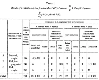
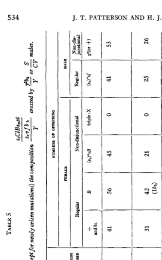
Results ofirradiation of Bar females (dose ‘‘t4”)(P1 cross: - 0 Xs,vf $;F1 cross: B
B
~ 0 X s , i f a t t d B $ ) .
S&f
NUMBER OF h-h CULTURES WITE YUThTION8 1N:
I
Lethal and semi-lethal -- 5(+2?) 6 6 -- 16(+2?) A B C
--
Total-
Lethal and Bemi- lethal 0 0 9 0 1 0 lo X)NDITION OF Pi‘EYAIES AT TIME OF IRRADIATION Visible --
--
0 Starved, virgin Fed, im- pregnated Fed, virgin 0 0 mrFmxlX R T l U 4 POINT MUTATION8
Vinible - 0 1 1 2 .__ SECTIONAL DI8LOCATION8 (CROSSINO OVER
REDUCED) Lethal - 0 0 0 0 - - Non- lethal - 0 U?) 0
’OINT YOTATIONf
SECTIONAL DISLOCATIONS
(cRo88INQ OVER
REDUCED)
Lethal Non-lethal
The factors of dosage for the so-called “t4” dose are as follows: filter, 1 mm aluminum;
peak voltage, 50 K.V.; milliamperes, 5; distance, 16 cm; duration, 48 minutes. In some cases the
milliamperage was doubled (that is, made 10) and the time cut in half, often, too, the distance was shortened and the time then reduced proportionately to the square of the distance; in all such cases the total energy is the same and the treatment is designated as “t4.” Treatments in the earlier experiments (1926 and most of 1927) were, however, given with a different machine from the later ones, and we have found that the “t4” treatment, involving the above factors, on the
old machine, belonging to Doctor DALTON RICHARDSON, was in reality about three times as
strong as the “t4” treatments involving the same factors, given later, on the new machine of the
same make (Victor, with Snook transformer) acquired by our own laboratory. In the present
paper, whenever the treatment was given on the old machine, it will be so stated-for example,
%(old machine)”--and when the machine is not designated, it may be understood that the
new machine was used. “t4(old machine),” which is the “t4” of the earlier papers, must there-
fore be understood to be the equivalent to our present “t12.”
I n the experiments of one of us, a somewhat different series of time factors has been used,
and the resultant dosages, all given on the new machine, have been designated in terms of “D,”
(PATTERSON 1929b). The dosage “D5” here referred to would be the same as “t6” (new machine),
and, in general, tl=Dl.OS, or Dl=t0.93.
We have found recently by means of dosimeter measurements that doses given only in terms
of theabovefactorsarefarfrom accurate. The “tl” on the new machine may represent a dosage
as high as 300 r units or as low as 150. Hereafter, where dosage is to be accurate, I units must
504 J. T. PATTERSON AND H. J. MULLER
males, and allowed to remain there for a week, laying the eggs that pro- duced the F1 females which were tested. The results of the tests of these
F1 females are summarized briefly in table 1. The minutae of the data from this experiment are not given here, since, as will be seen, the three groups showed no significant differences from one another, and since the chief interest of the work, from the standpoint of the present paper, attaches rather to the totals. These totals, for variations of different kinds, in the
X from the treated female germ cells, are to be compared with the corre- sponding totals from experiments in which spermatozoa were treated.
Reference to table 1 shows that, of the 18 to 20 mutations there listed in the X from the treated female germ cells, all but one doubtful one behaved
as point mutations. The exception was a case which was lost, due to sterility of the offspring, before it could be determined by subsequent breeding whether the chromosome abnormality was in the X of maternal or in that of paternal origin (the latter in this case having received the treatment also).6 Of the 11 or 12 mutations in the X from treated sperm, on the other hand, the number which contained distinct chromosome abnormalities was 2, or 3 if the doubtful case above alluded to is to be regarded as having been in the X of paternal origin. Only about a third as many of the sperm used had received treatment as of the eggs.
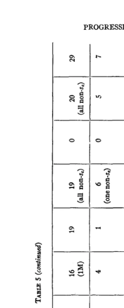
As the data relative to mutations in the X chromosomes from treated sperm are meagre in the above experiments, they may be supplemented by citation of an experiment in which the P1 cross was the reciprocal of the PI cross in the above experiment. Here Bar males were treated, and then crossed to untreated
“C1
B” females. The dosage (t13+) was, however, much higher than in the other experiment. Here, too, there were physiological differences between different groups of the treated flies,- in this case the difference was in temperature, one group being kept warm, the other cold, while treated-but as the two groups gave essentially simi- lar results, the data have been combined in table 2. It will be seen from this table that the total number of sectional changes is comparable with that of the point mutations, and that if anything like this ratio of the two classes had existed in the treated female cells of the experiment reported in table 1, the results obtained would surely have been different, despite the small total numbers o mutations there dealt with.Chromosome abnormalities do sometimes occur in the chromosomes of irradiated females,
however. In group A, for example, an F1 fly was found which proved to have a duplication on
one of its third chromosomes-a piece from the middle of the genetic map of the left arm, not
including the end, having become attached to the right end of an otherwise normal third chromo-
A B
Total
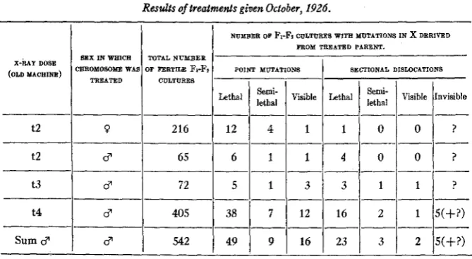
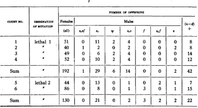
Further corroboration of the point in question may be obtained from the data of the original X-ray experiment, obtained in 1926 (MULLER 1927, 1928b), although these would, by themselves, hardly have been exten- sive enough, on the female side, to be conclusive. The figures pertinent
TABLE 2
S e V f h s,vjbi,
“CZB”
Resultsof irradialionojBarmales(dose 913”). P I cross:- 9 XB$;Fi C 7 0 S S : 7 9 x S c V j b b $ ) .
WMPERATURB OF P I MAIIEB ATTIMl OF IRRADIATION
6”L-2”C 34”Itl”C -___-
NUMBER OF FrFz CULTURES WITS MUTATIONS IN:
Lethal Visible
5 1 4
6 ~ _ _ _ - - TOTAL NUMBER OF FERTILE CUL- TURES FROM FL F E M A m S
Invisible
5
x DERIVED FROM PI MALE
x DERIVED
I
I11
1 1 9
FROM PI
FEMALE
____-
l(visib1e)
2 (visible)
3 (visible)
- Lethal 5 12 17 ___ __ 3 3 6 SECCIONAL DISLOCATIONS 36 62 0 0 0
_-
98to the matter a t issue are given in table 3. From the treated male cells, there is approximately the same proportion of sectional to point
TABLE 3
Reults of treatmenls given October, 1926.
NUMBER OF F ~ F I CULTURE8 WITS MUTATIONS IN x DERIVED FROM TREATED PARENT.
SXX IN WHlCS TOTAL NUMBER CSROMOSOME WAS OF FERTILE FPFI
TREATED CULTURES X - M Y OOSE
(OLD MACEINO) POINT MUTATIONS SECTIONAL DISLOCATIONS
Lethal
1 1
Visible !Invisible Lethal Visible-_____
12 4 1
t2 0 216
8 65
__- t2
t3
38
1
71
12t4
Sum 3
---
49
I
91
16506 J. T. PATTERSON AND H. J. MULLER
chromosome abnormalities, in proportion to the point mutations, there being only 1 to 17, whereas those from the treated males given the same dose show 4 to 8 and those from all treated males combined show 33 sec- tional to 75 point changes.
Taking these three experiments together, then, it seems safe to conclude that under certain conditions not nearly so many sectional changes can be obtained by treatment with X-rays, in proportion to the point mutations simultaneously obtained, as under other conditions. The decisive con- ditions seem, in the present experiments, to be somehow connected with the sex of the cells undergoing treatment, though whether the con- nection with sex is causal or accidental is as yet somewhat uncertain.
It is possible that the different average dosages given the males and females may also have played a part. But, be the basic determining conditions what they may, the important point here is that, by their means, one of these two processes may be influenced largely separately from the other. Accordingly, there must be some real difference be- tween the mechanisms whereby the sectional and point changes occur, and, though there may also be some feature or features common to the two mechanisms, as suggested by the fact that both can be initiated by X-rays, nevertheless there remains no reason to make the specific assumption that the difference between them is purely quantitative, rather than of some other nature.
I t is easy to conceive of ways in which the two processes might be related so that they would be affected in the manner found. For example, the breakage of a chromosome, as well as the change in composition of a
ment, might well be subject to different influences than the original process of inter-atomic rearrangement above postulated was subject to.
It might, for instance, be much more likely to occur when the chromo- somes were in a particular physiological condition, or when they were packed together tightly morphologically, as they are in the sperm-head. Under such circumstances, then, displacements of chromosome sections would be especially likely to occur, though the number of “point muta- tions” need not be correspondingly more frequent. We have sketched here, however, but one out of numerous possible interpretations. It would not, at the present stage of our knowledge, be profitable to speculate upon the matter in more detail, particularly since certain further tests bearing upon it can be made.
Iv.
THE PRODUCTION OF MULTIPLE ALLELOMORPHSIn the eyes of many geneticists, the most convincing line of evidence that has been brought against the (‘presence and absence’’ theory in general has been the phenomenon of multiple allelomorphism, with its attendant features. There is now evidence indicating that this same phenomenon, in all its details, can be induced by X-rays. If so, then the same series of arguments as have been found applicable in the case of this general question (MORGAN, STURTEVANT, MULLER and BRIDGES 1915, 1923, MULLER 1919,1920) now apply similarly against the idea of “presence and absence,” or(‘mutation by loss alone,” in the more specific case of the changes induced by X-rays. The experimental evidence concerning this matter will therefore be in place here.
The locus in Drosophila at which the greatest total number of “spon- taneous” mutations, and also the greatest number of different looking “spontaneous” mutations, have been detected, is that of white eye (MULL- ER 1920). Of the dozen or more different mutant allelomorphs of spon-
taneous origin known at this locus, white has been found by far the oftenest (over a dozen times, possibly two dozen), eosin several times, and most of the others just once in all the Drosophila work to date. I n the X-ray experiments, likewise, it has been this locus in which the most mutations have been discovered, and it is likewise found that most of the mutations induced at this locus have given rise to the allelomorph white. The latter has been induced by irradiation as a mutation in a germ cell on more than a
508 J. T. PATTERSON AND H. J. MULLER
allelomorphs at this locus have been found in our laboratory, after raying (MULLER 1928 a, c).
One of these allelomorphs is eosin, found by a graduate student, Miss CAMPBELL, in May, 1927, in an experiment directed by one of the authors to test the frequency of production of translocations. The eosin arose from the normal allelomorph, in a chromosome carrying also the mutant genes for scute, vermilion, and forked. A male
(PI)
bearing these genes had been heavily X-rayed (dosage, “ t4, old machine”) and mated to a femalehaving only normal genes in the X chromosome. In the male progeny
(Fz)
of one of the F1 females, there was the expected count so far as thecharacters that were supposed to have entered the cross were concerned, but the individuals carrying scute and vermilion were of a lighter eye color. By subsequent breeding, the gene responsible for this effect was separated from scute and vermilion, and was found to produce the peculiar color of eosin, in the sexually dimorphic fashion characteristic of the latter. Its locus also was detemined to be at about 2.0, with reference to scute, and when crossed with white it gave a light eosin color, as does the fa- miliar eosin. As there had been no eosin stock in the university except one which carried no other mutant genes, it is unreasonable to suppose that eosin could have crept into this particular combination with the expected genes, scute, vermilion, and forked, by contamination. The evidence is therefore complete that in this case eosin arose from the normal allelo- morph by mutation, after irradiation of the sperm.
A second allelomorph of white, found by one of the authors in the fall of 1928, is to all appearances identical with the known allelomorph, “apricot.” It arose in the experiment summarized in table 2, in a culture descended from the flies that had been kept a t the colder temperature
(6’ C) during treatment. The F1 female among whose progeny it was found had received from her mother an unradiated X chromosome with the genes, scute, vermilion, forked, bobbed, and from her father, a radiated X, containing Bar, in which a lethal inversion had just arisen somewhere in the right region, and the gene for apricot in the left. The male progeny, therefore, included only one non-crossover class (s, v
f
b b ) , the othernoncrossover class ( B ) dying. The former non-crossovers had the expected characteristics. There was, in addition to these non-crossovers, only one crossover male,due to the reduction of crossing over caused by the inversion, and to the fact that crossover males receiving the left end of the unradiated
for vermilion, forked, and bobbed (the unradiated right end), but it had a light lemon-like eye color, indicative of the new mutant gene (apricot) in the left-hand portion of it, derived from the radiated X. As a result of crosses between this male and normal females, numerous crossover males were obtained in
Fz
which carried the new mutant without the genesv f b b, and a pure stock was derived from these, in which i t was evident
that both males and females had the eye-color characteristic of the known apricot. Crosses with white resulted in an eye color of intermediate shade in the daughters; this showed that the new mutant was really an allelo- morph a t the locus in- question.
A probable third allelomorph was obtained by PATTERSON in 1929, from a cross between a radiated (D10) eosin singed male and a yellow female with attached X’s and a Y chromosome. The sons from such a cross, receiving their father’s X and mother’s Y, would ordinarily be eosin singed, like their father. The great majority were, but one appeared very much lighter in color than the rest. Through crossing, the new gene has been separated from singed, and a pure stock of it has been obtained, in which it is evident that the females are somewhat darker than the males, as is true of eosin, from which the new gene was derived, and of another known allelomorph called ivory; the flies are consistently lighter than eosins, however, and probably lighter than ivory. The fact that hetero- zygotes, carrying one dose of eosin and one of the new mutant, are of in- termediate color indicates that the case is one of allelomorphism rather than of modifying genes.
A probable fourth allelomorph arising in somatic tissue as a result of raying the allelomorph apricot in an embryonic stage will be referred to subsequently, in connection with the account of reverse mutations. ‘
Nine cases of mottled eyes have also been found by the authors
(MULLER 1928 a c, 1930). These are recessive to red and give inter- mediates when crossed with white and the other mutant allelomorphs of this locus. They vary through all the colors known in this series, and more, However, the mottleds, unlike the mutants of uniform color above described,do not behave as simple point m u t a t i o q b u t , without exception, involve breakages and reattachments of chromosome parts ; accordingly they do not furnish material for illustrating the principles here under discussion.
Another locus in which several “point mutations,” including different allelomorphs, have been induced by X-rays is that of forked bristles
(f).
510 J. T. PATTERSON AND H. J. MULLER
in experiments of one of us involving irradiation of mature spermatozoa, not designed specifically for studies of the mutability of this particular locus. In the first case (spring of 1927), males having the gene forsmall eye, but otherwise normal, were given the t4 (old machine) dose and mated to females carrying in one chromosome the “CJ?” combination and in the other sc v f ba. In a section of this experiment in which the males were held for six days after treatment, before mating, 243 F1 females inheriting the sc v f b b chromosome from the mother were produced. Among the latter, one was a typical forked which, on breeding, proved to be homozygous for the same, having a new gene for forked in the irradiated chromosome, with the gene for small eye, and no evidence of any X-chromosome ab- normality. Pure stock of the new forked had good viability and fertility, and exhibited the character in typical fashion.
I n another experiment (fall, 1928) males containing the gene for bobbed bristles (ba) and an inversion designated as “649” were given a t16 dose and crossed to yellow attached-X females. Among the 615 male offspring, one was a typical forked, which transmitted its mutant charac- ter to its offspring as a sex-linked recessive. Crosses with the old forked resulted in forked daughters;this showed the new gene to be allelomorphic to the old.
A third mutation in this locus occurred in the experiment shown in table 2 in the “warm” series. Among the 120 F1 females tested in this series, one which had received “ C J P from its mother and an irradiated Bar-containing X-chromosome from its father yielded sons all of which were both forked and Bar. Further study proved the new forked to be in the same locus as the old, but it was noticeable that in the new stock the forked character was not nearly as well marked as in the typical stock of the old forked. As it is unlikely that a modifier had happened to arise in just the same chromosome as the gene for forked itself, it is probable that there was here a different, and “weaker,” allelomorph,
“f”,”
such as has been found in some of the previous Drosophila work onmaterial not artificially irradiated. I n one of the following sections of the present paper, dealing with reverse mutations, another case is recorded of the origination of a “weakly forked” allelomorph (considerably weaker than the foregoing), and likewise of other mutations a t this locus, after irradiation, in experiments especially intended for the study of changes at this locus.
a distinctly different mutant allelomorph (shown in figure 1). This arose in an experiment in which wild-type adult males were given a t13 (D14) dose, and then mated to females having in one X chromosome the so
CzB
s, v t, complex and in the other s, v f bb. Four hundred thirty two fertile F1 females that had received C z B were bred in separate vials, the progeny (F,) being examined through the walls of the vials under the low power-of the binocular. In this method of examination, devised by C. P.OLIVER; the vial is held under the binocular in a horizontal position with
the stopper towards the light; the flies then congregate against the upper wall of the vial, next to the stopper, and any conspicuous visible mutations present in the males as a group can be readily detected (as well as the absence of males, indicating a lethal). Among the 432 cultures of the
FIGURE 1 .-The mutant “scutex” produced by irradiation.
type stated, one was found in which there were only a few males, and these were all of peculiar appearance, while all the females appeared to be scute. On anaesthetization and examination under higher power, it was found that the males were of a very extreme scute type, completely devoid of
bristles on the dorsal surface of the thorax and scutellum. The body color also appeared to be somewhat lighter, and the wings more transparent looking than in the normal, so that the whole fly had a delicate, flimsy appearance.
The females in the above cultures, on being bred to scute (attempts to breed or keep alive the males with the new character proving unsuccessful) gave rise to offspring all of which showed either scute or the new mutant. Hence either a new allelomorph of scute had arisen, or scute and an intensifier had arisen simultaneously. The latter inherently improbable
512 J. T. PATTERSON AND H. J. MULLER
assumption was made still less tenable by the crossover ratios, which showed the effect to be located at the left end of the X chromosome, like scute itself. The new allelomorph, which may be designated as “scutex”
(scz), was found to act as a semi-lethal, inasmuch as, in most cultures,
fewer then a tenth of the flies expected to show it actually hatch. I n combination with the old scute, the latter dominates, but .not com-
pletely: that is, there is on the average somewhat more reduction in
the number of thoracic bristles in the heterozygous sC2/sc females than in the homozygous s,/sc. As shown in the figure, the artist found postvert- ical and subhumeral bristles present, although these are absent on scute flies, which have a greater tendency to produce bristles in the other posi- tions. We have not been able to check up on this point in pure scutex, since the latter has recently been too inviable, but in compounds with scute we find the postverticals and subhumerals to be absent. If they were really present oftener in scutex than in scute it would be evident that these characters were not different merely in a quantitative way.6 They would be related non-quantitatively in a somewhat similar manner to that first found by MULLER to hold in the case of the allelomorphs of the truncate series (vortex, oblique, dumpy, lopped, thoraxate, truncate;
MULLER 1919, 1923).
Perusal of the above three cases of multiple allelomorphism, following upon irradiation, will bear out the contention that they exhibit all those characteristics which, in the case of the natural multiple allelomorphs, have been taken as evidence against the idea of mutationsin general consisting of losses. For one thing (l), the allelomorphs are surprisingly frequent if they represent only the last term-the most extreme possible cases-of close linkage between genes at essentially different loci. Cases of merely very close linkage, though occasionally found, are on such a view relatively much less frequent than they should be, and the advocate
of “presence and absence” is thereby forced to some such subsidiary hypothesis as the existence of intra-chromosomal groupings or “nests” of genes.
Secondly (2), the different mutant allelomorphs in any one of the three series affect the same general character (eye color, bristle conformation, or bristle distribution, as the case may be). There is no known or apparent
Since the above finding was made on scutex, the authors’ attention has been called to a cer-
tainly non-quantitative, and much more extensive series of allelomorphs of scute, arising after
irradiation, in experiments of SEREBROVSKY, DUBININ and their colleagues (SEREBBOVSKY,
reason why this should be true if the different allelomorphs in a series are simply losses of different completely linked (neighboring) genes, but it is readily understandable if they are different changes in one gene, that has become specialized to react chiefly, so far as visible characters are con- cerned, in the development of this particular character.
Thirdly ( 3 ) , whenever individuals with different mutant allelomorphs are crossed, instead of the normal type becoming reconstructed phaeno- typically in F1, as it is when individuals with recessive non-allelomorphic genes are crossed, the F1 shows the characteristics common to the mutants that were crossed, and, in any respect in which they are different, and both abnormal, it is no nearer to the normal than the allelomorph that is more normal in this respect. To explain this non-appearance of the normal in F1 on the “presence and absence” hypothesis (of different completely linked genes that become lost), requires the special assumption of the linked genes observing a precedence in regard to the order in which they are lost. On the presence and absence view, the less extreme allelomorph is considered to lose a gene, which may be designated as
“ A ” ; then the more extreme one, in order, on crossing with the other, to
yield a hybrid showing at least as much variation from normal as shown by the less extreme allelomorph, must likewise lack A , but it must lack some- thing else, “ B,” in addition, which distinguishes it from the less extreme
allelomorph. Thus, A can be lost by itself, but B is never lost unless A ,
which has the precedence, is lost with it. This assumption must be carried to considerable lengths in the case of the series of X-ray allelomorphs of white, where apricot must be considered to have lost A , eosin to have lost
B and A , the lighter mutant from eosin above mentioned to have lost
C, B, and A , and white to have lost
D,
C, B, and A simultaneously (ormany more, if the allelomorphs of spontaneous origin also are taken into account). On the other hand, the assumption of no suchhierarchy of genes is necessary on the view that the allelomorphs are merely different changes in one gene, which alter its reactivity to various extents, and in various ways. On this view the usual lack of reconstitution of the normal type, in crosses between two mutant allelomorphs, ceases to be paradoxical.
5 14 J. T. PATTERSON AND H. J. MULLER
consistent dominance of the normal allelomorph in these cases. (Apparent normals should arise, which did not have a sufficient number of elements to dominate.) A far more serious obstacle is encountered in the fact that, in the case of the white and probably the scute series, the members do not form a purely quantitative series a t all. Apricot, unlike eosin and the lighter variant from eosin, is alike in color in male and female; scute, though having far less extreme bristle reduction than scutex in most respects, nevertheless probably lacks certain bristles more often appearing in the latter. If there has been loss, then, the parts lost have been somehow differ- ent from one another, and yet these various parts were obviously related to one another in their functioning, in a much more intimate way than that in which genes in different loci are ordinarily related. The readiest method of explaining their peculiarly intimate relationships is to assume that they were chemically united. But we cannot tear off a piece of a molecule without healing the broken bond, either by a rearrangement of the remainder, or, as is far commoner, by the addition (substitution) of something else, large or small, in the place made vacant. I n either case, the idea of a pure loss becomes vitiated. I n truth, there is no theoretical reason left for assuming that a loss would be the exclusive method of change of the gene.
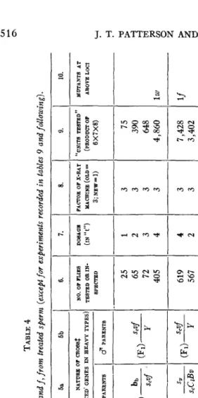
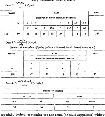
I n connection with these allelomorphic series, it is pertinent to put the question, “Were the different allelomorphs all really produced by the irradiation, or were only the commoner (usually the most extreme) allelomorphs so produced, and the others of ‘spontaneous’ origin in each case?” While this question cannot be given an absolute answer, it can be met in terms of strong probabilities. Thus, in the case of the white-locus series, in all the experiments on treated mature spermatozoa combined there was a total of somewhat less7 than 22,366 flies that would have served for the detection of white or one of its allelomorphs (table 4). Seven thousand two hundred fifteen of these were
F1
males derived from treated males crossed by attached-X females, and 15,151 were Fl females (de- rived from mothers with separate X’s) which were bred in individual cultures, and the male progeny of which were examined. I n this total there were 10 or 11 that carried white, and 1 that carried an allelomorph (apricot) as a heritable germinal mutation, and two males that carried white “fractionally” in some of the somatic and not in the germinal tissue. The “fractional” white mutations that were confined to the soma should probably be counted a t a value of a t least3
each. It is likely that there were parallel cases in which white occurred in the germinal tissue but inwhich the male was not bred because there were no somatic indications
of the mutation; the latter cases would, however, be less frequent than the former, since a t least one of the two eyes is usually derived from the same nucleus of the two-cell stage as is the germinal tissue. The use of the factor
3
will therefore serve to keep our results on the side of caution. Taking 13, accordingly, as the total number of whites, we find that white arose about once in 1800 sperm cells and a different allelomorph about once in twenty- three thousand sperm cells (in round numbers), in this work. I n all the previous Drosophila work on non-radiated material, up to 1925, it has been estimated that upwards of twenty million flies havebeenexamined (MORGAN, BRIDGES and STURTEVANT 1925). Nearly half of these must have been males in which white or one of its allelomorphs would have been pretty sure to be detected (and something like one one-hundredth must have been females subjected to the progeny test and similarly serviceable). Among the 10,000,000 thus available, white has been ob- served to originate only about two dozen times, a t the most, and all other allelomorphs of white, combined, not much over one and a half dozen times. This makes a frequency for white of about one in four hundred thousand times, and, for the other allelomorphs, of about one in six hundred thousand times. I n the total of less than 23,000 in the series of radiation experiments above referred to, there was, therefore, about 1 chance in 17 that white should have appeared at all, and 1 in approximately (17)13 that it should have appeared as often as 13 times, unless there had been some peculiarity in the conditions of the experiment responsible for producing it. In like manner, the chances that one of the other allelo- morphs, such as apricot, should have appeared once, would have been only1 in 26 if the material had not been somehow made especially mutable. It can readily be calculated from the above figures that, in all, the frequency of appearance of mutations a t the white locus in the irradiated sperm was of the order of magnitude of 200 times the corresponding frequency in non-radiated material. If, now, we weight the flies in all the experiments according to the dosage of radiation used upon the sperm,- which is legitimate, in view of the direct proportionality between dosage and mutation rate found by HANSON and HEYS and by OLIVER-We find that there would have been 1 detectable mutation of the normal allelo- morph of white to some mutant allelomorph or other among about 1,000
3. 4. METHOD DATE OF EXPERlMENT OF DE- 5a 5b NATURE OF CrrOsSS (TREATED’ GENES IN EEAVY TYPES‘
DOSAffE (IN
“t”) 1 2 3 4 4 2
PACPOR OF X-UY “UNITE TEBTED‘’ LDTAN’IS AT MACEINE (OLD= (PRODUCPOF ABOVE LOCI 3;NEw=I) 6X7X8) - 3 75 3 390 3 648 3 4,860 lw 3 7,428 lf 3 3,402
4 2 4
3 13,800 +w 3 7,940 lw 3 24,960
/lw \tw
TABLE 4 Summary of results with regard to loci of sc, w, and f, from treated sperm (except for experiments recorded in tables 9 and following).
- 2. - VORKER
-
1.~-
DESIQNATION 6. 7.I
8.I
9.I
IO. NO. OF FJJE8 TESTED OR IN- SPECTED 6 PARENTS25 65 72
405 ‘Lfirst”
-I
1-1---
“aged sperm”619 567
-I
++---.--
1,150 1,490
“fractional”
+
“SV’ (PI) - - y+ YY CY M1
fall, 19271
“1
(Pdy - 2,080“stability”
---
“649loci” “649 loci” NO. 2 649 (PI)
y
615 649 (P1)y
h
3
6
;
.-.
z
;
u
< b
'
a - 3
I II p G E
1 4 2 s
N I
I 0 2
&
si
s
m h l
I
2 010
0- 4
3-
1
2-
a- Q*.
2
'0-
* h r r )
* ") I I hl I x
* i
I C
h i *
4 l
",a
1
",j D l z i
I * 0 1I
N
m
hl .Im 4
9 1 3
--./
52% w v ) $3 hl-
s i
41,
3$1.
$1.
,h
5
$1" h 3+
3+I.
h
-
,--. c=2 -
5 , 2
VI
g g
. 3 &w
v
d
%
? Z
ve 5 2- 0
2 . 5 a b g , , a $
3 3 , .
5 % "
-
'= c=$ 2
6 2
% $
3 a v rG
v
! Jd
% + I
O H
1-
I I
,+I6
u .:cy
2
r'/d
1
::I i
/ =1
i l
G+G
2
j z
1 8 1 . / 2 1 29
3
44b
-pc: % l $ i
$3 1
j
G C o3
a
v v
Frr v a v A m hlhl h 01 L a
5 5
W-
2"/II
2 2 2 %1
=g1
=i
1
7a
i
.&
G .-d
u.d
d
2 6
3 .G .4
c
-
? F w F
!2
s.s
c,"
B l
g
-5
.-
20 E
c) c)
.e
z
*
E
2
DATE
OF
EXPERIMENT
METE0 OF
DE TEm101 TABLE 4 (continired 2. 5a 5b 6 7. 8. 0. 10 DEBIONATION WORHEl NO. OF FLIES TESTED OR IN- SPECTED
DOSAOE (IN
“6”) FACTOR OF X-RAY MACHINE (OLD 3; mw=C ‘UNITS TESTED” (PRODUCT OF 6 X7X8) MUTANTS AT AROYE MCI NATURE OF CROSS$ (TREATED OENES IN HEAVY TYPES) PARENTS
1
3 PARENTS (for locus of w only) “dosage” 0 8,444 3x 1 24,958 5w winter, 1928- (( 1929 (for locus of w only) “aging of males” H 2,967 8 1 23,736 3w winter,1928- 1929
factor even greater than that expressing the increase in the frequency of lethals a t the same dosage. (There is, however, more chance for errors in detection to affect the figures for white than those for lethals in untreated material). There can, therefore, be little doubt that the radiation was responsible for these mutations of the white locus.
Similar calculations can be made with respect to the mutations in the loci of forked and of scute. The results here will not be as accurate as in the case of the white locus because of the fact that these characters, being somewhat less conspicuous than those of the white series, are more apt to have been overlooked, especially in the non-radiated material. Neverthe- less, the chances of detection of both forked and scute are distinctly good; once an investigator has worked with them, he is not likely to overlook them. In all our work on the progeny of radiated sperm combined, includ- ing that reported in the subsequent sections of this paper, there have been 29,402 F1 flies in which a mutant gene for scute (received from treated sperm) would probably have been detected, and 5961 FI females in which, by progeny tests, scute would probably have been found. I n the sum- marized Drosophila work to 1925, scute was reported 4 times among the 10,000,000 males examined, and scutex not at all, among some 200,000 females whose male progeny were examined. These results would have given a chance of only 1 in about 71 of finding one scute mutation, or 1 in about 5000 of finding two of them, in a series of experiments of the magni- tude of the above irradiation experiments, while the chance of finding scutex in these experiments would be less than 1 in 33, and would really be too small to be reckonable from the data. I n all, the rate of mutation to scute found in sperm given a t12 (or t4, old machine) dose can be figured to have been about 250 times the rate in untreated cells.
520 J. T. PATTERSON AND H. J. MULLER
magnitude of the above irradiation experiments only 1 in 90, and the chance of finding two, one in 8,100. While the weaker allelomorphs are much more likely to be missed, still it can be seen that the odds must be greatly against finding one of these in our 11,881 flies either, unless some special influence were producing them. I n all, when the figures are corrected to appear as of the t12 dose, the mutation frequency to some allelomorph of forked found in the treated is over 300 times that found in the untreated individuals.
It is of course realized that in the case of this as of the other loci (white and scute) the mutations detected and reported in the untreated material represent only a fraction of those which occurred, since a special attempt was not made in most experiments to find the mutations in question, and since, even when they did occur, they were often ignored on account of the possibility of their having resulted from contamination. This probably accounts for the observed frequencies for the t12 dose being several hundred instead of about one hundred times those reported in the summar- ized untreated material (in view of the fact that the lethal frequency is raised only about one hundred- to one hundred fiftyfold). Nevertheless the reported frequencies of visibles in the untreated material are doubtless of the right order of magnitude at any rate, and so long as this is true, the conclusions reached from the above calculations would still hold, so high are the probabilities there arrived at.
v.
AN INDUCED MUTATION VISIBLY DIFFERENT FROM AKNOWN LOSS OF THE SAME LOCUS
In October, 1927,84 fertile males carrying a normal X chromosome (and a mutant combination including Star eye in one of their second chromo-
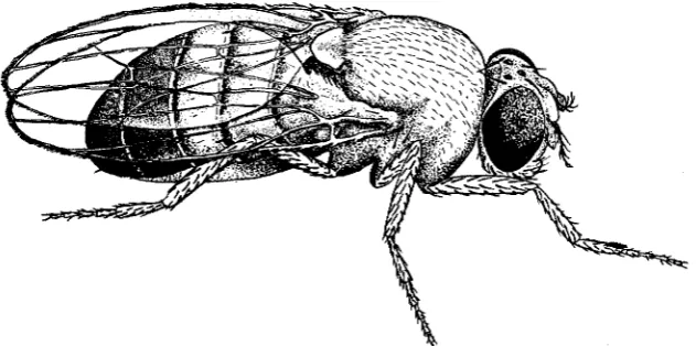
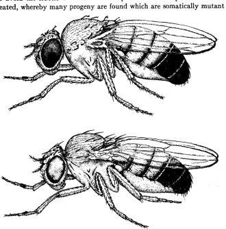
FIGURE 2.-Dominant “eyeless” (in fourth chromosome) produced by irradiation. Below is given pair of legs with sex combs of normal size, for comparison with the enlarged ones of the eye- less fly.
somes) were given the t4 dose (old machine) and, after being kept in isola- tion for 16 days, mated in individual cultures to yellow attached-X females heterozygous for Curly wing. After 4 days, the parents were transferred to
522 J. T. PATTERSON AND H. J. MULLER
to a third set ((‘brood 3”). Altogether, 2,080 F1 males were carefully ex- amined for mutations, and 437 F1 females (namely, all those of brood 1).
This count of males is shown on row 4, table 4. In the second brood, among the 18 F1 males appearing in the last culture (No. 84), there was one non-Star, non-Curly male which had one eye misshapen as if furrowed, and the other eye normal. The 11 males in brood 1 from the same parent were all normal. When the aberrant male was bred back to yellow at- tached-X females, it was found that the variation was transmitted as an autosomal dominant, to the daughters as well as to the sons, appearing in approximately half the members of both sexes. It usually affected both eyes rather strongly, causing a change in size and shape rather similar to that seen in the dominant second chromosome mutants “Lobe” and “Lobe,2” in the dominant third chromosome mutant Deformed, and in the recessive fourth chromosome mutant “ eyeless.” Besides the eye abnor-
mality, it produced, in the male, a hypertrophy of the sex combs, the latter being about doubled in size. Examples of the variation are shown in figure 2. Sometimes even more abnormal types are produced in this stock, the head becoming split or otherwise malformed, and developing peculiar protuberances and multiple antennae as is sometimes the case with the recessive eyeless.
As it was now highly probable that the new gene was a dominant allelo- morph of the known recessive “eyeless,” it was crossed to the latter. The
F1
flies containing the two genes in combination proved to have, on the average, a greater reduction in the size of the eye than was character- istic of either the homozygous recessive eyeless or the heterozygous new mutant, although they were seldom completely eyeless. It will then be assumed that the two genes are allelomorphic, and the new mutant will be designated as “Dominant eyeless” (euD).As all Dominant eyeless individuals tested proved to be heterozygous, even though both parents had carried the gene in question, i t appeared likely that it acted as a lethal when in the homozygous conditions. Crosses were made between it and the dominant minute bristle ( M r v ) , located in the same chromosome, which also is lethal when homozygous. The FI flies having the heterozygous combination euD/Mrv were next crossed to each other. This resulted in Fz all resembling their F I parents. In other words, a balanced lethal stock had been established, and there could be no doubt that homozygous euD was lethal. The combination of the two char- acters results in individuals that tend to be somewhat abnormal in bodily shape (broad and squat) but with no evidence of the rotated-abdomen condition which Mrv produces when combined with the recessive mutant of that name with which it is allelomorphic. The euD-Mrv combination flies have a low fertility, but, once numerous individuals are obtained, the stock can, with care, be perpetuated indefinitely.
Crosses of euD to bent and shaven show that it is allelomorphic to neither of the latter. Thus, of the four loci known in the tiny fourth chromosome- namely, that of bent, of shaven, of the allelomorphs Mrv and rotated abdomen (TCHETVERIKOFF)
,
and of eyeless-eVD shows allelomorphism to eyeless only. Hence it is unlikely that it involves a “deficiency” of a chromosome region, or any disturbance other than ordinary “point muta- tion.”A digression may be made at this point to call attention to the bearing of the above results on some previously published genetic maps of the fourth chromosome. In these (for example, in MORGAN’S Theory of the
Geme, 1928, p. 23) the known loci of chromosome I V are shown separated