DOI: 10.1534/genetics.109.106955
Spp382p Interacts with Multiple Yeast Splicing Factors, Including Possible
Regulators of Prp43 DExD/H-Box Protein Function
Shatakshi Pandit,
1Sudakshina Paul, Li Zhang,
2Min Chen, Nicole Durbin,
Susan M. W. Harrison and Brian C. Rymond
3Department of Biology, University of Kentucky, Lexington, Kentucky 40506-0225 Manuscript received July 1, 2009
Accepted for publication July 2, 2009
ABSTRACT
Prp43p catalyzes essential steps in pre-mRNA splicing and rRNA biogenesis. In splicing, Spp382p stimulates the Prp43p helicase to dissociate the postcatalytic spliceosome and, in some way, to maintain the integrity of the spliceosome assembly. Here we present a dosage interference assay to identify Spp382p-interacting factors by screening for genes that when overexpressed specifically inhibit the growth of a conditional lethalprp38-1spliceosome assembly mutant in thespp382-1suppressor background. Identified, among others, are genes encoding the established splicing factors Prp8p, Prp9p, Prp11p, Prp39p, and Yhc1p and two poorly characterized proteins with possible links to splicing, Sqs1p and Cwc23p. Sqs1p copurifies with Prp43p and is shown to bind Prp43p and Spp382p in the two-hybrid assay. Overexpression of Sqs1p blocks pre-mRNA splicing and inhibits Prp43p-dependent steps in rRNA processing. Increased Prp43p levels buffer Sqs1p cytotoxicity, providing strong evidence that the Prp43p DExD/H-box protein is a target of Sqs1p. Cwc23p is the only known yeast splicing factor with a DnaJ motif characteristic of Hsp40-like chaperones. We show that similar toSPP382,CWC23activity is critical for efficient pre-mRNA splicing and intron metabolism yet, surprisingly, this activity does not require the canonical DnaJ/Hsp40 motif. These and related data establish the value of this dosage interference assay for finding genes that alter cellular splicing and define Sqs1p and Cwc23p as prospective modulators of Spp382p-stimuated Prp43p function.
E
IGHT phylogenetically conserved DExD/H-boxproteins act at discrete steps to regulate the assembly, activation, and dissociation of the splicing
apparatus (reviewed in Brow 2002; Konarska and
Query2005; Linder2006). How these RNA-dependent
ATPases are temporally and functionally regulated remains poorly understood. One putative regulator, the 83-kDa Spp382/Ntr1 protein (henceforth referred
to by the Saccharomyces Genome Database standard
name, Spp382p), was discovered in a screen for mutants capable of suppressing defects in yeast spliceosome assembly (Panditet al.2006). Whilespp382null alleles
are lethal, partial loss of function suppresses mutations in several other splicing factors, including the genes encoding the essential spliceosomal proteins Prp8p and Prp38p. Spp382p is a spliceosomal protein that binds the Prp43p DExD/H-box protein to promote efficient dissociation of spliceosomal factors after completion of
splicing in vitro (Tsai et al. 2005). Some but not all
spp382 mutants also accumulate the excised intron product of splicingin vivo, ostensibly due to protection of the intron within a hyperstabilized spliceosome (Panditet al.2006; Tanakaet al.2007). The suppression
of spliceosome assembly defects byspp382mutation is
proposed to occur by impairing Spp382p-stimulated dissociation of kinetically impaired or otherwise
in-efficient spliceosomes by Prp43p (Panditet al. 2006).
Consistent with this hypothesis, prp43 mutations also
suppress spliceosome assembly defects in a manner that, within limits, is inversely proportional to the residual ATPase activity of Prp43p (Panditet al.2006). In this
light, it is possible that the Spp382 and Prp43 proteins are components of the ‘‘discard pathway’’ for spliceo-some dissociation predicted by the kinetic proofreading
model of DExD/H-box protein function (Burgesset al.
1990; Konarskaand Query2005).
Recently, several groups made the surprising obser-vation that the Prp43p splicing factor is also required for
ribosome biogenesis. Mutations in PRP43 inhibit 35S
pre-rRNA cleavage and limit downstream steps in this
processing pathway (Lebaronet al.2005; Combset al.
2006; Leeds et al. 2006). Prp43p is $10-fold more
abundant than the splicing-restricted DExD/H-box proteins (e.g., Brr2p, Prp2p, Prp5, Prp16, Prp22p, and Supporting information is available online athttp://www.genetics.org/
cgi/content/full/genetics.109.106955/DC1.
1Present address: Department of Cellular and Molecular Medicine, University of California at San Diego, La Jolla, California 92093.
2Present address:Center for Food Safety and Applied Nutrition, U.S. Food and Drug Administration, College Park, MD 20740-3835.
3Corresponding author:Biology Department, 675 Rose St., University of Kentucky, Lexington, KY 40506-0225. E-mail: [email protected]
Prp28p; see Ghaemmaghamiet al.2003) and, consistent
with dual function in splicing and rRNA processing, nuclear Prp43p is enriched in the nucleolus (Huhet al.
2003). Furthermore, proteins and small RNAs acting exclusively in splicing or in rRNA biogenesis copurify with Prp43p, supporting its direct contribution to both processes (Hoet al.2002; Lebaronet al.2005; Combs
et al.2006; Gavinet al.2006; Kroganet al.2006; Leeds
et al. 2006). While it is not certain how Prp43p is partitioned within the cell, its association with Spp382p appears critical for recruitment to the postcatalytic spliceosome and for stimulation of the intrinsic Prp43p helicase activity (Tsai et al. 2005; Boon et al. 2006;
Pandit et al.2006; Tanaka et al. 2007). A structurally
related protein, Pxr1p, interacts with Prp43p (Lebaron
et al.2005) and is required for efficient rRNA processing (Guglielmi and Werner 2002). Pxr1p may serve a
parallel role for Prp43p recruitment and activation within the rRNA processing apparatus but direct evi-dence for such function is lacking.
Here we describe a genetic approach to identify factors
that interact withSPP382and function in spliceosome
dynamics. Specifically, we describe a dosage interference assay to find genes that when overexpressed inhibit the growth of a yeast strain in which the
temperature-sensitive prp38-1 spliceosome assembly mutation (Xie
et al.1998) is suppressed by thespp382-1suppressor allele (Panditet al.2006). We identify multipleGAL1-effector
genes that preferentially inhibit growth of theprp38-1
spp382-1 double mutant compared with either single-mutant host or a wild-type yeast strain. Consistent with the goal of this screen, most genes cause splicing inhibition with galactose induction.
Among the recovered genes are SQS1 and CWC23,
which encode proteins that purify from yeast in multi-subunit protein complexes containing Prp43p but have
unknown functions (Lebaron et al. 2005; Panditet al.
2006). Sqs1p is a nonessential 87-kDa protein that, similar to the Prp43p interacting proteins Pxr1p and Spp382p, possesses the glycine-rich G-patch motif common to a
subset of RNA binding proteins (Aravindand Koonin
1999).CWC23encodes a 33-kDa protein with a canonical
DnaJ motif characteristic of Hsp40-like activators of Hsp70 chaperones (Walshet al.2004; Voset al.2008). Cwc23p
interacts with Spp382p in the two-hybrid assay and by affinity selection (Panditet al. 2006). The genetic and
biochemical results presented here establish Sqs1p and Cwc23p as Spp382p-interacting proteins with contribu-tions to RNA processing distinct from what might be expected on the basis of their protein motif characteristics.
MATERIALS AND METHODS
RNA analysis:Low-density yeast cultures (OD600 nm,0.5)
were collected by centrifugation and washed once in sterile water and twice in Li buffer (100 mmLiOAc, 10 mmTris, pH
7.5, 1 mmEDTA). The cell pellets were then disrupted in
one-fiftieth the original culture volume of Li buffer plus 100ml of phenol:chloroform:isoamyl alcohol (PCI) at 50:49:1 by shak-ing with sterile glass beads for 4 min in a Mini-Beadbeater (Biospec Products). The RNA was purified by two additional PCI extractions followed by ethanol precipitation. Northern blot analysis of the pre-mRNA and spliceosomal snRNAs was conducted with random prime labeled probes (Promega, Madison, WI) prepared from cloned copies of the indicated DNAs that were hybridized and washed under standard conditions (Sambrooket al.1989). A 59-end-labeled
oligonu-cleotide probe was used to detect the 35S rRNA precursor, 23S and 20S intermediates (oligo rRNA 004 59-CGGTTTTAATTG TCCTA-39), and the 27SA2 intermediate (oligo rRNA 003 59 -TGTTACCTCTGGGCCC-39) (Kos and Tollervey 2005).
Hybridization with the oligonucleotide probes was done overnight in 63 SSC, 53Denhardt’s solution, and 1% SDS at 43°. The blots were washed in 13SSC, 0.5% SDS at 43°
(rRNA 0003) or 36°(rRNA 004).
For RNase H digestion, 1 mm of oligo(dT) 15-mer (In-tegrated DNA Technologies) or a control oligo (SNR30, 59-GAAACTGCTCGTAGTCTGACG-39) was incubated in 13
digestion buffer [20 mmTris-HCl (pH 7.5), 20 mmKCl, 10 mm
MgCl2, 0.1 mmEDTA, 0.1 mmDTT] at 55°for 10 min and then
slowly cooled to 37°. Five units of Escherichia coliRNase H (United States Biochemical, Cleveland) were added and the reaction was incubated at 37°for 60 min prior to gel electro-phoresis and Northern analysis. The membrane transfers were analyzed with a Typhoon 9600 Phosphorimager and Image-Quant 5.2 software (GE Corporation).
CWC23 and SQS1 mutagenesis and gene overexpression:
Site-specific mutations were introduced by inverse PCR using mutagenic oligonucleotides (sequences available upon re-quest) with Expand Long Template polymerase (Roche Biochemicals) on CWC23in a YCplac22 backbone (Gietz
and Sugino1988) containing the open reading frame and
300 bp 59- and 100 bp 39-flanking sequence or as a cloned open reading in the pACT and PAS2 two-hybrid vectors (Durfeeet al.1993).SQS1was mutated similarly except that
theGAL1fusion clone in BG1805 (Gelperinet al.2005) was
used for expression. To facilitate downstream analysis, a unique Not1 recognition sequence was included at the mutagenesis site. All mutant constructs were sequenced prior to use. Functional analysis of the cwc23 mutants was con-ducted after removal of a wild-typeCWC23allele on aURA3 -based vector by plasmid shuffle. Two-hybrid analysis was conducted in the PJ69-4A yeast strain ( Jameset al.1996) and
growth scored at 30° on medium lacking adenine or on histidine-deficient medium supplemented with 20 mm
3-aminotrizaole.
Overexpression studies were conducted usingGAL1fusion constructs (Open Biosystems) in the URA3-based plasmid BP1805 (Gelperin et al. 2005) transformed into strain
MGD353 46D (a ura3-52 trp1-289 leu2-112, 113 his cyh2r), ts192 (a prp38-1 ura3-52 trp1-289 leu2-112, 113 his cyh2r), SP302 (a ura3D0 trp1-289 leu2 D0 his3 D1 spp382TKAN pYCplac111-spp382-1), or the double mutant (aprp38-1 ura3-52 trp1-289 leu2-112 113 his spp382-1 cyh2r) as described in Pandit et al. (2006). The plate cultures were grown on
synthetic defined medium or nutrient-rich, yeast extract peptone (YP) agar (Kaiseret al. 1994). Liquid cultures were
yeast previously transformed with GAL1-SQS1. The plate growth assays were performed at 30°for 3–15 days as indicated in the figure legends.
RESULTS
Dosage interference of spp382-1-based suppression: Prp38p is an essential component of the U4/U6.U5 tri-snRNP particle (Blantonet al.1992; Xieet al. 1998).
At restrictive temperatures, the prp38-1
temperatusensitive (ts) mutation allows for efficient snRNP re-cruitment but greatly impedes spliceosome activation at
the stage of U4 snRNP release (Xie et al. 1998). This
splicing defect and corresponding ts growth defect can be partially relieved by the recessivespp382-1suppressor allele (Panditet al. 2006). To identify potential
inter-acting factors we sought genes that when overexpressed specifically inhibit growth in this genetically sensitized
prp38-1 spp382-1 suppressor background on
nutrient-rich YP medium (Figure 1 and see materials and
methods). In principle, such dosage effectors might
either act by antagonizing the suppressor function of
spp382-1or more directly inhibit splicing by sequestra-tion of other splicing factors. We scored a candidate
set of 140 GAL1-promoter fusion genes that encode
most splicing-related factors in this library collection (Gelperinet al.2005), other RNA-associated proteins,
and randomly selected controls (supporting
informa-tion,Figure S1).
Predictably, complementation by wild-type SPP382
blocks growth of theprp38-1 spp382-1double mutant at
35°as this reveals the underlying temperature sensitivity
of the unsuppressedprp38-1mutation (Figure 2A). Of
the remainingGAL1-gene fusions tested, 10 show strong
growth impairment on galactose medium when com-pared to the empty vector control transformant. These encode the established splicing factors Prp8p, Prp9p, Prp11p, Prp39p, and Yhc1p; a DExD/H-box protein involved in mRNA decapping, Dhh1p; mitochondrial proteins Yml6p and Mrp13p; and the product of an uncharacterized open reading frame, YDR230W. Two
other genes with possible links to splicing, SQS1 and
CWC23 (see below), also showed markedly impaired
growth. Overexpression ofPRP38or the knownprp38-1
dosage suppressor, SPP381 (Lybarger et al. 1999),
caused modest growth inhibition in the prp38-1
spp382-1 background, similar to that seen with GAL1-PRP43 or with GAL1-BET2 that encodes the Bet2p subunit of the yeast geranylgeranyltransferase. Other than the exceptions noted below, none of these genes significantly reduce the growth of wild-type yeast or the
spp382-1orprp38-1single mutants on the nutrient-rich galactose medium.
Consistent with a dosage effect, downregulation of
theGAL1promoter on glucose medium largely reverses
theGAL1-PRP9,GAL1-PRP11, andGAL1-MRP13growth
defects in the prp38-1 spp382-1 background at 35°
(Figure 2A). This is not true for most transformants, however, suggesting that even modestly elevated expres-sion of the effector gene may impair growth. Of all genes
testing positive, only MRP13 and SQS1 greatly inhibit
growth of theprp38-1 spp382-1suppressor strain at room temperature on YP-galactose medium. As expected,
increased expression of PRP38and the previously
de-scribedSPP381dosage suppressor ofprp38-1(Lybarger
et al.1999) permits growth of the ts lethalprp38-1single mutant at the restrictive growth temperature. Increased levels of a Spp382p-associated protein, Ntr2p (Tsaiet al.
2005), had no obvious effect on the prp38-1 spp382-1
double mutant although it did improve the slow growth
of the spp382-1 single mutant (Figure 2A). Finally, we
find that all transformants revert to the original growth state after plasmid removal (data not shown).
Unlike what is seen here, overexpression ofSQS1was
previously shown to inhibit the growth and cell cycle progression of wild-type yeast on synthetic defined
medium (Stevenson et al. 2001). We explored this
apparent discrepancy and found that in contrast to the
results obtained on nutrient-rich medium, SQS1
over-expression does indeed impair wild-type yeast growth on synthetic defined medium (Figure 2B). Growth studies conducted in liquid medium indicate a four- to
fivefold increase in generation time with GAL1-SQS1
Figure 1.—Schematic of the dosage interference assay.
expression (data not shown). The medium-dependent
difference in yeast sensitivity is not limited to SQS1
expression, however, as overexpression ofPRP8,PRP9,
CWC23,YDR230W,DHH1,MRP13,YML6, andSPP382
also reduces growth of wild-type yeast on defined
medium at 23° or 35°, although inhibition in the
prp38-1 spp382-1 background remains comparatively more severe. The basis of this media-dependent growth difference was not investigated.
Enhanced gene expression inhibits pre-mRNA
splic-ing:Although theGAL1-effector genes might interfere
with any essential cellular process, the hypersensitivity of theprp38-1 spp382-1background suggests specificity to the splicing pathway. To investigate this possibility we assayed the splicing efficiency ofprp38-1 spp382-1 trans-formants after growth in galactose-based defined me-dium. The yeast cultures were shifted to 37°for the final 2 hr of an 8-hr galactose induction to increase the sensitivity for detecting splicing defects. A Northern transfer of RNA isolated from each of these strains was hybridized with probes against the intronless U2 snRNA to show equal sample loading and general RNA integrity in each sample (Figure 3A). Equivalent results were obtained after hybridization with a probe specific for the
intronlessADE3mRNA (data not shown).
At 37°, the prp38-1 spp382-1 double mutant shows
residual splicing impairment and unprocessedRPS17A
pre-mRNA is clearly visible in the vector-only control
transformant (Figure 3 and see Pandit et al. 2006),
consistent with its slow growth compared with wild-type
yeast (Panditet al.2006 and data not shown). The most
severe reduction in RNA abundance is seen withSQS1
expression as both mRNA and pre-mRNA are almost undetectable after 8 hr of gene induction, preventing an estimate of splicing efficiency (but see below).
Overexpression ofPRP8,CWC23,PRP9,PRP11,BET2,
YDR230W, DHH1, NTR2, MRP13, YHC1, PRP39, and
SPP382 showed clear evidence of splicing inhibition compared with the vector control (measured as de-creased mRNA/pre-mRNA ratios). Expression of the
GAL1-driven PRP43, YML6, and SPP381 genes or the
complementing PRP38 gene reduced splicing less
severely or not at all. Similar results were obtained when
anACT1probe was used to monitor splicing efficiency
(data not shown). Other thanYML6, which shows a very
strong growth defect but comparatively modest splicing impairment, the splicing and growth inhibition assay results correlate reasonably well. Splicing is much more efficient for all strains when monitored at temperatures
between 23° and 30° where only SQS1, BET2, and
MRP13 show obvious reductions of fully processed
mRNA after 8 hr ofGAL1induction (Figure 4B). While
inhibited splicing may contribute to reduced mRNA levels at lower temperatures, other factors, such as lower rates of transcription or mRNA turnover due to direct or indirect affects ofGAL1-effector gene expression, have not been ruled out.
The pre-mRNA that accumulates with splicing in-hibition often appears as broad band or doublet. The upper band of the doublet is generally of low abundance
Figure2.—Impact of gene overexpression on yeast growth. (A) Thespp382-1suppressedprp38-1spliceosome assembly mutant
but can be $25% of the total pre-mRNA (e.g., PRP8,
PRP43, DHH1, MRP13, YHC1, and the empty vector control). We used RNase H and oligo(dT) to investigate whether differential poly(A) tail length contributes to this pre-mRNA spread. After treatment, a downward shift
is seen with the400-nt matureRPS17AmRNA in the
GAL1-PRP43sample when compared with the untreated sample or when an unrelated control oligonucleotide is used (Figure 3C). In contrast to the comparatively
uniform shift of theRPS17A mRNA band with poly(A)
tail removal, RNase H treatment converts the broad
unsplicedRPS17Aprecursor into a much more sharply
focused800-nt band indicating significant poly(A) tail length heterogeneity in this pre-mRNA. Taken together, these data show that multiple genes inhibit yeast growth and pre-mRNA processing when overexpressed in the
splicing sensitizedprp38-1 spp382-1genetic background
and reveal construct-specific poly(A) tail length
differ-ences afterGAL1induction.
SQS1 overexpression inhibits rRNA processing: While the function of Sqs1p is unknown, its presence
in pre-rRNA processing complexes (Gavinet al.2006;
Krogan et al. 2006) suggests possible involvement in
this pathway. An ethidium bromide stain of the mem-brane presented in Figure 3A shows comparable levels
of mature 25S and 18S rRNA after 8 hr ofGAL1-effector
gene expression (Figure 4A). We could not rule out a rRNA processing defect based on the mature 25S and 18S rRNA levels, however, since the inhibited growth
observed afterGAL1-SQS1induction will limit dilution
of the stable rRNA by cell division even if rRNA
biogenesis is blocked. To address this possibility, we rehybridized the Northern blot membrane with oligo-nucleotide probes specific for the unprocessed
pre-rRNA and diagnostic pre-rRNA intermediates (Kos and
Tollervey2005); a diagram of the processing pathway
and the probe description is found inFigure S2). Many
of the samples show modest general reductions in the 35S rRNA precursor and processing intermediates relative to the vector control, possibly due to reduced
Figure3.—Splicing impairment with GAL1 gene expression. (A) RNA iso-lated from the indicated prp38-1 spp382-1transformants after growth on galactose medium for 8 hr (the initial 6 hr at room temperature and the final 2 hr at 37°). The top panel shows hy-bridization for the intron-bearing RPS17A pre-RNA (P) and processed mRNA (M). The mRNA/pre-mRNA in-dicator of splicing efficiency is in paren-theses (the values forSQS1were too low to measure and are reported as not done, ND). Normalization for loading and general RNA destabilization is pro-vided by hybridization for the intronless U2 snRNA. (B) RNA was isolated as in A except that yeast were grown continu-ously at 23°. The mRNA/pre-mRNA ra-tio is noted above the lanes. (C) RNA from yeast that express GAL1-PRP43 was resolved before (no oligo) and after treatment with RNase H and an oligonu-cleotide complementary to the poly(A) tail [oligo(dT)] or a sequence not pre-sent in the RPS17Atranscript (control oligo). The unprocessed RPS17A pre-cursor dimer (Pre-mRNA, brackets) and mRNA are shown at the left.
Figure 4.—GAL1-SQS1expression inhibits rRNA
rRNA transcription in response to impaired cell growth
(Figure 4B). In contrast, overexpression ofSQS1causes
23S intermediate levels to increase while the levels of the 23S-derived 20S rRNA intermediate and the indepen-dently derived 27SA2 intermediate are more severely
reduced than seen with the otherGAL1-effector
con-structs. Similar changes are reported when the Dbp4p DExD/H-box protein is depleted or when components of the small ribosomal subunit processome (SSU) are removed and are consistent with inhibition of cleavages at pre-rRNA sites A0, A1, and A2 (Gallagheret al.2004;
Kosand Tollervey2005).
Prp43p is a target for Sqs1p-based cytotoxicity: In addition to Sqs1p, four other G-patch proteins are annotated in the MIPS Comprehensive Yeast Genome Database: Spp382p, Pxr1p, Spp2p, and the product of the uncharacterized open reading frame, YLR271W
(Figure S3). The position of this motif within each
protein varies greatly and there is limited sequence homology within this protein set outside of the G-patch
domain. Of these genes, only GAL1-SQS1 expression
strongly inhibits splicing within 8 hr of transfer to galactose medium in wild-type yeast (Figure 5A). The strikingly elevated pre-mRNA levels seen here contrast with the loss of both pre-mRNA and mRNA signal in
prp38-1 spp382-1, reflecting the greater sensitivity of the splicing sensitized double mutant. This sensitivity ex-tends to the rRNA processing pathway as well since, in contrast to theprp38-1 spp382mutant, we detect little or
no change in the rRNA processing with GAL1-SQS1
expression (Figure 5A and data not shown), strongly suggesting that inhibited splicing most likely accounts
for the growth impairment observed with GAL1-SQS1
expression in wild-type yeast. Although too much Sqs1p is toxic and blocks splicing, the absence of Sqs1p appears phenotypically neutral; haploid yeast bearing asqs1TKANnull allele grow as well as wild-type yeast and
efficiently process pre-mRNA at temperatures between
19°and 37°(Figure 5B and data not shown).
It seemed likely that the inhibition of splicing and
ribosomal RNA biogenesis caused by GAL1-SQS1
ex-pression results from the sequestration of one or more essential processing factors into inactive complexes with Sqs1p. Prp43p appeared an obvious candidate since this
protein can be purified with Sqs1 (Gavin et al. 2006;
Krogan et al. 2006) and is required for both splicing
and rRNA biogenesis. If Prp43p is a major target for Sqs1p, then increased Prp43p abundance might be expected to mitigate the toxic impact of elevated Sqs1p levels. Indeed, transformation of yeast with a second
Figure 5.—GAL1-SQS1
inhibits pre-mRNA splicing in wild-type yeast and in-teracts with Prp43p. (A) Northern analysis of RNA isolated from wild-type yeast transformed GAL1 -driven copies of the five yeast G-patch protein genes (SPP2,SPP382,SQS1,PXR1, and YLR271W) and an empty vector. The cultures were grown on galactose medium for 8 hr (the initial 6 hr at room temperature and the final 2 hr at 37°; equivalent results were ob-tained when the cultures were grown continuously at 30°). The positions of the RPS17A precursor (P) and spliced mRNA (M) and the mRNA/pre-mRNA ratio are noted (top panel) and the 23S and 20S ribo-somal RNA intermediates are shown (bottom panel). (B) Northern analysis of RNA isolated from wild-type yeast (WT), thesqs1TKANstrain, and the splicing-impairedspp382-6mutant (Panditet al.2006)
grown continuously at 23°( ) or after a 2-hr shift to 37°(1). The positions ofRPS17Apre-mRNA, mRNA, andADE3 mRNA are indicated at the left. The mRNA/pre-mRNA ratio is in parentheses. (C) Growth of yeast transformed with the indicated con-structs on medium that induces (galactose) or represses (glucose)GAL1expression for 3 days at 30°(except for theGAL1-SQS1 pPRP43double transformant and its matchedGAL1-SQS1single transformant that incubated for 15 days). (D) Two-hybrid assays conducted on medium lacking histidine supplemented with 20 mm3-aminotriazole for reporter gene activation (Y2H Reporter)
copy ofPRP43expressed from its natural promoter on a
single-copy plasmid improves growth of theGAL1-SQS1
yeast (Figure 5C). We note, however, that the enhanced
growth is modest and that additional increases inPRP43
expression did not further reduce Sqs1p toxicity, sug-gesting that other cellular targets of Sqs1p may exist. Artificially increased levels of the Prp43p activator,
Spp382p or the Pxr1 protein do not reduce
GAL1-SQS1toxicity (M. Chenand B. C. Rymond, unpublished
results).
Spp382p is proposed to interact with Prp43p through
its G-patch motif (Tanakaet al.2007). Since Spp382p,
Pxr1p, and Sqs1p all possess the G-patch domain and interact with Prp43p, the simplest interpretation of the
splicing and rRNA processing defects seen with
GAL1-SQS1expression is that Sqs1p competes with Spp382p
and Pxr1p for Prp43p association through this common
element (i.e., the G-patch domain). Consistent with
direct binding competition, we find that full-length constructs of Prp43p and Sqs1p interact in the two-hybrid assay; Sqs1p also interacts with Spp382p in this assay (Figure 5C). However, the simple model of com-petitive binding of Prp43p through the Sqs1p G-patch motif appears incorrect since a complete deletion of this motif from Sqs1p does not detectably inhibit its associ-ation with Prp43p (or with Spp382p) (Figure 5D).
Furthermore, the GAL1-sqs1DG-patch construct is as
inhibitory to growth (Figure 5C) and splicing (M. Chen
and B. C. Rymond, unpublished results) asGAL1-SQS.
And, as seen withGAL1-SQS1, growth inhibition due to
GAL1-sqs1DGinduction is reduced by increased Prp43p expression (data not shown). Thus, while Prp43p interacts with Sqs1p and is inhibited by excess Sqs1p, the Prp43p–Sqs1p association does not require the G-patch domain in common with the Prp43p binding partners Pxr1p and Spp382p.
Cwc23p is needed for efficient splicing but does not act as a canonical Hsp40 protein in the requisite step of pre-mRNA processing: CWC23 encodes a 283-amino-acid protein that interacts with Prp43p by the two-hybrid assay and is the only spliceosomal protein observed to biochemically copurify with Spp382p at high stringency (Panditet al. 2006). Cwc23p was previously shown to
possess a strong match to the DnaJ consensus motif (or J domain) roughly positioned between amino acids 12 and 93. This motif is used by bacterial DnaJ (or eukaryotic Hsp40) proteins to activate the ATPase activity of a companion Hsp70 protein during
macro-molecular assembly and disassembly events (Walsh
et al. 2004; Vos et al. 2008). Null mutants of CWC23
were previously described as lethal (Giaeveret al.2002)
or viable but temperature and cold sensitive (Tizon
et al.1999). We were able to isolate slow-growing yeast
withoutCWC23by 5-fluorootic acid (5-FOA) selection
against a plasmid-borne wild-type allele in acwc23TKAN
chromosomal background lacking the entire CWC23
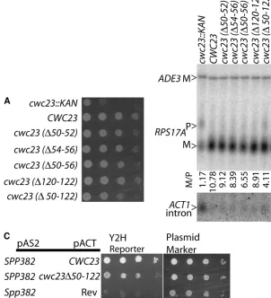
protein coding sequence (Figure 6A). While growth is
impaired at 30°, thecwc23TKANmutant does not show
much greater growth inhibition at lower (19°) or higher
(37°) temperatures (data not shown) relative to
wild-type yeast.
Given its tight physical association with the Spp382 splicing factor, a role for Cwc23p in pre-mRNA process-ing seemed likely. Indeed, we find that splicprocess-ing is very
poor in thecwc23TKANnull mutant compared with an
otherwise isogenic control strain (Figure 6B). Primer extension analysis of RNA isolated from this mutant confirms that the slowly migrating RNA band labeled pre-mRNA in Figure 6B is unspliced transcript and not
the similarly sized lariat intermediate (S. Pandit and
B. C. Rymond, unpublished observation). In addition to
reduced splicing efficiency, we note that the level of
excised intron product increases in the cwc23TKAN
deletion mutant. This apparent inconsistency (less RNA spliced but more intron present) is characteristic of mutations in the splicing apparatus defective in intron release from the spliceosome [e.g.,spp382(Panditet al.
2006)] or subsequent degradation of the released intron [e.g.,dbr1(Chapmanand Boeke1991)].
The only known role of the DnaJ motif is in the activation of Hsp70 chaperones during substrate pre-sentation. Since at least two Hsp70 proteins, Ssa2 and Ssa4, can be recovered with Cwc23 in the native yeast spliceosome (e.g., Wanget al.2003), it seemed possible
that the splicing and intron release defects of the
cwc23TKANmutant might result from reduced protein
chaperone activity. To test this, we mutated the canon-ical J domain by removal of the HPD tripeptide required
for chaperone function (Walshet al.2004). Cwc23p is
unusual in having a pair of nearly adjacent HPD repeats
within the J domain (i.e., 50HPDKHPD56) as well as a
third HPD sequence 29 amino acids downstream of the J domain (120HPD122). We removed each HPD
ele-ment singly (D50–52,D54–56,D120–122), removed the
first two HPD elements together (D50–56), or removed
all protein coding sequence between the first and last
HPD elements (D50–122) and assayed each construct for
the ability to support growth and splicing. For each mutant, three alanine codons replaced the deleted sequence. Surprisingly, each Cwc23p derivative sup-ported efficient growth and pre-mRNA splicing (Figure 6, A and B). Only the largest deletion, which removes
much of the DnaJ motif and 25% if the entireCWC23
protein coding sequence, shows a minor decrease in splicing efficiency. This Cwc23D50-122 derivative con-tinues to interact with Spp382p in the two-hybrid assay although with somewhat decreased efficiency (Figure 6C). On the basis of these observations, we conclude that similar to Spp382p, Cwc23p is critical for efficient cellular splicing and intron metabolism but that these functions do not require Cwc23p to act as a typical Hsp40-like chaperone.
Finally, while they are capable of exacerbating the
no evidence for direct interaction of Cwc23p or Sqs1p with Prp38p. Neither protein was reported in the U4/
U6.U5 tri-snRNP particle with Prp38p (Stevens and
Abelson1999; Stevenset al.2001) and we find that
full-length constructs of Cwc23p and Sqs1p fail to interact with Prp38p by the two-hybrid method (data not shown). In addition, when selected under conditions that release
Prp38p from the tri-snRNP particle (Xie et al. 1998),
Prp38p copurifies as a three-component complex with the Spp381p and Snu23p splicing factors without Cwc23p or Sqs1p (Figure S4).
DISCUSSION
Spp382p recruits and activates the Prp43 DExD/H-box protein to recycle splicing factors upon completion of the spliceosome cycle. In addition, diminished Spp382p or Prp43p activity suppresses splicing defects associated with theprp38-1,prp38-2,prp19-1,prp8-1, and
prp8-2 spliceosome assembly mutants (Pandit et al.
2006; S. Pandit and B. C. Rymond, unpublished
results), suggesting that this complex may also stimulate the disassembly of defective splicing complexes that differ in subunit composition from the postcatalytic spliceosome. Here we describe a genetic approach to find modifiers of Spp382p activity by seeking genes that
antagonize the growth of aspp382-1suppressedprp38-1
spliceosome assembly mutant when overexpressed. While the gene set examined was purposely biased to-ward splicing factors, most had no impact on growth.
However,10% of theGAL1-effector plasmids did show
growth inhibition upon induction and, consistent with the goals of this screen, most showed enhanced splicing defects when expressed at elevated temperature.
Fur-thermore, two of the identified genes,SQS1andCWC23,
are shown to have physical and genetic properties linked to Spp382p-dependent Prp43p activation and function.
While we have not studied the modes of splicing inhibition in detail, it seems likely that many of the splicing factors testing positive in this assay (i.e., Prp8, Prp9p, Prp11p, Yhc1p, and Prp39p) act by sequestering other spliceosomal components in nonproductive com-plexes. This situation might be exacerbated in the prp38-1 spp382-prp38-1background as this strain has both a primary
defect in spliceosome maturation due toprp38-1and a
second mutation that inhibits the recycling of spliceo-somal complexes (i.e.,spp382-1). The sequestration of splicing factors might antagonize Spp382-1p suppressor function or directly inhibit mRNA processing in this
splicing compromised background. For instance,
GAL1-SPP382presumably acts by direct complementation of
Figure 6.—Cwc23p is required for efficient
pre-mRNA splicing but its DnaJ motif is not crit-ical. (A) Growth of wild-type yeast (CWC23), the cwc23null mutant (cwc23TKAN), and yeast
spp382-1, revealing the conditional lethal defect of
prp38-1. Increased Prp43p levels might also antagonize
spp382-1 suppression although the constitutively high
levels of Prp43p (Ghaemmaghami et al. 2003) may
diminish the magnitude ofGAL1-PRP43 contribution.
Other genes may antagonize spp382-1 suppression by
actively promoting spliceosome disassembly. For in-stance, it is conceivable that excessive amounts of an early-acting splicing factor (e.g., the U1 snRNP protein, Yhc1p) might stimulate the dissociation of a late-acting
factor (e.g., the U6 snRNP) through competition for a
common protein or RNA binding sequence [the
pre-mRNA 59-splice site (Duand Rosbash2002)]. Dhh1p,
which stimulates mRNA decapping and decay (Coller
and Parker 2005), presumably acts by a mechanism
related to RNA stability.
Four genes without obvious connections to RNA processing were found to inhibit growth when overex-pressed in the double-mutant background. This might reflect indirect exacerbation of cellular processes ren-dered inefficient by the intrinsically poor splicing in this mutant and would include mitochondrial protein
synthesis or function (YML6, MRP13, and perhaps
YDR230) (Dimmer et al.2002) and vesicular transport
(BET2). Such indirect contributions would not explain
the greater splicing defect observed after GAL1
in-duction, however. An alternative possibility is that the artificially high levels of some effector proteins may reduce the expression or function of an authentic splicing factor. In this light, the identification of the
Bet2p b-subunit of geranylgeranyltransferase is
espe-cially curious since two other components of this path-way, the Bts1p geranylgeranyl diphosphate synthase and
the Bet4p a-subunit of the geranylgeranyltransferase,
were previously found as dosage suppressors with another splicing mutant (i.e., clf1D2) (Vincent et al.
2003). Geranylgeranyl substrates include various Ras,
Rho, Rab, and Rac signaling proteins (Dogbo et al.
1988; Kuzuguchi et al.1999). While the biochemical
basis of these genetic interactions needs additional study, it is enticing to speculate that geranylgeranyl addition may influence signal transduction pathway(s) shown to promote changes in splicing efficiency (Pleiss
et al.2007; see Lynch2007).
Beyond splicing defects, the expression of someGAL1
promoter fusion constructs alters pre-mRNA tail lengths, suggesting mechanistic differences in the RNA processing defects,e.g., short poly(A) tails with GAL1-CWC23expression and equal amounts of short and long withGAL1-PRP43. This observation is consistent with the hyperadenylation of nuclear retained unprocessed
pre-mRNA (Hilleren et al. 2001; Jensen et al. 2001;
Hammell et al. 2002), although the possibility of
inhibited poly(A) addition or enhanced metabolism for pre-mRNA with shorter poly(A) tails has not been ruled out. Finally, while we present these effects as due to simple overexpression, each construct is expressed with
a 19-kDa C-terminal affinity tag (Gelperinet al.2005) that
might interfere with normal function and inhibit splicing. But even in such cases, the elucidation of the underlying mode of splicing inhibition may add to our understand-ing of spliceosome homeostasis and function.
Like the Prp43p-binding proteins, Spp382p and
Pxr1p, Sqs1p has one copy of the 45-amino-acid
G-patch motif that is common to a subset of RNA
processing factors (Aravindand Koonin1999). Of the
five G-patch proteins annotated in the MIPS
Compre-hensive Yeast Genome Database (http://mips.gsf.de/
genre/proj/yeast/), three are essential (Spp2p,
Spp382p) or nearly essential (Pxr1p) and assist DExD/ H-box factors in RNA processing. Spp2p functions with Prp2p in splicing (Silvermanet al.2004) while Spp382p
and Pxr1p act with Prp43p in splicing and ribosome
biogenesis, respectively (Guglielmiand Werner2002;
Tsai et al. 2005; Boon et al. 2006; Pandit et al. 2006;
Tanakaet al.2007). Pxr1p and Spp382p also influence
telomerase activity and genomic stability (Lin and
Blackburn 2004; Herrmann et al. 2007), suggesting
additional contributions to nuclear function. The bi-ological roles for Sqs1p and the Ylr271W product are
unknown but because single (Giaever et al. 2002) or
combined (S. Panditand B. C. Rymond, unpublished
results) null mutants of these genes are viable, neither one makes a critical contribution to cellular biochemistry under standard conditions.
GAL1-SQS1 induction is shown here to poison the Prp43p-dependent processes of rRNA biogenesis and pre-mRNA splicing. When Sqs1p is expressed at phys-iological levels, it copurifies with Prp43p in complexes containing splicing and rRNA biogenesis factors (Gavin et al.2006; Kroganet al.2006) and a recent
screen for interactions among nonessential yeast genes provides evidence for possible Sqs1p function
in ribosome biogenesis or activity (Decourty et al.
2008). These observations, the Prp43p–Sqs1p two-hybrid interaction and the buffering effect of in-creased Prp43p expression in Sqs1p toxicity, make it likely that Prp43p is an intracellular binding partner of Prp43p. If so, direct competition between Sqs1p and the structurally similar Spp382p or Pxr1p for Prp43p association is an attractive possibility to account for the observed Sqs1p toxicity and RNA processing defects. Splicing rather than rRNA processing appears to be the
primary target of inhibition when GAL-SQS1 is
components of the exon junction complex (Nielsen
et al.2009).
Mutational studies suggest that the G-patch element in Spp382p is needed to stimulate Prp43p RNA helicase activity (Panditet al.2006; Tanakaet al.2007) and in
Spp2p to recruit Prp2p to the spliceosome (Silverman
et al.2004). While these observations suggest a protein-recognition interface, the G-patch motif has also been proposed to serve as an RNA-binding function (Bauerova-Zabranska et al.2005) and such function
has not been ruled out for any of the yeast G-patch proteins. Surprisingly, the removal of the Sqs1p G-patch motif did not reduce its ability to bind Prp43p or change the growth inhibition seen when overexpressed, ruling out a simple G-patch competition model for Prp43p as-sociation. As we were not able to detect convincing conservation outside of G-patch domains, Sqs1p likely binds Prp43p in a manner distinct from Spp382p or Pxr1p. Nevertheless, it remains likely that association of Spp382p, Pxr1p, and perhaps Sqs1p with Prp43p con-tributes to the functional partitioning of Prp43p be-tween the splicing and rRNA processing pathways.
Cwc23p was first found as a copurifying protein with
the Cef1p splicing factor (Ohi et al. 2002) and was
subsequently identified in other spliceosomal prepara-tions (Wanget al.2003; Chenet al.2007). Alternatively
reported as essential (Giaever et al. 2002) or
non-essential (Tizon et al.1999), we find the cwc23TKAN
null mutant to be viable but sickly. This stain is also genetically unstable, giving rise to more rapidly growing suppressors, an observation perhaps relevant to the
diversity of cwc23 phenotypes reported (S. Pandit,
M. Chen and B. C. Rymond, unpublished results).
Cwc23p interacts by the two-hybrid assay with Spp382p and is the only splicing factor seen to copurify with Spp382p at 450 mmNaCl (Panditet al.2006), raising the
possibility that Cwc23p may assist Spp382p in the re-cruitment, activation, or dissociation of Prp43p. Indeed, we find that thecwc23TKANmutant not only is splicing
defective but also accumulates excess excised introns, a characteristic of impaired spliceosome disassembly that
is observed with certain spp382 and prp43 mutants
(Martinet al.2002; Panditet al. 2006).
Twenty-two Hsp40p/DnaJ-like proteins are present in yeast (Walshet al.2004), with Cwc23p, a class C or III
subfamily member (Stirlinget al.2006; Voset al.2008)
and the only DnaJ protein observed in the yeast spliceosome. At least two DnaJ proteins, DNAJC8 and DNAJC13, copurify with the mammalian spliceosome but neither one has been studied for function in splicing ( Juricaet al.2002; Chenet al.2007). Removal
of much of the DnaJ motif from yeast Cwc23p has little obvious consequence on splicing, growth, or this protein’s interaction with Spp382p. Consequently, while important for efficient splicing, Cwc23p does not pro-vide an obligate Hsp40-like cochaperone function with spliceosome-associated Hsp70 proteins [e.g., Ssa2p and
Ssa4p (Wanget al.2003)] or use this canonical J domain
in an unexpected manner to stimulate another ATPase activity necessary for splicing (e.g., Prp43p). It remains possible, however, that Cwc23p stimulates a cellular Hsp70p needed for the processing of pre-mRNA from only certain genes or under special environmental
conditions when splicing efficiency is stressed (Yost
and Lindquist1991; Vogelet al.1995; Brackenand
Bond1999).
We thank Mingxia Zhang and Carly Joehl for valuable technical assistance and Daipayan Banerjee for helpful comments while this work was in progress. Support was provided by the National Institutes of Health award GM42476 to B.C.R.
LITERATURE CITED
Aravind, L., and E. V. Koonin, 1999 G-patch: a new conserved
do-main in eukaryotic RNA-processing proteins and type D retrovi-ral polyproteins. Trends Biochem. Sci.24:342–344.
Bauerova-Zabranska, H., J. Stokrova, K. Strisovsky, E. Hunter, T.
Rumlet al., 2005 The RNA binding G-patch domain in retroviral
protease is important for infectivity and D-type morphogene-sis of Mason-Pfizer monkey virus. J. Biol. Chem. 280: 42106– 42112.
Blanton, S., A. Srinivasanand B. C. Rymond, 1992 PRP38
enco-des a yeast protein required for pre-mRNA splicing and mainte-nance of stable U6 small nuclear RNA levels. Mol. Cell. Biol.12:
3939–3947.
Boon, K. L., T. Auchynnikava, G. Edwalds-Gilbert, J. D. Barrass,
A. P. Droopet al., 2006 Yeast ntr1/spp382 mediates prp43
func-tion in postspliceosomes. Mol. Cell. Biol.26:6016–6023. Bracken, A. P., and U. Bond, 1999 Reassembly and protection of
small nuclear ribonucleoprotein particles by heat shock proteins in yeast cells. RNA5:1586–1596.
Brow, D. A., 2002 Allosteric cascade of spliceosome activation.
Annu. Rev. Genet.36:333–360.
Burgess, S., J. R. Coutoand C. Guthrie, 1990 A putative ATP
binding protein influences the fidelity of branchpoint recogni-tion in yeast splicing. Cell60:705–717.
Chapman, K. B., and J. D. Boeke, 1991 Isolation and
characteriza-tion of the gene encoding yeast debranching enzyme. Cell65:
483–492.
Chen, Y. I., R. E. Moore, H. Y. Ge, M. K. Young, T. D. Leeet al.,
2007 Proteomic analysis of in vivo-assembled pre-mRNA splic-ing complexes expands the catalog of participatsplic-ing factors. Nu-cleic Acids Res.35:3928–3944.
Coller, J., and R. Parker, 2005 General translational repression by
activators of mRNA decapping. Cell122:875–886.
Combs, D. J., R. J. Nagel, M. Ares, Jr. and S. W. Stevens,
2006 Prp43p is a DEAH-box spliceosome disassembly factor es-sential for ribosome biogenesis. Mol. Cell. Biol.26:523–534. Decourty, L., C. Saveanu, K. Zemam, F. Hantraye, E. Frachon
et al., 2008 Linking functionally related genes by sensitive and quantitative characterization of genetic interaction profiles. Proc. Natl. Acad. Sci. USA105:5821–5826.
Dimmer, K. S., S. Fritz, F. Fuchs, M. Messerschmitt, N. Weinbach et al., 2002 Genetic basis of mitochondrial function and mor-phology in Saccharomyces cerevisiae. Mol. Biol. Cell13:847–853. Dogbo, O., A. Laferriere, A. D’Harlingue and B. Camara,
1988 Carotenoid biosynthesis: isolation and characterization of a bifunctional enzyme catalyzing the synthesis of phytoene. Proc. Natl. Acad. Sci. USA85:7054–7058.
Du, H., and M. Rosbash, 2002 The U1 snRNP protein U1C
recog-nizes the 59splice site in the absence of base pairing. Nature419:
86–90.
Durfee, T., K. Becherer, P. L. Chen, S. H. Yeh, Y. Yang et al.,
1993 The retinoblastoma protein associates with the protein phosphatase type 1 catalytic subunit. Genes Dev.7:555–569. Gallagher, J. E., D. A. Dunbar, S. Granneman, B. M. Mitchell, Y.
pre-rRNA processing are linked by specific SSU processome compo-nents. Genes Dev.18:2506–2517.
Gavin, A. C., P. Aloy, P. Grandi, R. Krause, M. Boesche et al.,
2006 Proteome survey reveals modularity of the yeast cell ma-chinery. Nature440:631–636.
Gelperin, D. M., M. A. White, M. L. Wilkinson, Y. Kon, L. A. Kung et al., 2005 Biochemical and genetic analysis of the yeast proteome with a movable ORF collection. Genes Dev.19:2816– 2826.
Ghaemmaghami, S., W. K. Huh, K. Bower, R. W. Howson, A. Belle et al., 2003 Global analysis of protein expression in yeast. Na-ture425:737–741.
Giaever, G., A. M. Chu, L. Ni, C. Connelly, L. Riles et al.,
2002 Functional profiling of the Saccharomyces cerevisiae ge-nome. Nature418:387–391.
Gietz, R. D., and A. Sugino, 1988 New yeast-Escherichia coli shuttle
vectors constructed with in vitro mutagenized yeast genes lacking six-base pair restriction sites. Gene74:527–534.
Guglielmi, B., and M. Werner, 2002 The yeast homolog of human
PinX1 is involved in rRNA and small nucleolar RNA maturation, not in telomere elongation inhibition. J. Biol. Chem.277:35712– 35719.
Hammell, C. M., S. Gross, D. Zenklusen, C. V. Heath, F. Stutz et al., 2002 Coupling of termination, 39processing, and mRNA export. Mol. Cell. Biol.22:6441–6457.
Herrmann, G., S. Kais, J. Hoffbauer, K. Shah-Hosseini, N.
Bruggenolteet al., 2007 Conserved interactions of the
splic-ing factor Ntr1/Spp382 with proteins involved in DNA double-strand break repair and telomere metabolism. Nucleic Acids Res.35:2321–2332.
Hilleren, P., T. McCarthy, M. Rosbash, R. Parker and T. H.
Jensen, 2001 Quality control of mRNA 39-end processing is
linked to the nuclear exosome. Nature413:538–542.
Ho, Y., A. Gruhler, A. Heilbut, G. D. Bader, L. Moore et al.,
2002 Systematic identification of protein complexes in Saccha-romyces cerevisiae by mass spectrometry. Nature415:180–183. Huh, W. K., J. V. Falvo, L. C. Gerke, A. S. Carroll, R. W. Howson
et al., 2003 Global analysis of protein localization in budding yeast. Nature425:686–691.
James, P., J. Halladayand E. A. Craig, 1996 Genomic libraries and
a host strain designed for highly efficient two-hybrid selection in yeast. Genetics144:1425–1436.
Jensen, T. H., K. Patricio, T. McCarthyand M. Rosbash, 2001 A
block to mRNA nuclear export in S. cerevisiae leads to hyperade-nylation of transcripts that accumulate at the site of transcrip-tion. Mol. Cell7:887–898.
Jurica, M. S., L. J. Licklider, S. R. Gygi, N. Grigorieffand M. J.
Moore, 2002 Purification and characterization of native
spli-ceosomes suitable for three-dimensional structural analysis. RNA8:426–439.
Kaiser, C., S. Michaelisand A. Mitchell(Editors), 1994 Methods in Yeast Genetics. Cold Spring Harbor Laboratory Press, Cold Spring Harbor, NY.
Konarska, M. M., and C. C. Query, 2005 Insights into the
mecha-nisms of splicing: more lessons from the ribosome. Genes Dev.
19:2255–2260.
Kos, M., and D. Tollervey, 2005 The putative RNA helicase Dbp4p
is required for release of the U14 snoRNA from preribosomes in Saccharomyces cerevisiae. Mol. Cell20:53–64.
Krogan, N. J., G. Cagney, H. Yu, G. Zhong, X. Guo et al.,
2006 Global landscape of protein complexes in the yeast Sac-charomyces cerevisiae. Nature440:637–643.
Kuzuguchi, T., Y. Morita, I. Sagami, H. Sagamiand K. Ogura,
1999 Human geranylgeranyl diphosphate synthase. cDNA clon-ing and expression. J. Biol. Chem.274:5888–5894.
Lebaron, S., C. Froment, M. Fromont-Racine, J. C. Rain, B. Monsarrat et al., 2005 The splicing ATPase prp43p is a component of multiple preribosomal particles. Mol. Cell. Biol.25:9269–9282.
Leeds, N. B., E. C. Small, S. L. Hiley, T. R. Hughesand J. P. Staley,
2006 The splicing factor Prp43p, a DEAH box ATPase, func-tions in ribosome biogenesis. Mol. Cell. Biol.26:513–522. Lin, J., and E. H. Blackburn, 2004 Nucleolar protein PinX1p
reg-ulates telomerase by sequestering its protein catalytic subunit in an inactive complex lacking telomerase RNA. Genes Dev. 18:
387–396.
Linder, P., 2006 Dead-box proteins: a family affair–active and
pas-sive players in RNP-remodeling. Nucleic Acids Res. 34:4168– 4180.
Lybarger, S., K. Beickman, V. Brown, N. Dembla-Rajpal, K. Morey et al., 1999 Elevated levels of a U4/U6.U5 snRNP-associated protein, Spp381p, rescue a mutant defective in spliceosome mat-uration. Mol. Cell. Biol.19:577–584.
Lynch, K. W., 2007 Regulation of alternative splicing by signal
trans-duction pathways. Adv. Exp. Med. Biol.623:161–174.
Martin, A., S. Schneiderand B. Schwer, 2002 Prp43 is an
essen-tial RNA-dependent ATPase required for release of lariat-intron from the spliceosome. J. Biol. Chem.277:17743–17750. Nielsen, K. H., H. Chamieh, C. B. Andersen, F. Fredslund, K.
Hamborget al., 2009 Mechanism of ATP turnover inhibition
in the EJC. RNA15:67–75.
Ohi, M. D., A. J. Link, L. Ren, J. L. Jennings, W. H. McDonaldet al.,
2002 Proteomics analysis reveals stable multiprotein complexes in both fission and budding yeasts containing Myb-related Cdc5p/Cef1p, novel pre-mRNA splicing factors, and snRNAs. Mol. Cell. Biol.22:2011–2024.
Pandit, S., B. Lynnand B. C. Rymond, 2006 Inhibition of a
spliceo-some turnover pathway suppresses splicing defects. Proc. Natl. Acad. Sci. USA103:13700–13705.
Pleiss, J. A., G. B. Whitworth, M. Bergkesseland C. Guthrie,
2007 Rapid, transcript-specific changes in splicing in response to environmental stress. Mol. Cell27:928–937.
Sambrook, J., E. F. Fritschand T. Maniatis, 1989 Molecular Clon-ing: A Laboraory Manual.Cold Spring Harbor Laboratory Press, Cold Spring Harbor, NY.
Silverman, E. J., A. Maeda, J. Wei, P. Smith, J. D. Beggset al.,
2004 Interaction between a G-patch protein and a spliceosomal DEXD/H-box ATPase that is critical for splicing. Mol. Cell. Biol.
24:10101–10110.
Stevens, S. W., and J. Abelson, 1999 Purification of the yeast
U4/U6.U5 small nuclear ribonucleoprotein particle and identification of its proteins. Proc. Natl. Acad. Sci. USA 96:
7226–7231.
Stevens, S. W., I. Barta, H. Y. Ge, R. E. Moore, M. K. Younget al.,
2001 Biochemical and genetic analyses of the U5, U6, and U4/ U6 x U5 small nuclear ribonucleoproteins from Saccharomyces cerevisiae. RNA7:1543–1553.
Stevenson, L. F., B. K. Kennedyand E. Harlow, 2001 A large-scale
overexpression screen in Saccharomyces cerevisiae identifies pre-viously uncharacterized cell cycle genes. Proc. Natl. Acad. Sci. USA98:3946–3951.
Stirling, P. C., S. F. Bakhoum, A. B. Feigl and M. R. Leroux,
2006 Convergent evolution of clamp-like binding sites in di-verse chaperones. Nat. Struct. Mol. Biol.13:865–870.
Tanaka, N., A. Aronovaand B. Schwer, 2007 Ntr1 activates the
Prp43 helicase to trigger release of lariat-intron from the spliceo-some. Genes Dev.21:2312–2325.
Tizon, B., A. M. Rodriguez-Torres and M. E. Cerdan, 1999
Disruption of six novel Saccharomyces cerevisiae genes reveals that YGL129c is necessary for growth in non-fermentable carbon sources, YGL128c for growth at low or high temperatures and YGL125w is implicated in the biosynthesis of methionine. Yeast
15:145–154.
Tsai, R. T., R. H. Fu, F. L. Yeh, C. K. Tseng, Y. C. Lin et al.,
2005 Spliceosome disassembly catalyzed by Prp43 and its associated components Ntr1 and Ntr2. Genes Dev. 19:2991– 3003.
Vincent, K., Q. Wang, S. Jay, K. Hobbs and B. C. Rymond,
2003 Genetic interactions with CLF1 identify additional pre-mRNA splicing factors and a link between activators of yeast ve-sicular transport and splicing. Genetics164:895–907.
Vogel, J. L., D. A. Parselland S. Lindquist, 1995 Heat-shock
pro-teins Hsp104 and Hsp70 reactivate mRNA splicing after heat in-activation. Curr. Biol.5:306–317.
Vos, M. J., J. Hageman, S. Carra and H. H. Kampinga,
2008 Structural and functional diversities between members of the human HSPB, HSPH, HSPA, and DNAJ chaperone fami-lies. Biochemistry47:7001–7011.
Walsh, P., D. Bursac, Y. C. Law, D. Cyrand T. Lithgow, 2004 The
Wang, Q., K. Hobbs, B. Lynnand B. C. Rymond, 2003 The Clf1p
splicing factor promotes spliceosome assembly through N-terminal tetratricopeptide repeat contacts. J. Biol. Chem. 278: 7875– 7883.
Xie, J., K. Beickman, E. Otteand B. C. Rymond, 1998 Progression
through the spliceosome cycle requires Prp38p function for U4/U6 snRNA dissociation. EMBO J.17:2938–2946.
Yost, H. J., and S. Lindquist, 1991 Heat shock proteins affect RNA
processing during the heat shock response of Saccharomyces cer-evisiae. Mol. Cell. Biol.11:1062–1068.
Supporting Information
http://www.genetics.org/cgi/content/full/genetics.109.106955/DC1
Spp382p Interacts With Multiple Yeast Splicing Factors, Including
Possible Regulators of Prp43 DExD/H-Box Protein Function
Shatakshi Pandit, Sudakshina Paul, Li Zhang, Min Chen, Nicole Durbin,
Susan M. W. Harrison and Brian C. Rymond
S. Pandit et al. 2 SI
Open reading frames fused to the GAL1 promoter used in this study. ORF Gene Name Alias
YBL026W LSM2 "SMX5, SNP3" YBL074C AAR2
YBR055C PRP6 "RNA6, TSM7269" YBR065C ECM2 SLT11
YBR102C EXO84 USA3 YBR119W MUD1 "U1-A, U1A" YBR152W SPP381
YBR188C NTC20
YBR237W PRP5 RNA5 YCL018W LEU2
YCL037C SRO9 YCR046C IMG1 YDL030W PRP9
YDL043C PRP11 RNA11 YDL084W SUB2
YDL087C LUC7 "EXM2, EPE1" YDL160C DHH1
YDL175C AIR2
YDL209C CWC2 NTC40 YDR007W TRP1
YDR037W KRS1 GCD5 YDR088C SLU7 SLT17 YDR163W CWC15
YDR225W HTA1 "H2A1, SPT11" YDR230W Dubious ORF
YDR235W PRP42 "MUD16, SNU65" YDR240C SNU56 MUD10
YDR243C PRP28 YDR283C GCN2 AAS1
YDR364C CDC40 "PRP17, SLT15, SLU4" YDR381W YRA1 SHE11
YDR416W SYF1 NTC90
YDR432W NPL3 "MTR13, MTS1, NOP3, NAB1" YDR473C PRP3 RNA3
YEL026W SNU13 YER006W NUG1 YER009W NTF2 YER013W PRP22
YER029C SMB1 "Sm B, SmB" YER107C GLE2 RAE1
YER112W LSM4 "SDB23, USS1" YER146W LSM5
YFL003C MSH4
YFL017W-A SMX2 "SNP2, YFL018W-A, SmG" YFR005C SAD1
YGL030W RPL30 "L30, L32, YL38, rp73" YGL049C TIF4632 eIF4G2
YGL120C PRP43 YGL122C NAB2 YGL128C CWC23
YGL147C RPL9A "L8A , L9A YL11, rp24" YGL238W CSE1 KAP109
YGR013W SNU71 YGR075C PRP38 YGR084C MRP13 YGR091W PRP31 YGR129W SYF2 NTC31 YGR162W TIF4631 eiF4G1 YGR204W ADE3
YGR278W CWC22
S. Pandit et al. 3 SI
YHR156C LIN1 SNU40
YHR165C PRP8 "DBF3, DNA39, RNA8, SLT21, USA2" YIL061C SNP1 "U1 70K, U1-70K"
YIR005W IST3 SNU17 YIR009W MSL1 YIB9 YJL080C SCP160
YJL124C LSM1 SPB8
YJL203W PRP21 SPP91 YJR022W LSM8
YJR050W ISY1 "UTR3, NTC30" YKL012W PRP40
YKL074C MUD2 YKL095W YJU2 CWC16 YKL173W SNU114 GIN10 YKL214C YRA2
YKR022C NTR2 YKR086W PRP16 YLL036C PRP19 PSO4 YLR016C PML1
YLR116W MSL5 BBP
YLR117C CLF1 "SYF3, NTC77" YLR132C Uncharacterized ORF YLR147C SMD3 "SLT16, Sm D3" YLR183C TOS4
YLR226W BUR2 CST4
YLR249W YEF3 "TEF3, Sm D2" YLR275W SMD2
YLR293C GSP1 "CNR1, CST17" YLR298C YHC1 U1C
YLR347C KAP95 RSL1 YLR409C UTP21
YLR424W SPP382 "CCF8, NTR1" YLR438C-A LSM3 "SMX4, USS2" YLR453C RIF2
YML025C YML6 YML029W USA1 YML046W PRP39 YML117W NAB6 YMR049C ERB1
YMR116C ASC1 CPC2 YMR213W CEF1 NTC85 YMR216C SKY1
YMR240C CUS1 YMR268C PRP24 YNL005C MRP7 YmL23 YNL118C DCP2 PSU1 YNL147W LSM7
YNL189W SRP1 "KAP60, SCM1" YNL210W MER1
YNL224C SQS1 YNL245C CWC25 YNL251C NRD1 YNL286W CUS2
YNR011C PRP2 RNA2 YNR053C NOG2 NUG2
YOL133W HRT1 "RBX1, ROC1, HRT2" YOL139C CDC33 "TIF45, eIF4E" YOR128C ADE2
YOR148C SPP2
YOR159C SME1 Sm E
YOR167C RPS28A "S28A , S33A , YS27 " YOR202W HIS3
S. Pandit et al. 4 SI
YPL151C PRP46 NTC50 YPL169C MEX67
YPL178W CBC2 "MUD13, SAE1, CBP20" YPL190C NAB3 HMD1
YPL213W LEA1
YPL235W RVB2 "TIP49B, TIH2, TIP49" YPR057W BRR1
YPR082C DIB1 SNU16 YPR094W RDS3
YPR101W SNT309 NTC25 YPR161C SGV1 BUR1 YPR176C BET2
YPR178W PRP4 RNA4 YPR182W SMX3 Sm F
S. Pandit et al. 5 SI
FIGURE S2.—A schematic of the yeast rRNA biogenesis. This image show the parallel processing of the 35S precursor ribosomal RNA into the mature 25S, 18S and 5.8S rRNA products through two alternative paths based on initial cleave at the A3 (left) or A0 (right) site is shown (see (GRANNEMAN et al. 2006; KOS and TOLLERVEY 2005) for related discussion). The placement of the hybridization probes used for the detection of the 35 S precursor and the 23S and 20S (probe 004) and 27SA2 (probe 003) intermediates is indicated on the first line.
35S
18S D
20S
5’ ETS ITS1 ITS2 3’ ETS
C2 C1
E B2B0
18S 25S
A0 A1 D A2A3 B1L B1S 5.8S C2 C1 E B2 18S 25S
A0 A1 D A2A3 B1L B1S 5.8S 27SA2 C2 C1 E B2 25S A3 B1L B1S 5.8S 32S C2 C1 E B2
18S D A2A3 25S
B1L B1S 5.8S 33S C2 C1 E B2 18S 25S
A1 D A2A3 B1L B1S 5.8S 27SBs C2 C1 E 25S 5.8S C2 C1 E 25S A3 B1L B1S 5.8S C2 C1 E 25S B1L B1S 5.8S 27SA3 E
5.8S C1 25S
26S 7Ss C2 C1 E 25S B1S 5.8S 27SL E B1S
5.8S C1 25S
26S 7SL
Primary Transcript
Adapted from: M. Kos, and D. Tollervey, 2005. Mol Cell 20:53-64; S. Granneman et. al, 2006. Mol Cell Biol 26:1183-94. 003
004
B0 cleavage
D cleavage
A1 cleavage
Exonuclease E-C2 & C2-C1 C2 cleavage
A0 cleavage first
Exonuclease B2-B0
A3 cleavage B1L cleavage
Exonuclease A3-B1s
Exonuclease E-C2 & C2-C1 C2 cleavage
5.8S 25S
5.8Ss 25S
25S B1S
5.8S
5.8SL 25S
18S
18S A3 cleavage first
18S
A0 A1 D A2
23S
18S
A1 D A2
22S
18S D A2 21S
A2 cleavage A0 cleavage
A1 cleavage
A2 cleavage
Probes Hybridization
S. Pandit et al. 6 SI
FIGURE S3.—Primary sequence alignment of the G-patch domains. The Saccharomyces MIPS database
((http://mips.gsf.de/genre/proj/yeast/) (2009) was used to identify five annotated G-patch proteins in baker’s yeast using
InterProScan Version 7.1 (InterPro domain = IPR000467). The alignment of the G-patch was done using Multalin (CORPET 1988) with a consensus sequence included beneath the aligned sequences. In parentheses after each protein name is the amino acid domain used for alignment followed by the length of the entire protein.
Spp382p (61-108/708) ..TYGIGAKL LSSMGYVAGK GL...GKDG. SGITTPIETQ SRPMHNAGLG MFSN Pxr1p (25-72/271) ..TSRFGHQF LEKFGWKPGM GL...GLSPM NSNTSHIKV. SIKDDNVGLG AKLK Sqs1p (720-767/767)..NENIGRRM LEKLGWKSGE GL...GIQGN KGISEPIFA. KIKKNRSGLR HSES Ylr271w (41-87/274) ..IMPRGYKM MENMGYKEGE TL...G.SNE SALKEPIKV. EINTKRRGIR AEKP Spp2p (100-149/185)VPVEEFGDAL LRGMGWESDS EQDSKGDKTQ SRNKDVSNVS QIHPDGLGIG .... Consensus ..te.fG.kl LekmGwksGe gL...G.sg. sgikepIkv. sIk.dn.Glg a...
Spp382p (61-108/708) ..TYGIGAKL LSSMGYVAGK GL...GKDG. SGITTPIETQ SRPMHNAGLG MFSN Pxr1p (25-72/271) ..TSRFGHQF LEKFGWKPGM GL...GLSPM NSNTSHIKV. SIKDDNVGLG AKLK Sqs1p (720-767/767)..NENIGRRM LEKLGWKSGE GL...GIQGN KGISEPIFA. KIKKNRSGLR HSES Ylr271w (41-87/274) ..IMPRGYKM MENMGYKEGE TL...G.SNE SALKEPIKV. EINTKRRGIR AEKP Spp2p (100-149/185)VPVEEFGDAL LRGMGWESDS EQDSKGDKTQ SRNKDVSNVS QIHPDGLGIG .... Consensus ..te.fG.kl LekmGwksGe gL...G.sg. sgikepIkv. sIk.dn.Glg a...
Sup 3
S. Pandit et al. 7 SI
No T
ag
Pr
p38-T
AP
Spp381-T
AP
Snu23-T
AP
<
<
<
<
<
<
Snu23p Prp38p Snu23-CBP Prp38-CBP Spp381p Spp381-CBP 150 kDa
100
75
50
37
25
20
<
55 kD backgroundFIGURE S4.—Prp38p, Spp382p, and Snu23p form a heteromeric complex without stably bound Cwc23p or Sqs1p. The indicated epitope tagged (TAP) yeast strains were metabolically labeled with Trans 35S-label (ICN) for 4 hours at 30C and then broken with glass beads in buffer A (10 mM HEPES, pH 7.9, 10 mM KCl, 200 mM NaCl, 10% glycerol, 0.5 mM dithiothreitol, 0.5 mM phenylmethylsulfonyl fluoride, 2 mM benzamide, 0.5% NP-40) and the lysate cleared by centrifugation at 14,000 X g. Sequential protein A agarose and calmodulin agarose affinity (TAP) selection was done as previously modified (WANG et al. 2003) except that the stringency of the wash steps was increased to 450 mM NaCl. The proteins were resolved on a SDS 10% polyacrylamide gel and imaged using a Typhoon 9600 phosphorimager (GE Healthcare). The positions of the molecular weight markers and the 55 kDa background band are noted at the left.
Three protein bands, Prp38p, Spp382p, and Spp381p co-purify independent of the protein used for selection. The band identities were confirmed by immune detection with anti-Prp38p antibody (Rymond, unpublished) and anti-CBP antibody (Santa Cruz Biotechnology) and by mass spectroscopy with a Deca Quadruopole ion trap mass spectrometer (ThermoElectron, Waltham, MA after extract scale-up using unlabeled sample prepared in an identical manner. CBP indicates the residual calmodulin binding peptide present in the TAP-tagged target protein after cleavage by TEV protease. Note: The Spp381 protein labels poorly with the 35S met, cys mixture but resolves as a band that migrates just below the dark background band of
approximately 55 kDa present in the untagged lane. We previously reported the anomalous electrophoretic migration of this 34 kDa protein detected using the small (9 amino acid) HA epitope (LYBARGER et al. 1999). Spp381-CBP migrates above the 55 kDa background band but is not well resolved on this gel system. Both Snu23p and Spp381p also interact with Prp38p in the two-hybrid assay (LYBARGER et al. 1999; UETZ et al. 2000).
CORPET,F., 1988 Multiple sequence alignment with hierarchical clustering. Nucleic Acids Res 16: 10881-10890.
GELPERIN,D.M., M.A.WHITE, M.L.WILKINSON, Y.KON, L.A.KUNG et al., 2005 Biochemical and genetic analysis of the yeast proteome with a movable ORF collection. Genes Dev 19: 2816-2826.
GRANNEMAN,S., K.A.BERNSTEIN, F.BLEICHERT and S.J.BASERGA, 2006 Comprehensive mutational analysis of yeast DEXD/H box RNA helicases required for small ribosomal subunit synthesis. Mol Cell Biol 26: 1183-1194. KOS,M., and D.TOLLERVEY, 2005 The Putative RNA Helicase Dbp4p Is Required for Release of the U14 snoRNA from
Preribosomes in Saccharomyces cerevisiae. Mol Cell 20: 53-64.
LYBARGER,S., K.BEICKMAN, V.BROWN, N.DEMBLA-RAJPAL, K.MOREY et al., 1999 Elevated Levels of a U4/U6.U5 snRNP-Associated Protein, Spp381p, Rescue a Mutant Defective in Spliceosome Maturation. Mol Cell Biol 19: 577-584. UETZ,P., L.GIOT, G.CAGNEY, T.A.MANSFIELD, R.S.JUDSON et al., 2000 A comprehensive analysis of protein-protein
interactions in Saccharomyces cerevisiae. Nature 403: 623-627.