Subjective orientation perception in hemispatial neglect:
Effects of internal and external mediators of subjective space perception
and of feedback-based perceptual training
Dissertation
at the Graduate School of Systemic Neuroscience
Ludwig-Maximilian-University Munich
Submitted by
MSc Johanna Funk
from Munich, Germany
Supervisors:
Dr. Kathrin Finke
Prof. Dr. Hermann Mülle
r
Declaration
I hereby confirm that this work submitted for assessment „Subjective orientation perception in hemispatial neglect: Effects of internal and external mediators of subjective space perception and of feedback-based perceptual training” is my own and is expressed in my own words, except where otherwise stated. Any uses made of the work of other authors (e.g., ideas, findings, tests, text) are properly acknowledged at the point of their use. A full list of the references employed has been included.
This dissertation has not already been accepted for any degree, and is also not being concurrently submitted for any other degree.
Munich, January 2012
Acknowledgements
This dissertation would not have been written without the contribution of many people. I am very grateful for that, and I thank the people who contributed directly to my PhD project, or who provided logistic help, support and encouragement.
My deepest gratitude is to my first advisor, Dr. Kathrin Finke who supported me during the whole PhD period, and on each individual level of my work. Kathrin already accompanied my university career from the first semester on and was one of the teachers who deepened my interest in cognitive Neuroscience. I have been fortunate to have an advisor who gave me freedom and encouragement to explore things on my own, and at the same time her competent guidance wherever necessary. Her patience and support helped me to overcome many difficult situations and finish this dissertation.
I am grateful to my second advisor, Prof. Dr. Hermann Müller, for supporting me during my Master and PhD studies. He accepted me as a student of the Neuro-cognitive Psychology masters program, which deepened my knowledge in cognitive Neuroscience and research methods and finally paved the way to my PhD project. I would like to thank Hermann for his insightful comments and constructive advice at various stages of my studies and research.
Prof. Dr. Georg Kerkhoff, my external advisor and mentor accompanied my university career from my first semester until today. His infectious enthusiasm for his research field inspired me to delve more deeply into the subject of cognitive Neuroscience. Georg supervised my first research project and my diploma thesis and encouraged me to apply for the Neuro-cognitive Psychology program. He accompanied my studies and research in a supportive and encouraging way and gave me great and invaluable support for my PhD project.
I would like to acknowledge my external collaborators, Kathrin Utz, Silvia Sollfranck, Stefan Reinhart, Mareike Kardinal, Alisha Rosenthal and Caroline Kuhn, for contributing to my research or revising my manuscripts. I am furthermore grateful to Rudolf Preger from the neurological clinic Kipfenberg for the permission to recruit patients, and Günter Neumann from Kipfenberg and Udo Münßinger from the Hospital Bogenhausen for their support in the recruitment of patients.
Most importantly, none of this would have been possible without the patience of my family, who has been a constant source of support and strength during the last years. I would like to express my gratitude to my parents for their encouragement and financial support and to my husband and daughter who anchored me in many situations.
List of publications
Funk, J., Finke, K., Mueller, H.J., Preger, R., & Kerkhoff, G. (2010a). Systematic biases in the tactile perception of the subjective vertical in patients with unilateral neglect, and the influence of upright vs. supine posture. Neuropsychologia, 48(1), 298-308.
Funk, J., Finke, K., Mueller, Utz, K.S., & Kerkhoff, G. (2010b). Effects of lateral head inclination on multimodal spatial orientation judgments in neglect: Evidence for impaired spatial orientation constancy. Neuropsychologia, 48(6), 1616-1627.
Funk, J., Finke, K., Mueller, H.J., Utz, K.S., & Kerkhoff, G. (2011). Visual context modulates the subjective vertical in neglect: Evidence for an increased Rod-and-Frame-Effect. Neuroscience, 173, 124-134.
Abstract
Table of Contents
Acknowledgements...iii
List of publications...v
Abstract...vi
Table of contents...vii
Chapter 1: General Introduction...1
1.1. Cortical pathways for visual perception: The dorsal and ventral visual system...2
1.1.1. Differential views on the dorsal visual system...3
1.1.2. Space processing and multimodal integration in parietal cortex...4
1.1.3. Orientation representation in parietal cortex...6
1.2. Neuropsychological disorders after parietal lesions...7
1.2.1. Visuospatial Disorders...8
1.2.2. Hemispatial Neglect...9
1.2.3. Deficits in subjective verticality perception...11
1.3. Rationale of the project...13
1.3.1. Hypotheses...14
1.4. References...17
Chapter 2: Studies on individual research questions...25
2.1. Systematic biases in the tactile perception of the subjective vertical in patients with unilateral neglect, and the influence of upright vs. supine posture...26
2.1.1. Abstract...27
2.1.2. Introduction...28
2.1.3. Methods...34
2.1.4. Results...39
2.1.5. Discussion...46
2.1.6. References...55
2.2. Effects of lateral head inclination on multimodal spatial orientation judgments in neglect: Evidence for impaired spatial orientation constancy...61
2.2.2. Introduction...63
2.2.3. Methods...70
2.2.4. Results...76
2.2.5. Discussion...87
2.2.6. References...95
2.3. Visual context modulates the subjective vertical in neglect: Evidence for an increased Rod-and-Frame-Effect...101
2.3.1. Abstract...102
2.3.2. Introduction...103
2.3.3. Methods...109
2.3.4. Results...115
2.3.5. Discussion...120
2.3.6. References...128
2.4. Effects of feedback-based visual line orientation discrimination training in visuospatial disorders after stroke...133
2.4.1. Abstract...134
2.4.2. Introduction...135
2.4.3. Methods...138
2.4.4. Results...145
2.4.5. Discussion...150
2.4.6. References...154
Chapter 3: General Discussion...159
3.1. Synopsis of results...160
3.2. Evidence for a supramodal orientation deficit in neglect...161
3.2.1. Impaired spatial orientation constancy in neglect...162
3.3. Putative mechanisms of plasticity following visuospatial training...164
3.3.1. Clinical relevance of training and transfer effects...165
3.4. Conclusion...166
3.5. Future directions...167
1.1. Cortical pathways for visual perception: The dorsal and ventral visual system
(e.g., position, orientation, motion direction and velocity, for reviews, see e.g. Desimone & Ungerleider, 1989; Maunsell & Newsome, 1987). Clinical studies in brain-damaged patients have shown that also in humans, there are specific clinical syndromes produced by occipitotemporal and occipitoparietal lesions depending on the respective module damaged by the lesion. Syndromes occurring after occipitotemporal lesions include e.g. visual object agnosia, prosopagnosia, and achromatopsia (e.g., Newcombe et al., 1987; for review, see Farah, 2004), whereas those occurring after occipitoparietal lesions include e.g. visuospatial neglect, apraxia, optic ataxia, akinotopsia, and disorders of spatial perception and cognition (e.g., Eglin et al., 1989; Vaina et al., 1990; Zihl et al., 1991; for review, see Newcombe & Ratcliff, 1989).
1.1.1. Differential views on the dorsal visual system
A re-interpretation of the two visual systems by Goodale and Milner (Goodale & Milner, 1992; Milner & Goodale, 2008) suggests that the two streams are not only specialized with regard to the kind of information they process, but rather with regard to the purpose of information processing: they assume that in the ventral stream, information about a range of object parameters is transformed for perceptual purposes, whereas in the dorsal stream (some of the same and some different) object parameters are transformed for the control of actions. In this view, the parietal cortex is processing visuospatial information for mediating visually guided (e.g., grasping) movements, but not visuospatial perception for recognition.
damage are often accompanied by visuomotor impairments, they cannot be fully accounted for by them.
On the basis of new anatomical data as well as a reconsideration of functional and clinical data, Rizolatti and Matelli (2003) tried to integrate the different views on the the functional role of the dorsal visual stream and proposed that the dorsal stream (and its recipient parietal areas) form two distinct functional systems: the dorso-dorsal stream, serving the ‘on-line’ control of actions, and the ventro-dorsal stream involved in space perception and the understanding of actions. Reviewing recent imaging and lesion studies, Husain and Nachev (2007) argued that inferior parietal regions (that were typically suggested to contribute to vision-for-action and/or spatial functions) also have non-spatial functions which are neither ‘dorsal’ nor ‘ventral’ in nature, such as sustaining attention or detecting salient events. Considering the diversity of functions attributed to the dorsal stream and its associated brain areas, it still remains necessary to further specify the construct ‘dorsal stream’ and the neuroanatomy underlying the specific sub-modules of this construct.
1.1.2. Space processing and multimodal integration in parietal cortex
sub-module of visuospatial perception which is highly specialized for the processing of clearly defined aspects of spatial information (for reviews, see e.g., Colby & Goldberg, 1999; Rizolatti & Matelli, 2003), including different aspects of motion processing (MT and MST), extrapersonal (MIP) and peripersonal / perioral (VIP) spatial information processing, and eyemovement- (LIP) or grasp-related spatial representation (AIP).
1.1.3. Orientation representation in parietal cortex
The assumption of the parietal cortex as the anatomical substrate of a supramodal spatial referece frame is supported by findings indicating the existence of multimodal (e.g. Duhamel, et al., 1998; Graziano & Gross, 1995) and ‘axis-orientation-selective’ (Sakata et al., 1997) neurons in the parietal cortex. Based on single-cell recordings in the monkey parietal cortex, Sakata et al. (1997; 1999) identified neurons in the lateral bank of the caudal intraparietal sulcus and in the anterior intraparietal sulcus which are sensitive to orientation information, though these cells might support different aspects of orientation perception with regard to intentional orientation discrimination as opposed to the visual guidance of hand actions. In humans, lesion and imaging studies support the view that the (right) parietal cortex plays a dominant role in spatial orientation perception, but also indicate that several brain regions contribute to visual orientation discrimination. Early patient studies reported that deficits in visual orientation discrimination frequently occur after right hemisphere lesions, typically affecting the (temporo-) parietal lobe and/or the basal ganglia, less frequently also after left frontal lesions (Benton et al., 1975; Kim et al., 1984). A recent patient study analyzing the lesion sites critical for line orientation discrimination showed that failure was most strongly associated with lesions in the right posterior parietal region (Tranel et al., 2009).
demonstrated that the caudal part of the intraparietal sulcus was activated more strongly in an orientation discrimination task than when surface orientation was used to guide reaching. Based on these findings and on their own observations in patients with parietal damage, Riddoch et al. (2004) proposed that orientation information is coded by different brain areas in parallel, but for different purposes. They suggest that the ventral system is concerned with an implicit coding of orientation into shapes (for pattern recognition) while the dorsal system is concerned with a more explicit coding of orientation.
1.2. Neuropsychological disorders after parietal lesions
and awareness. Accordingly, lesions restricted to the superior part in humans therefore often lead to disturbances of visuomotor control (such as optic ataxia). On the other hand, spatial neglect was attributed to lesions of the inferior part of the parietal lobe which is assumed to deal with abstract spatial processing based on input from the ventral stream, allowing the formation of perceptual and cognitive representations. However, the basic pathophysiological principles leading to spatial neglect are still an issue of debate. In an effort to resolve this debate, Karnath (1997) suggested that exploratory and goal-directed behavior in space do not share the same neural mechanisms. Karnath argued that space representation in the inferior parietal lobe most probably serves as a matrix for spatial exploration and orienting in space whereas visuomotor processes (involved e.g. in reaching for objects) are rather located in the superior parietal lobe. Accordingly, spatial neglect would typically occur after inferior parietal lesions, while optic ataxia would typically occur after superior parietal lesions. Although there is converging evidence for the anatomical dissociations within the parietal lobe, critical lesion sites inducing spatial neglect remain debated (see literature reviewed below). Since spatial neglect is characterized by a large heterogeneity in clinical manifestations, which overlaps with or comprises visuospatial and –constructive deficits, hemispatial neglect and visuospatial disorders are even more difficult to disentangle. Therefore, the criteria for diagnosing neglect and for differentiating between neglect and other, e.g. visuospatial, disorders have been challenged by some authors recently (e.g., Karnath & Rorden, in press).
1.2.1. Visuospatial Disorders
hemisphere (30-50%; Jesshope et al., 1991) lead to impairments in the perception of visual space as well as acting and orienting in space. Typically, right hemisphere lesions cause not only more frequent, but also more severe deficits compared to left hemisphere lesions (e.g., Kim et al., 1984). Deficits RBD patients show include line orientation judgements (Benton et al., 1975; De Renzi et al., 1971; Kim et al., 1984), line bisection / subjective straight ahead (e.g., Ferber & Karnath, 1999), size, distance and position estimation (e.g., Milner & Harvey, 1995; Tartaglione et al, 1981, 1983), clock reading / drawing (Freedman et al., 1994), and block design performance (Young et al., 1983).
After large MCA infarctions, visuospatial and -constructive disorders often occur simultaneously with spatial neglect. However, the two disorders can also occur independently of each other. Visuospatial disorders (and also spatial neglect) are often accompanied by substantial deficits in mobility and ADL functions (activities of daily living), e.g. dressing or transfer from bed to chair (Jesshope et al., 1991; Kaplan & Hier, 1984). Furthermore, these deficits are significant predictors of the course of disease, as they show adverse effects on therapy outcome of the patients and, thus, delay and impair their recovery. Patients with large, right hemispheric lesions show the poorest outcome in neurorehabilitation, which is at least partly due to anosognosia, the unawareness of the deficits. Anosognosia is an essential problem for the therapeutic success, since it is often little noticed but associated with problems in ADL functions. Therefore, information about the quality and magnitude of the visuospatial disorder is of great importance in the therapy of such disorders.
1.2.2. Hemispatial Neglect
et al., 1996), show unilateral spatial representational deficits (Bisiach & Luzatti, 1978; Bisiach et al., 1981) and frequently display a reduced use of their contralesional extremities (Laplane & Degos, 1983). Most of the current models of neglect focus on the explanation of deficits in the horizontal plane. Such deficits are apparent, for example, as left-sided omissions in visual search, reading, writing and drawing tasks, as deficits in (horizontal) size perception in the contralesional hemispace (Milner & Harvey, 1995; Milner et al., 1993), as a compression of contralesional hemispace (Gainotti & Tiacci, 1971; Nichelli et al., 1989) or even both hemispaces (Halligan & Marshall, 1991), as rightward deviations in line bisection and in pointing straight ahead, and as a deviation of space representation toward the ipsilesional hemispace (Karnath, 1997). However, numerous studies have demonstrated deficits in visuospatial perception and visuomotor performance that cannot result solely from impairments restricted to the horizontal plane. These include impairments in visual orientation discrimination and position estimation (Tartaglione et al., 1981, 1983; Taylor & Warrington 1973; Warrington & James 1967) as well as deficits in the judgment of the subjective visual vertical (SVV) and horizontal (SVH; Howard, 1982; Lenz, 1944), and judgments of oblique line orientations (Benton et al., 1975; De Renzi et al., 1971; Kim et al., 1984).
anatomical correlate of spatial neglect (as a consistent and uniform disorder), but rather started to identify dissociable functional components of the disorder and their anatomical correlates. In a recent pioneer study, Verdon et al. (2010) identified coherent profiles of co-varying deficits in a statistical factorial analysis of neglect performance and examined the neural correlates of these distinct profiles using a statistical voxel-based lesion-symptom mapping method. This analysis revealed three main factors (explaining 82% of the variance) suggesting distinct components related to perceptive/visuo-spatial, exploratory/visuo-motor, and allocentric/object-centred aspects of spatial neglect which were linked to specific neural correlates for each component, including the right inferior parietal lobe, the right dorsolateral prefrontal cortex, and deep temporal lobe regions. If such a multi-componential model of neglect based on factorial analyses proves valid in further studies, future research might not deal with ‚the’ neglect syndrome in the current sense any longer.
1.2.3. Deficits in subjective verticality perception
unilateral hemispheric lesions. MRI analyses revealed that the most impaired patients had lesions centering on the human homologue of the monkey parieto-insular-vestibular cortex (PIVC; Grüsser et al., 1990). In a recent study with 80 stroke patients, Pérennou et al. (2008) found that patients with right hemisphere lesions showed CCW visual (in 55% of the subjects), tactile (32.5%) and postural (42%) tilts in the frontal plane. Since especially parietal lesions caused marked visual and tactile tilts in the frontal plane, the authors concluded that the right parietal cortex is crucially involved in the elaboration of an internal supramodal model for verticality perception.
1.3. Rationale of the present project
A better understanding of the factors that mediate different aspects of spatial biases in visuospatial disorders and hemispatial neglect following (right) parietal brain lesions is important not only for obtaining a clearer picture of the nature and the underlying mechanisms of the spatial deficits but also for identifying potentially successful intervention schemes. Recent research revealed effective internal and external modulators of spatial deficits. Studies on the effectiveness of modulations of internal mediators of spatial deficits have used neck muscle vibration (e.g., Schindler et al., 2002), transcutaneous electroneural stimulation (TENS; Pizzamiglio et al., 1996), postural modulations (Karnath et al., 1998; Pizzamiglio et al., 1995), prism adaptation (e.g., Rossetti et al., 1998; Saevarsson et al., 2009; Vankilde & Habekost, 2010), and vestibular stimulation (Karnath, 1994); those on modulations of external (contextual) factors have employed optokinetic stimulation (e.g., Kerkhoff, 2000; Mattingley et al., 1994b) and cueing (e.g., Butter & Kirsch, 1995; Lin et al., 1996). This research enabled the development of the most applied neglect-therapies for spatially biased behaviour such as, for instance, extinction, the unawareness of contralesional stimuli, or motor neglect.
1995; Kerkhoff, 2000; Lin et al., 1996; Mattingley et al., 1994b), on SV judgments in RBD neglect has been carried out to date. Also, research investigating the effectiveness of therapeutic interventions for visuospatial disorders does not include reports on the effects of feedback-based orientation training on SV perception and orientation discrimination so far. The present project aimed at conducting a comprehensive set of experiments in order to obtain a clearer picture of the nature and the underlying mechanisms of the spatial deficits in RBD neglect and to identify potential mediators of subjective space perception and potentially successful intervention schemes. Therefore, we investigated whether and how spatial orientation deficits in RBD neglect are modulated (1) by internal factors mediating the perception of verticality, including body posture and lateral head orientation, (2) by visual context as an external mediator of space perception and (3) by the systematic feedback-based training of visual line orientations.
1.3.1. Hypotheses
more references. By contrast, in neglect patients, the processing of gravitational input is impaired (i.e., asymmetric; e.g. Pizzamiglio et al., 1995) and gravitational information cannot be used as the predominant ’intrinsic’ reference for the perception of the upright to the same extent as in healthy subjects. Therefore, it is suggested that RBD neglect patients show a pathological weighting of information integrated in parietal cortex to generate a subjective percept of space. With regard to manipulations of internal and external mediators of subjective space perception, they are assumed to display a much stronger impact of e.g. somatosensory or visual contextual information on subjective space perception.
Following this line of argumentation, we developed several hypotheses: (1) RBD neglect patients (but not brain-damaged control patients without neglect or healthy controls) generally exhibit a systematic visuospatial orientation deficit; that is, they generally display a substantial CCW tilt of their SV and other visual orientation judgements. (2) Axis orientation performance is differently modulated by internal (body posture, lateral head orientation) and external (visual context) modulators of subjective space perception in RBD neglect patients compared to control patients and healthy controls: SV judgements of all participants might generally vary as a consequence of the respective manipulation; however, performance of neglect patients should be far more strongly biased compared to all control groups, since these patients are assumed to be pathologically biased by internal and external cues as they cannot rely on gravitational information to the same extent as controls.
1.4. References
Andersen, R.A., Essick, G.K., & Siegel, R.M. (1985). The encoding of spatial location by posterior parietal neurones. Science, 230, 456-458.
Andersen, R.A., & Mountcastle, V.B. (1983). The influence of the angle of gaze upon the excitability of the light sensitive neurons of the posterior parietal cortex. Journal of Neuroscience, 3, 532-548.
Antonucci, G., Guariglia, C., Judica, A., Magnotti, L., Paolucci, S., Pizzamiglio, L., & Zoccolotti, P. (1995). Effectiveness of neglect rehabilitation in a randomized group study. Journal of Clinical and Experimental Neuropsychology, 17, 383-389.
Bender, M., & Jung, R. (1948). Abweichungen der subjektiven optischen Vertikalen und Horizontalen bei Gesunden und Hirnverletzten. European Archives of Psychiatry and Clinical Neuroscience, 181, 193-212.
Benton, A.L., Hannay, H.J., & Varney, N.R. (1975). Visual perception of line direction in patients with unilateral brain disease. Neurology, 25, 907-910.
Bisiach, E., Capitani, E., Luzatti, C., & Perani, D. (1981). Brain and conscious representation of outside reality. Neuropsychologia, 19, 543-551.
Bisiach, E., & Luzatti, C. (1978). Unilateral neglect of representational space. Cortex, 14, 129-133.
Bisiach, E., Pizzamiglio, L., Nico, D., & Antonucci, G. (1996). Beyond unilateral neglect. Brain, 119, 851-857
Brandt, Th., Dieterich, M., & Danek, A. (1994). Vestibular Cortex Lesions Affect the Perception of Verticality. Annals of Neurology, 35, 403-412.
Bremmer, F., Klam, F., Duhamel, J.-R., Ben Hamed, S., & Graf, W. (2002). Visual-vestibular interactive responses in the macaque ventral intraparietal area (VIP). European Journal of Neuroscience, 16, 1569-1586.
Brotchie, P.R., Andersen, R.A., Snyder, L.H., & Goodman, S.J. (1995). Head position signals used by parietal neurones to encode locations of visual stimuli. Nature, 375, 232-235. Butter, C.M., & Kirsch, N. (1995). Effect of lateralized kinetic visual cues on visual search in
patients with unilateral spatial neglect. Journal of Clinical and Experimental Neuropsychology, 17, 856-867.
Colby, C.L., & Goldberg, M. (1999). Space and attention in parietal cortex. Annual Review of Neuroscience, 22, 319-349.
Courtney, S.M., Ungerleider, L.G., Keil, K., & Haxby, J.V. (1996). Object and spatial visual working memory activate seperate neural systems in human cortex. Cerebral Cortex, 6(1), 39-49.
De Renzi, E., Faglioni, P., & Scotti, G. (1971). Judgment of spatial orientation in patients with focal brain damage. Journal of neurology, neurosurgery, and psychiatry, 34, 489-495.
De Renzi, E. (1982). Disorders of space exploration and cognition. Wiley, New York.
Desimone, R., & Ungedeider, L.G. (1989). Neural mechanisms of visual processing in monkeys. In F. Boller, & J. Grafman (Eds.), Handbook of Neuropsychology, Vol. 2. New York: Elsevier.
Duhamel, J.R., Colby, C.L., & Goldberg, M.E. (1998). Ventral intra-parietal area of the macaque: congruent visual and somatic response properties. Journal of Neurophysiology, 79, 126-136.
Eglin, M., Robertson, L.C., & Knight, R. T. (1989). Visual search performance in the neglect syndrome. Journal of Cognitive Neuroscience, 1, 372–385.
Faillenot, I., Sunaert, S., Van Hecke, P., & Orban, G. A. (2001). Orientation discrimination of objects and gratings compared: An fMRI study. European Journal of Neuroscience, 13, 585–596.
Farah, M. J. (2004). Visual Agnosia. Cambridge, MA: MIT Press.
Ferber, S., & Karnath, H.O. (1999). Parietal and occipital lobe contributions to perception of straight ahead orientation. Journal of neurology, neurosurgery, and psychiatry, 67, 572-578.
Freedman, M., Leach, L., Kaplan, E., Shulman, K. I., & Delis, D. C. (1994). Clock drawing: A neuropsychological analysis. New York: Oxford University Press.
Gainotti, G., Tiacci, C. (1971). The relationships between disorders of visual perception and unilateral spatial neglect. Neuropsychologia, 9(4), 451-458.
Goodale, M.A., & Milner, A.D. (1992). Separate visual pathways for perception and action. Trends in Neurosciences, 15(1), 20-25.
Grüsser, O.J., Pause, M., & Schreiter, U. (1990). Vestibular neurones in the parieto-insular cortex of monkeys (macaca fascicularis): visual and neck receptor responses. Journal of Physiology, 430, 559-583.
Halligan, P.W., & Marshall, J.C. (1991b). Spatial compression in visual neglect: a case study. Cortex, 27, 623-629.
Haxby, J.V., Grady, C.L., Horwitz, B., Ungerleider, L.G., Mishkin, M., Carson, R.E., & Herscovitch, P. (1991). Dissociation of object and spatial visual processing pathways in human extrastriate cortex. Proceedings of the National Academy of Sciences of the United States of America, 88, 1621-1625.
Hier, D.B., Mondlock, J., Caplan, L.R. (1983). Recovery of behavioral abnormalities after right hemisphere stroke. Neurology, 33, 345-350.
Howard, I.P. (1982). Human visual orientation. New York: Wiley.
Husain, M., & Nachev, P. (2007). Space and the parietal cortex. Trends in Cognitive Science, 11(1), 30-36.
Jesshope, H.J., Clark, M.S., Smith, D.S. (1991). The Rivermead Perceptual Assessment Battery: its application to stroke patients and relationship with function. Clinical Rehabilitation, 5, 115-122.
Kaplan, J., & Hier, D.B. (1982). Visuospatial deficits after right hemishpere stroke. The American Journal of Occupational Therapy, 36(5), 314-321.
Karnath, H.O. (1994). Subjective body orientation in neglect and the interactive contribution of neck muscle proprioceptive and vestibular stimulation. Brain, 117, 1001-1012.
Karnath, H.-O. (1997). Spatial orientation and the representation of space with parietal lobe lesions. Philosophical Transactions of the Royal Society, London B, 352, 1411 - 1419. Karnath H.-O., Fetter M., & Niemeier M. (1998). Disentangling gravitational, environmental,
and egocentric reference frames in spatial neglect. Journal of Cognitive Neuroscience, 10, 680–690.
Karnath, H.O., Ferber, S., & Himmelbach, M. (2001). Spatial awareness is a function of the temporal not the posterior parietal lobe. Nature, 411, 950-953.
Karnath, H.O., Fruhmann-Berger, M., Kuker, W., & Rorden, C. (2004). The anatomy of spatial neglect based on voxelwise statistical analysis: a study of 140 patients. Cerebral Cortex, 14, 1164-1172.
neglect: a cross-over study. Restorative Neurology & Neuroscience, 12, 27-40.
Kerkhoff, G. (1999). Multimodal spatial orientation deficits in left-sided visual neglect. Neuropsychologia, 37, 1387-1405.
Kerkhoff, G. (2000). Multiple perceptual distortions and their modulation in patients with left visual neglect. Neuropsychologia, 38(7), 1073-86.
Kerkhoff, G. (2001). Spatial hemineglect in humans. Progress in Neurobiology, 63, 1-27. Kerkhoff, G., & Zoelch, C. (1998). Disorders of visuo-spatial orientation in the frontal plane
in patients with visual neglect following right or left parietal lesions. Experimental Brain Research, 122, 108-120.
Kim, Y., Morrow, L., Passafiume, D., & Boller, F. (1984). Visuoperceptual and visuomotor abilities an locus of lesion. Neuropsychologia, 22(2), 177-185.
Laplane, D., & Degos, J.D. (1983). Motor neglect. Journal of Neurology, Neurosurgery, and Psychiatry, 46, 152-158.
Lenz, H. (1944). Raumsinnesstörungen bei Hirnverletzungen. Deutsche Zeitschrift für Nervenheilkunde, 157, 22-64.
Lin, K.C., Cermak, S.A., Kinsbourne, M., & Trombly, C.A. (1996). Effects of left-sided movements on line bisection in unilateral neglect. Journal of the International Neuropsychological Society, 2, 404-411.
Mattingley, J.B., Bradshaw, J.L., Bradshaw, J.A. (1994). Horizontal visual motion modulates focal attention in left unilateral spatial neglect. Journal of Neurology, Neurosurgery and Psychiatry, 57, 1228-1235.
Maunsell, J.H.R., and Newsome, W.T. (1987). Visual processing in monkey extrastriate cortex. Annual Review of Neuroscience, 10, 363–401.
Meerwaldt, J.D., & Van Harskamp, F. (1982). Spatial disorientation in right-hemisphere infarction. Journal of Neurology, Neurosurgery and Psychiatry, 45, 586-590.
Mesulam, M.M. (1999). Spatial attention and neglect: parietal, frontal, and cingulate contributions to the mental representation and attentional targeting of salient extrapersonal events. Philosophical Transactions of the Royal Society of London. Series B, Biological Sciences, 354, 1325-1346.
Milner, A.D., & Goodale, M.A. (1995). The visual brain in action. Oxford: Oxford University Press.
Milner, A.D., & Harvey, M. (1995). Distortion of size perception in visuospatial neglect. Current Biology, 5, 85-89.
Milner, A.D., Harvey, M., Roberts, R.C., & Forster, S.V. (1993). Line bisection errors in visual neglect: misguided action or size distortion? Neuropsychologia, 31(1), 39-49. Mishkin, M., Ungerleider, L.G., & Macko, K.A. (1983). Object vision and spatial vision: two
cortical pathways. Trends in Neurosciences, 6, 414-417.
Mort, D.J., Malhotra, P., Mannan, S.K., Rorden, C., Pambakian, A., Kennard, C., & Husain, M. (2003). The anatomy of visual neglect. Brain, 126, 1986-1997.
Newcombe, R., Ratcliff, G., & Damasio, H. (1987). Dissociable visual and spatial impairments following right posterior cerebral lesions: clinical, neuropsychological and anatomical evidence. Neuropsychologia, 25(1b), 149-161.
Newcombe, F. & Ratcliff, G. (1989). Disorders of visuo-spatial analysis. In F Boller, & J Grafman (Eds.), Handbook of Neuropsychology. New York: Elsevier.
Ng, V.W., Eslinger, P.J., Williams, S.C., Brammer, M.J., Bullmore, E.T., Andrew, C.M., Suckling, J., Morris, R.G., & Benton, A.L. (2000). Hemispheric preference in visuospatial processing: a complementary approach with fMRI and lesion studies. Human brain mapping, 10(2), 80-86.
Nichelli, P., Rinaldi, M., & Cubelli, R. (1989). Selective spatial attention and length representation in normal subjects and in patients with unilateral spatial neglect. Brain and cognition, 9(1), 57-70.
Pérennou, D. (2006). Postural disorders and spatial neglect in stroke patients: A strong association. Restorative Neurology and Neuroscience, 24(4-6), 319-334.
Pérennou, D., Mazibrada, G., Chauvineau, V., Greenwood, R., Rothwell, J., Gresty, M.A., & Bronstein, A.M. (2008). Lateropulsion, pushing and verticality perception in hemispheric stroke: a causal relationship? Brain, 131, 2401-2413.
Perenin, M.T. (1997). Optic ataxia and unilateral neglect: clinical evidence for dissociable spatial functions in posterior parietal cortex. In H.O. Karnath, & P. Thier (Eds.), Parietal lobe contributions to orientation in 3D space. Berlin: Springer.
Pizzamiglio, L., Vallar, G., & Doricchi, F. (1995). Gravity and hemineglect. NeuroReport, 7, 370 –371.
Pohl, W. (1973). Dissociation of spatial discrimination deficits following frontal and parietal lesions in monkeys. Journal of Comparative and Physiological Psychology, 82(2), 227-239.
Riddoch, M.J., Humphreys, G.W., Jacobson, S., Pluck, G., Bateman, A., & Edwards, M. (2004). Impaired orientation discrimination and localisation following parietal damage: on the interplay between dorsal and ventral processes in visual perception. Cognitive Neuropsychology, 21(6), 597-623.
Rizolatti, G., & Matelli, M. (2003). Two different streams form the dorsal visual system: anatomy and functions. Experimental Brain Research, 153, 146-157.
Rossetti, Y., Rode, G., Pisella, L., Farné, A., Boisson, D., & Perenin, M.T. (1998). Prism adaptation to a rightward optical deviation rehabilitates left hemispatial neglect. Nature, 98, 166-169.
Sack, A.T., Hubl, D., Prvulovic, D., Formisano, E., Jandl, M., Zanella, F.E., Maurer, K., Goebel, R., Dierks, T., & Linden, D.E.J. (2001). The experimental combination of rTMS and fMRI reveals the functional relevance of parietal cortex for visuospatial functions. Cognitive Brain Research, 13, 85-93.
Saevarsson, S., Kristjánsson, Á., Hildebrandt, H., & Halsband, U. (2009). Prism adaptation improves visual search in hemispatial neglect. Neuropsychologia, 47(3), 717-725.
Saj, A., Honoré, J., Davroux, J., Coello, Y., & Rousseaux, M. (2005). Effect of posture on the perception of verticality in neglect patients. Stroke, 36, 2203-2205.
Saj, A., Honoré, J., & Rousseaux, M. (2006). Perception of the vertical in patients with right hemispheric lesion: Effect of galvanic vestibular stimulation. Neuropsychologia, 44, 1509-1512.
Saj, A., Honoré, J., Bernati, T., Richard, C., & Rousseaux, M. (2008). Reducing rightward bias of subjective straight ahead in neglect patients by changes in body orientation. Journal of Neurology, Neurosurgery, and Psychiatry, 79(9), 991-996.
Sakata, H., Taira, M., Kusunoki, M., Murata, A., & Tanaka, Y. (1997). The parietal association cortex in depth perception and visual control of hand action. Trends in Neuroscience, 20, 350-357.
Schindler, I., Kerkhoff, G., Karnath, H.O., Keller, I., & Goldenberg, G. (2002). Neck muscle vibration induces lasting recovery in spatial neglect. Journal of Neurology, Neurosurgery & Psychiatry, 73, 412-419.
Shikata, E., Hamzei, F., Glauche, V., Koch, M., Weiller, C., Binkofski, F., et al. (2003). Functional properties and interactions of the anterior and posterior intraparietal areas in humans. European Journal of Neuroscience, 17, 1105–1110.
Snyder, L.H., Batista, A.P., & Andersen, R.A. (1997). Coding of intention in the posterior parietal cortex. Nature, 386, 167.
Taira, M., Kawashima, R., Inoue, K, & Fukada, H. (1998). A PET study of axis orientation discrimination. Neuroreport, 9(2), 283-288.
Tartaglione, A., Benton, A.L., Cocito, L., Bino, G., & Favale, E. (1981). Point localization in patients with unilateral brain damage. Journal of Neurology, Neurosurgery and Psychiatry, 44, 935-941.
Tartaglione, A., Cocito, L., Bino, G., Pizio, N., & Favale, E. (1983). Further evidence for asymmetry of point localization in normals and unilateral brain damaged patients. Neuropsychologia, 21, 407-412.
Taylor, A.M., & Warrington, E.K. (1973). Visual discrimination in patients with localized cerebral lesions. Cortex, 9, 82-84.
Tranel, D., Vianna, E., Manzel, K., Damasio, H. & Grabowski, T. (2009). Neuroanatomical correlates of the Benton Facial Recognition Test and Judgement of Line Orientation Test. Journal of Clinical and Experimental Neuropsychology, 31(2), 219-233.
Ungerleider, L.G., & Haxby, J.V. (1994). ‚What‘ and ‚where‘ in the human brain. Current Opinion in Neurobiology, 4(2), 157-165.
Ungerleider, L.G., & Mishkin, M. (1982). Two cortical visual systems. In D.J. Ingle, R.J.W. Mansfield, & M.S. Goodale (Eds.), The Analysis of Visual Behavior. Cambridge, MA: MIT Press.
Ungerleider, L.G., Courtney, S.M., & Haxby, J.V. (1998). A neural system for human visual working memory. Proceedings of the National Academy of Sciences of the United States of America, 95, 883-890.
Utz, K.S., Keller, I., Artinger, F., Stumpf, O., Funk, J., & Kerkhoff, G. (2011). Multimodal and multispatial deficits of verticality perception in hemispatial neglect. Neuroscience, 188, 68-79.
“biological motion” and “structure from motion” perception in a patient with impaired motion mechanisms: a case study. Visual Neuroscience, 5, 353–369.
Vallar, G., & Perani, D. (1986). The anatomy of unilateral neglect after right hemisphere stroke lesions. A clinical/CT-scan correlation study in man. Neuropsychologia, 24, 609-622.
Vandenberghe, R., Dupont, P., De Bruyn, B., Bormans, G., Michiels, J., Mortelmans, L., & Orban, G.A. (1996). The influence of stimulus location on the brain activation pattern in detection and orientation discrimination. A PET study of visual attention. Brain, 119, 1263-1276.
Vangkilde, S., & Habekost, T. (2010). Finding Wally: Prism adaptation improves visual search in chronic neglect. Neuropsychologia, 48(7), 1994-2004.
Verdon, V., Schwartz, S., Lovblad, K.O., Hauert, C.A., & Vuilleumier, P. (2010). Neuroanatomy of hemispatial neglect and ist functional components: a study using voxel-based lesion-symptom mapping. Brain, 133, 880-894.
Von Cramon, D., & Kerkhoff, G. (1993). On the cerebral organization of elementary spatial perception. In B. Gulyas, D. Ottoson, & P. Roland (Eds.), Functional oranisation of the human visual cortex. Oxford: Pergamon.
Warrington, E.K., & James, M. (1967). Disorders of visual perception in patients with localised cerebral lesions. Neuropsychologia, 5, 253-266.
Weinberg, J., Piasetsky, E., Diller, L., & Gordon, W. (1982). Treating perceptual organization deficits in nonneglecting RBD stroke patients. Journal of Clinical and Experimental Neuropsychology, 4, 59-74.
Yelnik, A. P., Lebreton, F. O., Bonan, I. V., Colle, F., Meurin, F. A., Guichard, J. P., & Vicaut, E. (2002). Perception of Verticality After Recent Cerebral Hemispheric Stroke. Stroke, 33, 2247-2253.
Young, G.C., Collins, D., & Hren, M. (1983). Effect of pairing scanning training with block design training in the remediation of perceptual problems in left hemiplegics. Journal of Clinical Neuropsychology, 5, 201-212.
2.1. Systematic biases in the tactile perception of the subjective vertical in patients with
unilateral neglect and the influence of upright vs. supine posture
2.1.1. Abstract
Patients with right hemisphere lesions often show contralesional neglect. Recent research focused on deficits beyond the typical neglect symptoms observed in the horizontal plane. Studies investigating deficits in the frontal and sagittal plane revealed impairments in the judgment of the subjective vertical. Systematic deviations in the subjective vertical have been demonstrated in the visual and tactile modality, indicating a supramodal spatial orientation deficit. Further, the magnitude of deviations appears to be manipulable by modulations of body posture. The present study investigated the subjective tactile vertical (STV) in neglect patients in the frontal and sagittal plane and its dependence on posture. Neglect patients and healthy controls performed tactile-spatial judgments of axis orientations in supine and upright posture. Neglect patients displayed a marked variability as well as a systematic tilt in their STV judgments. The STV was tilted counterclockwise in the frontal and backward in the sagittal plane. This tilt was larger in severe compared to moderate neglect patients, while it was not evident in healthy subjects. Our results support previous evidence and indicate a multisensory spatial orientation deficit in neglect patients which is related to neglect severity. Further, we found that performance of neglect patients deteriorated in supine compared to upright posture. This finding conflicts with the suggestion of a performance benefit in supine posture due to reduced (asymmetric) gravitational input. The negative effect of supine posture on the spatial bias in neglect is discussed with respect to a presumably further reduced intrinsic alertness state in the typically hypo-aroused neglect patients.
2.1.2. Introduction
Neglect is a supramodal neurological disorder characterized by a complex syndrome of sensory, motor and representational deficits (for a review, see Kerkhoff, 2001). Patients with hemispatial neglect fail to detect or respond to stimuli in their contralesional hemispace (Bisiach, Pizzamiglio, Nico, & Antonucci, 1996), show unilateral spatial representational deficits (Bisiach & Luzatti, 1978; Bisiach, Capitani, Luzatti, & Perani, 1981) and frequently display a reduced use of their contralesional extremities (Laplane & Degos, 1983). By definition, for diagnosing neglect, these deficits must not be primarily due to sensory, motor or cognitive/emotional impairments. Neglect frequently occurs after infarctions in the territory of the right (less often of the left) middle cerebral artery (Vallar, 1993), causing lesions which center on the inferior parietal cortex (BA 40, 7; Mort et al., 2003; but see Karnath, Ferber, & Himmelbach, 2001, for a different view).
All of these current models of neglect focus on the explanation of deficits in the horizontal plane. Such deficits are apparent, for example, as left-sided omissions in visual search, reading, writing and drawing tasks, as deficits in (horizontal) size perception in the contralesional hemispace (Milner & Harvey, 1995; Milner, Harvey, Roberts, & Forster, 1993), as a compression of contralesional hemispace (Gainotti & Tiacci, 1971; Nichelli, Rinaldi, & Cubelli, 1989) or even both hemispaces (Halligan & Marshall, 1991), as rightward deviations in line bisection and in pointing straight ahead, and as a deviation of space representation toward the ipsilesional hemispace (Karnath, 1997).
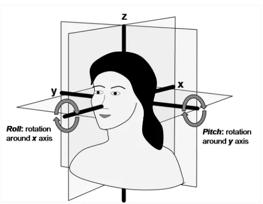
However, numerous studies have demonstrated deficits in visuospatial perception and visuomotor performance in patients with right-sided parietal lesions that cannot result solely from impairments restricted to the horizontal plane (for a review, see De Renzi, 1982). These include impairments in visual orientation discrimination and position estimation (Warrington & James 1967; Taylor & Warrington 1973; Tartaglione, Benton, Cocito, Bino, & Favale, 1981; Tartaglione, Cocito, Bino, Pizio, & Favale, 1983) as well as deficits in the judgment of the subjective visual vertical (SVV) and horizontal (SVH; Howard, 1982; Lenz 1944; De Renzi, Faglioni, & Scotti, 1971), and judgments of oblique line orientations (Kim, Morrow, Passafiume, & Boller, 1984; Benton, Hannay, & Varney, 1975). Such deficits in the judgment of the principal axes represent abnormalities in visuospatial perception in another spatial plane: the frontal (or roll) plane (see figure 1).
oblique orientation. The deficits were not a general consequence of brain damage because patients with left or right hemisphere lesions, but without neglect, performed at the level of healthy control subjects. Furthermore, Yelnik and colleagues (2002) showed that deviations of the SVV do not primarily depend on the localization and size of the underlying lesion, but are rather correlated with the severity of spatial neglect. In summary, these findings indicate a severe disturbance in the representation of space in the frontal plane in neglect patients which is related to the severity of neglect and, thus, does not seem to constitute an epiphenomenon, but rather one of the core deficits of these patients.
Figure 1: Schematic illustration of the three orthogonal planes defining three-dimensional space: frontal or roll plane, sagittal or pitch plane and horizontal or yaw plane.
Saj, Honoré, Bernati, Coello, and Rousseaux (2005) extended the assessment of spatial representations into a third, the sagittal (or pitch) plane (see figure 1). Neglect patients were presented with a luminous bar which they had to rotate and orient in a vertical position, either in the roll plane or in the pitch plane. The CCW tilt of the SVV in the roll plane was replicated. Furthermore, in the sagittal plane, too, neglect patients showed a systematic deviation from the true vertical, that is, a significant backward tilt of the SVV (hence towards the observer).
A further experiment of the Kerkhoff (1999) study was the first to demonstrate influences of body posture, specifically, lateral head inclination, on visual and tactile orientation judgments in a neglect patient (but not in a healthy control subject). The orientation deficits of the patient were significantly aggravated by a tilt of the head to the left by 25°, and significantly reduced by a comparable rightward tilt. This finding suggests that deficits in spatial orientation judgments are significantly modulated by gravitational input in neglect patients.
visuohaptic subjective vertical in the roll plane. Neglect patients and healthy control subjects oriented a bar into a subjectively vertical position in four different body posture conditions: (1) sitting with plantar sole support, (2) sitting without plantar sole support, (3) sitting with legs extended on a support, and (4) supine body position. It is important to note that the graviceptive information was varied via the modulation of the otholitic input only in the supine body position. In all other postures, only the somatosensory information changed, but the graviceptive input was kept stable. It was found that the body posture had a significant effect on SVV judgments in the neglect patients, but not in the control subjects. The CCW tilts of neglect patients decreased from position (1) through positions (2) and (3) to position (4), whereby the supine body position differed significantly from the three sitting positions. These data indicate that the progressive change of body posture and the accompanying modulation of graviceptive input and somatosensory afferences resulted in a progressive reduction of the bias in the subjective perception of verticality in neglect patients. Positive effects of lying body position on line bisection deviations (Pizzamiglio, Vallar, & Doricchi, 1995) and of a backward-tilted body position on exploration bias (Karnath, Fetter, and Niemeier, 1998) have also been documented in neglect.
Pizzamiglio et al., 1995). In supine position, the influence of otholitic input on judgments of orientations in space is reduced due to a decreasing sensitivity of the utricles with increasing backward tilt of the head, leading to deteriorations of orientation judgments (Howard, 1982). In patients with spatial neglect, a reduction of the influence of pathologically disturbed graviceptive input in the lying body position might therefore reduce the (in this case disrupting) effect of graviceptive information on the perception and representation of space (Pizzamiglio et al., 1995; Saj et al., 2005b; Saj et al., 2008).
The supramodal spatial perception and representation impairments of neglect patients do not only affect the horizontal, but also other spatial planes (e.g., Kerkhoff & Zoelch, 1998; Saj et al., 2005a). Although different potential mechanisms might underlie deviations of the subjective vertical in these planes, the tilts in all planes can be interpreted as manifestations of a generally deficient representation of spatial information. The severity and variability of the tilts of the principal axes were shown to correlate with that of spatial neglect in both the visual and the tactile modality (Kerkhoff, 1999). For the SVV (Saj et al., 2005b), it has been furthermore documented that the deviations are influenced by body posture.
upright body posture, we expected vestibular input to play a dominant role in verticality perception, whereas in the lying body posture, the impact of vestibular input was expected to be reduced and that of somatosensory input to be decisive (e.g., Anastasopoulos, Bronstein, Haslwanter, Fetter, &, Dichgans, 1999; Howard, 1982). Since the processing of vestibular information is deficient in patients with spatial neglect (e.g., Lafosse, Kerckhofs, Troch, Santens, & Vandenbussche, 2004; Pérennou, 2006; Pizzamiglio et al., 1995), the systematic tilt of the STV was expected to be reduced in lying compared to upright body posture. From these considerations, we derived the following hypotheses: (1) Neglect patients show larger STV deviations than healthy control subjects. (2) The neglect patients’ tilt is systematic: it is CCW in the frontal plane and backwards in the sagittal plane. (3) Neglect patients show reduced STV tilts in the lying compared to an upright body posture. (4) There is a significant positive correlation between the severity of neglect and the size and variability of the STV tilt.
2.1.3. Methods
Subjects
months, S.D. = 1.7 months). The age of the neglect patients ranged from 33 to 78 years, mean age was 57 years (S.D. = 12.0 years). The healthy control group consisted of ten subjects who were age-matched to the neglect patients (31-73 years; mean = 56.1 years; S.D. = 12.9 years) and ten younger subjects (age 20-29; mean = 25.4 years; S.D. = 3.1 years). To check for possible confounds due to age effects in the healthy control subjects, separate ANOVAs with the factors group (young vs. old control), plane (roll, pitch) and posture (supine, upright), were calculated for each of the STV parameters preceding the analysis of interest. Since these ANOVAs did not reveal a significant effect of age or any interaction with other factors, healthy control subjects (age-matched and younger controls) were combined in one control group for further analyses.
Neglect tests
Tactile-spatial tests
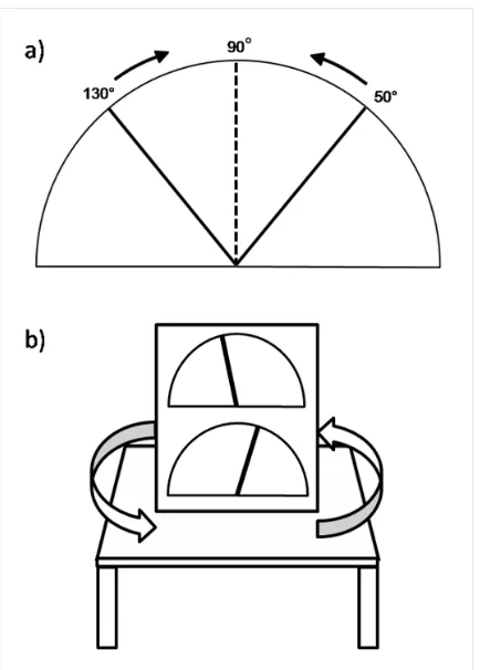
The subjective tactile vertical (STV) was measured in an angle-fitting procedure in two spatial planes (frontal and sagittal) and two body postures (upright and supine). Subjects were presented with two metal rods (length: 15 cm, width: 4 mm) attached to a plate (height: 50 cm, width: 40 cm). The plate contained two rods (an upper and a lower one) in order to allow subjects to accomplish the task using the rod which was easier to reach for them. The experimental plate was mounted on a bench (34 cm high) vertically 0.5 m in front of the patient. The plate could be rotated on the bench in the horizontal plane, which permitted the STV in the participants’ frontal and sagittal plane to be tested. The rod was rotatable on a semi-circle in the plate plane. Along the radius of the rotatable rod, a scale comprising 180° (horizontal left: 0°, vertical: 90°, horizontal right: 180°) allowed for the measurement of the angle of the rod in steps of 1° (see figure 2).
tactilely explore the whole experimental setup, especially the angles of the plate and the bench. In each condition, the apparatus was centered relative to the mid-sagittal plane of the participant, and the distance between the participant and the rod (0.5 m) was kept similar.
Figure 2: Experimental setup. 1a) Subjects adjusted a rod to their STV, starting at 50° or 130°. 1b) The setup contained two rods (an upper and a lower one) and subjects accomplished the task using the rod that was easier to reach for them. The rods were attached to a plate which was mounted on a bench and could be rotated on the bench in the transverse plane, permitting testing in the frontal and sagittal plane.
respectively, counter-clockwise (CCW) from the true vertical. Between blocks, participants had the opportunity to take a short break. Time for STV adjustment was not limited, in order to enable patients to compensate for any motor or attention deficits. STV angles in degrees were registered by the experimenter on a protocol sheet.
2.1.4. Results
Subjective tactile vertical - parameters
As a measure of the central tendency of STV deviation from the true vertical, constant errors were computed: individual STV adjustments were subtracted from 90° and averaged across all trials within a condition. This parameter displays possible systematic CW or CCW tendencies of the STV in each experimental condition (CW deviations: negative sign; CCW deviations: positive sign). Additionally, mean error size (=unsigned errors) was computed as a measure of the amplitude of the deviation. Mean error size was calculated via the absolute values of the deviations, thus disregarding their directions. Furthermore, the interval of uncertainty was determined, in terms of the complete range within which the subject considered the displayed rod as being exactly vertical (biggest – smallest STV value).
Condition sequence and starting position
Neither the sequence of the conditions nor the starting position did have an effect on any STV parameter (all P > 0.10). There were no significant interactions between condition sequence or starting position and any other factor (P > 0.10), except one: for the intervals of uncertainty, there was a significant four-way interaction between plane, posture, group and starting position (F(1, 38) = 5.49; P < 0.05). This interaction was based on larger intervals of uncertainty of neglect patients in the pitch plane when the starting position was directed towards the subject, especially in the upright posture. Interestingly, in the roll plane, starting position did not have an effect on STV parameters, neither did it interact with the factor subject group, i.e. the direction of movement did not influence the performance of neglect patients. Since the observed interaction is not related in any way to our hypothesis on effects and interactions between the factors of interest, starting position and condition sequence were not included in the further analysis of the data.
Size of deviations
Figure 3: Mean intervals of uncertainty (and associated SEM) of neglect patients and healthy controls for both body postures in the roll and pitch plane. C, healthy control subjects; N, patients with neglect; performance in the roll plane is shown in black, performance in the pitch plane is shown in grey; solid lines indicate performance in upright posture, dashed lines indicate performance in supine posture.
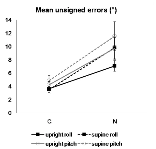
Separate mixed (2x2x2) ANOVAs with the factors group (neglect, control), plane (roll, pitch) and posture (supine, upright) were calculated for mean unsigned errors and intervals of uncertainty. ANOVAs revealed a significant ‘group’ effect on the intervals of uncertainty [F(1, 38) = 11.37; P < 0.01] and on the mean error size [F(1, 38) = 13.15; P < 0.01], i.e. neglect patients showed significantly larger intervals of uncertainty and mean unsigned errors in their STV judgments than healthy controls. For the intervals of uncertainty, there was no further main effect (P > .30) or significant interaction (P > .30). However, for the mean unsigned errors, there was a significant main effect of plane [F(1, 38) = 8.20; P < 0.01], a significant main effect of posture [F(1, 38) = 5.53; P < 0.05] and a by trend significant interaction between group and posture [F(2, 38) = 3.63; P < 0.07]. In general, subjects showed significantly larger unsigned errors in the pitch plane compared to the roll plane and in supine compared to upright posture. The difference between posture conditions was more pronounced in the neglect patients compared to healthy controls, i.e. neglect patients performed significantly worse in supine compared to upright posture [t(19) = 2.36; P < 0.05], while healthy controls did not show such a significant difference (P > 0.60).
Direction of tilt
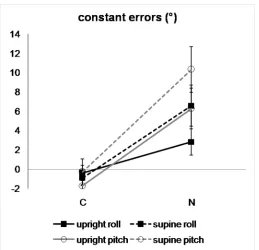
Figure 5:Mean constant errors (and associated SEM) of neglect patients and healthy controls for both body postures in the roll and pitch plane. C, healthy control subjects; N, neglect patients; performance in the roll plane is shown in black, performance in the pitch plane is shown in grey; solid lines indicate performance in upright posture, dashed lines indicate performance in supine posture.
subject in both planes (across postures). As can be seen, constant errors of healthy subjects do not differ systematically from zero into any direction, whereas constant errors of most neglect patients differ systematically from zero in a positive direction, with larger deviations in the severe neglect patients. Additional one sample t-tests comparing the constant errors of each group to zero deviation (i.e. the objective vertical) were calculated to demonstrate that differences in constant errors between neglect patients and healthy controls were due to significant CCW deviations of neglect patients’ subjective verticals. T-tests showed that constant errors of healthy subjects did not differ significantly from zero [t(19) = -0.93; P > 0.35]. By contrast, constant errors of neglect patients revealed a significant positive deviation from zero indicating a CCW tilt of their STV judgments [t(19) = 3.95; P < 0.01].
Relation between neglect severity and STV
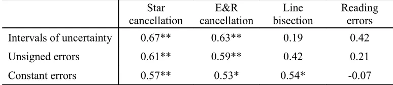
Hypothesis 4 predicted a relationship between the severity of the neglect and the size of the STV deviations. To evaluate whether the tactile-spatial deficits observed in our study were related to neglect, we correlated the individual STV parameters of each neglect patient (constant errors, mean unsigned errors and intervals of uncertainty averaged across postures and planes) with the composite neglect severity score. Spearman rank correlations revealed no significant correlation with neglect severity for the constant errors (r = 0.21; P > 0.15) and the mean unsigned errors (r = 0.21; P > 0.15). The correlation between the interval of uncertainty and the neglect severity index tended to be significant (r = 0.33; P < 0.09). To further analyze the relation between typical neglect performance in individual screening tests and performance in our tactile-spatial task, we computed Pearson correlations between the STV parameters of the neglect patients (constant errors, mean unsigned errors and intervals of uncertainty averaged across postures and planes) and the patients’ performance in the individual screening tests (line bisection, star cancellation, E&R cancellation and reading). Table 2 displays the Pearson correlation coefficients. As can be seen all three STV parameters were significantly correlated with performance in both cancellation tasks whereas line bisection performance was correlated only with constant errors and reading performance was not correlated with any parameter.
Star cancellation E&R cancellation Line bisection Reading errors
Intervals of uncertainty 0.67** 0.63** 0.19 0.42
Unsigned errors 0.61** 0.59** 0.42 0.21
Constant errors 0.57** 0.53* 0.54* -0.07
Further, to compare performance of all subjects in the roll and the pitch plane, Pearson correlations were computed to assess correlations between STV parameters in the two planes, i.e. each parameter in the roll plane (averaged across posture conditions) was correlated with the same parameter in the pitch plane. Correlations between STV parameters in the roll and pitch plane were highly significant (all r > 0.75; all P < 0.01) indicating that subjects showed highly comparable patterns of results in the two planes.
2.1.5. Discussion
Recently, deficits beyond the horizontal plane have been described in neglect patients that include impairments also in the frontal (roll) and the sagittal (pitch) plane (e.g., Saj et al., 2005; Kerkhoff & Zoelch, 1998; Kerkhoff, 1999). CCW deviations of the SVV and STV in the roll plane and backwards directed deviations of the SVV in the pitch plane (Kerkhoff & Zoelch, 1998; Kerkhoff, 1999; Saj et al., 2005a) were shown to be related to neglect severity (Kerkhoff & Zoelch, 1998; Kerkhoff, 1999). Further, Saj et al. (2005b) found that SVV deviations were modulated by the patients’ body posture.
Support for a multimodal spatial orientation deficit in neglect
The first and second hypotheses were clearly supported by our data: Neglect patients displayed a marked variability (larger intervals of uncertainty) and increased mean error amplitude (larger unsigned errors) as well as a systematic tilt (positive constant errors) in their STV judgments. Neglect patients’ STV judgments varied within a wider range than those of healthy controls, and the ranges and mean error amplitudes tended to be larger for patients with severe compared to those with moderate neglect. Also, the patients took substantially longer to make their STV judgments. This pattern mirrors neglect patients’ uncertainty regarding the judgment of verticality and more generally spatial orientation.
Besides larger STV ranges, the neglect patients displayed systematic CCW tilts in the roll plane and even larger backward tilts in the pitch plane. Healthy subjects, by contrast, did not exhibit systematic STV tilts. Patients with severe neglect tended to display larger systematic tilts than patients with moderate neglect. CCW tilts of the STV in neglect patients have already been found by Kerkhoff (1999), and backward tilts of the SVV in the pitch plane have been demonstrated by Saj and colleagues (2005a). However, our data for the first time reveal backward tilts of the subjective tactile vertical in the pitch plane in neglect patients. We found that not only in the frontal, but also in the sagittal plane, neglect patients show systematic tilts of the subjective tactile vertical in the same direction as their subjective visual vertical. This finding of identical tilts in the visual and tactile modalities supports the assumption of a multisensory or even supramodal spatial orientation deficit in neglect patients (Kerkhoff, 1999), suggesting that damage to certain, multimodal neurons is responsible for the deficits in the perception and representation of the principal spatial axes (that become manifest in identical tilts in different modalities). According to the recent findings by Pérennou et al. (2008), the anatomical substrate of this supramodal or transmodal tilt (in their terminology) is found in the right parietal cortex. This suggests that the right parietal cortex is involved in the elaboration of an internal model of verticality, as well as in the distribution of spatial attention towards the contralesional hemispace; that is, the same brain region is involved in the processing of spatial information in different spatial planes.
Moreover, cells in the posterior parietal cortex have been reported to contribute to the representation of space by integrating multimodal afferent (and reafferent) information via ‘gain-field modulation’ (Andersen, Essick, & Siegel, 1985). For instance, parietal areas 7a and LIP (lateral intraparietal) receive visual signals as well as eye position signals, with cells’ receptive fields being modulated by eye position (Andersen & Mountcastle, 1983; Andersen et al., 1985). Approximately half of these cells also have gain fields for the head, including efference copies of motor signals, but also vestibular signals and neck proprioceptive signals (Brotchie et al., 1995; Snyder et al., 1997), to account for head movements in space. This is in line with the view that systematic tilts of the coordinate systems can be caused not only by lesions of the parietal cortex, but also by damage to other parts of a complex system underlying the representation of space, including lesions of the central vestibular pathways (brain stem, thalamus, or vestibular cortex), as well as sensory pathways and regions involved in visuospatial disturbances such as (right) parietal lesions.
Effect of posture on STV tilt
compared to upright posture. However, the comparisons between intervals of uncertainty, mean error size and constant errors of neglect patients in supine and upright posture did not reveal improved performance (i.e., reduced STV tilts) in the supine position. On the contrary, neglect patients showed significantly larger constant errors and unsigned errors in the supine compared to upright posture. This pattern of results is at variance with our expectation of a reduced bias in a lying position. It seems to be in direct opposition to the findings of Saj and colleagues (2005b), and also in conflict with several other studies which have documented similar effects of head and body position on performance in visual-spatial tasks in neglect patients (e.g., Pizzamiglio et al., 1995; Karnath et al., 1998).
One explanation for this inconsistence might be that the effect of posture on tilts of the subjective vertical is based on differential mechanisms in the visual and tactile domains. However, our finding that the subjective tactile vertical is tilted in the same direction as the subjective visual vertical (as documented in comparable studies of the SVV) does not support this view. Furthermore, Kerkhoff (1999) demonstrated that tilts of the subjective vertical do not only occur in a unimodal, but also a crossmodal task, indicative of a supramodal/central deficit in the representation of space underlying tilts of the subjective vertical.