Article
1
Effects of Pet Insects on Cognitive Function Among
2
the Elderly: An fMRI Study
3
Ji-Yeon Park2, Hae-Jin Ko1,2*, A-Sol Kim1,3, Ha-Na Moon3, Hye-In Choi2, Jin-Hee Kim2, Yongmin
4
Chang4, Seong-Hyun Kim5
5
1 Department of Family Medicine, School of Medicine, Kyungpook National University, Daegu, Korea
6
2 Department of Family Medicine, Kyungpook National University Hospital, Daegu, Korea
7
3 Department of Family Medicine, Kyungpook National University Chilgok Hospital, Daegu, Korea
8
4 Department of Molecular Medicine, School of Medicine, Kyungpook National University, Daegu, Korea
9
5 Division of Applied Entomology, National Academy of Agricultural Science, Rural Development
10
Administration, Jeongju, Korea
11
* Correspondence: [email protected]; Tel.: +82-53-200-6578
12
13
14
Abstract: (1) Background: Animal-assisted therapy has positive effects on cognitive function,
15
depression, performance ability, and social functioning in elderly patients. The aim of this study
16
was to evaluate the effects of rearing pet insects on the cognitive function of healthy elderly
17
participants, with fMRI (functional magnetic resonance imaging) being used for this purpose. (2)
18
Methods: Community-dwelling elderly women (≥60 years) with normal cognitive function were
19
enrolled during April 2015. They were randomized at a 1:1 ratio into two groups: insect-rearing
20
and control (n=16) groups, with the insect-rearing group being further classified into two groups
21
for analysis according to the subjects’ scores in the Wisconsin Card Sorting Test, WCST) at the first
22
fMRI: insect-rearing group I with a relatively high score (n=13), and insect-rearing group II with a
23
relatively low score (n=6). All subjects were educated on a healthy lifestyle for better cognitive
24
function at every visit, and the insect-rearing groups received and reared crickets as pet insects.
25
The fMRI was performed at baseline and after 8 weeks using the WCST as a stimulus. The WCST
26
consisted of two variations, a high level baseline (HLB) and semi-WCST version. (3) Results: There
27
were no significant differences in the baseline characteristics among the three groups. There was a
28
significant difference accuracy of the HLB–semi-WCST (p<0.05) in insect-rearing group II after 8
29
weeks from the baseline test. In the fMRI analysis involving the WCST reaction test, increased
30
activation was observed in the right dorsal lateral prefrontal cortex and parietal cortex in
31
insect-rearing group II when the semi-WCST, rather than the HLB, was performed. There were no
32
significant differences in the other groups. (4) Conclusion: The rearing of pet insects as an
33
animal-assisted therapy is cost-effective, easy, and occupies little space. In this study, it showed
34
positive effects on executive functions and performance improvement in elderly women. Further
35
larger studies on the effects of pet insects on cognitive function are warranted.
36
Keywords: cognitive function; pet insects; animal-assisted therapy; Wisconsin Card Sorting Task;
37
functional magnetic resonance imaging; elderly women
38
39
.
40
1. Introduction
41
People aged 65 or older make up 14.2% of the South Korean population, and South Korea is one
42
of the countries where the population is aging rapidly [1]. Elderly patients have a high prevalence of
43
comorbidity, and many of them take multiple medications, that is, polypharmacy is common, with
44
an increased risk of medication-related complications, which results in a subsequent poor quality of
45
life. Furthermore, medical expenses for the elderly constitute a third of the total medical expenses in
46
South Korea, which results in a burden not only for individuals but also for the nation [1]. There is
47
therefore a need to find noninvasive and economical treatment tools for the elderly, and physicians
48
should play an important role in finding these tools.
49
Various studies have tried to use animal-assisted therapy (AAT) to treat physical and mental
50
illnesses, and it has shown some positive results, especially in the elderly. These positive results
51
include improvements in cognitive function, depression, performance ability, and social function
52
[2-4]. AAT also showed beneficial results in elderly people with psychiatric disorders (dementia,
53
depression, and schizophrenia), as well as in relatively healthy elderly subjects [2-7]. AAT is a
54
noninvasive and relatively safe intervention that has been shown to have good potential as a useful
55
alternative therapy for neuropsychotherapy. A diverse range of species have been used for AAT;
56
however, most studies have involved mammals such as dogs and cats [2]. Insects could be used as a
57
species of mediators, and pet insect-associated therapy is a new area. The area related insects have
58
recently been expanded to not only agriculture, but also other areas, because the demand for the
59
culture insect industry is growing. For example, the educational curriculum for children(elementary
60
school students) has included natural organisms such as insects for science classes and activities [8].
61
The rearing of insects costs less than that of animals and occupies less space, and they are
62
relatively easy to care for. It is therefore expected that they can be used for therapeutic interventions
63
in the elderly. A previous study reported the possibility of the beneficial effects of pet insects on
64
cognitive function and depression scores in the community-dwelling elderly [9]. However, this
65
study had a limitation in that it depended on questionnaires to verify cognitive function. Functional
66
magnetic resonance imaging (fMRI) measures brain activation using changes in deoxyhemoglobin in
67
specific regions in reaction to stimuli [10, 11]. In particular, evaluation of visuospatial working
68
memory tasks is an effective tool for assessing cognitive performance [12, 13]. To the best of our
69
knowledge, there is no study using fMRI to assess the effect of AAT on cognitive function. Therefore,
70
this study aimed to use fMRI to evaluate the effects of rearing pet insects on the cognitive
71
functioning of elderly subjects.
72
2.Methods
73
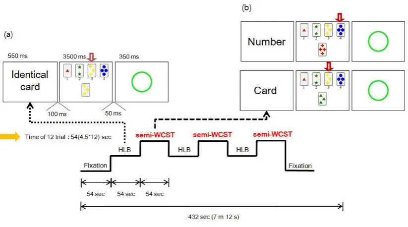
2.1. Subject Eligibility
74
Community-dwelling right-handed elderly women (≥60 years) with normal cognitive function
75
(Mini-Mental State Examination [MMSE] score ≥24) were enrolled during April 2015 through a
76
community center in South Korea. All subjects were asked to submit written consent to participate
77
in the research. Subjects were eligible for the study if they were relatively healthy and consented to
78
participate in the study. Subjects were excluded if they met any of the following exclusion criteria:
79
(i)decreased activities of daily living due to severe physical disease, (ii)metal substances in the body
80
that could affect the fMRI such as dental implants, artificial joints, or pacemakers, (iii) the taking of
81
psychiatric medication or a history of psychiatric disease [14], (iv)severely impaired cognitive
function (MMSE score <24) or a clinical diagnosis of dementia, (v)claustrophobia, (vi)
83
contraindications for MRI[15], and (vii)withdrawal of consent to participate in the study.
84
The protocol was approved by the Institutional Review Board of Kyungpook National
85
University Hospital and was conducted in compliance with research ethics (protocol No. KNUH
86
2015-04-032).
87
2.2. Screening
88
All recruited subjects were initially screened and their demographic and medical information
89
was collected. A questionnaire was conducted to determine the following: age, smoking status,
90
drinking status, regular exercise, past medical history (hypertension, diabetes, dyslipidemia, and
91
history of stroke), and education level, any of which could possibly affect cognitive function.
92
In the first visit to hospital, the subjects’ height, weight, waist circumference, blood pressure, and
93
pulse rate were measured. All measurements were performed by the same trained person according
94
to standardized protocols. Body height and weight were measured with a reliable digital
95
height–weight scale. Subjects stood straight without shoes and wore light clothing. Waist
96
circumference (WC) was measured at the middle of the body between the lower line of the ribs and
97
the upper line of the pelvis. Body mass index (BMI) was calculated as weight in kg divided by
98
height squared in meters. Blood pressure and pulse rate were measured using an autonomic blood
99
pressure monitor, with the subjects sitting quietly for 5minutes before measurements. Blood
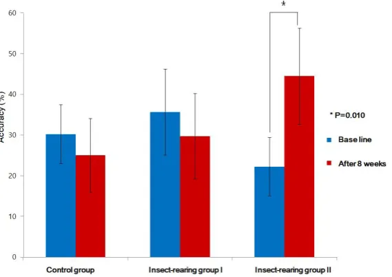
100
pressure was measured twice, with a brief break in between.
101
The MMSE was used to assess the baseline cognitive function of the subjects at screening. The
102
MMSE is a 30-point test that evaluates orientation to time (5 points), orientation to place (5 points),
103
memory registration (3 points), attention and calculation (5 points), memory recall (3 points),
104
language (8 points), and copying ability (1 point) [16]
105
2.3. Randomization and Study Procedure
106
The subjects were selected through the screening process and randomly allocated into two
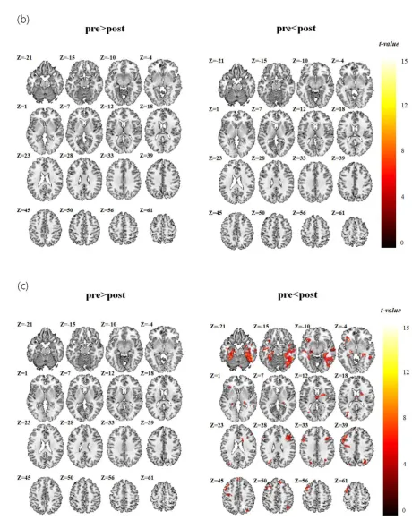
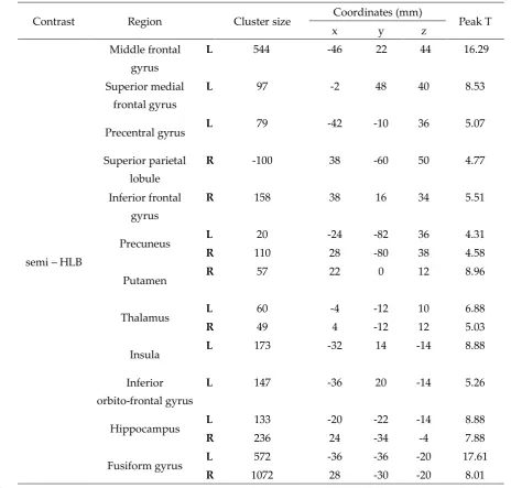
107
groups in a 1:1 ratio: the insect-rearing group and control group. All subjects underwent the same
108
tests during fMRI imaging (see below) at baseline and after 8 weeks.
109
The oriental garden cricket (Teleogryllus emma) was selected as a pet insect for the insect-rearing
110
group, in consultation with insect experts. The reasons for choosing the crickets were, first, the
111
crickets are common in East Asia, and the chirping and appearance of crickets would be familiar to
112
elderly Koreans; second, the connection between crickets and farm life can create nostalgia in the
113
elderly, which can manifest as an affection for insects [17]; third, their small size(26–40mm) means
114
there is little space limitation for insect rearing; and, finally, they can be raised indoors at room
115
temperature and are relatively easy to care for because they are omnivorous [18]. The insect-rearing
116
group received pet insects (4–5 crickets, a uniform distribution of males and females) in a cage and
117
then reared them. Sufficient fodder and all tools necessary for rearing were provided by the
118
researchers. In addition, the researchers provided appropriate training on how to keep the pet
119
insects. Every week, the research assistant confirmed the study subjects’ compliance and conducted
120
telephone counseling to encourage the cricket rearing.
121
The control group was compensated for the possible auditory effects of pet crickets by the
122
receipt of a CD containing meditative music with the natural chirping sound of the crickets, and the
123
research assistant conducted telephone counseling every week to ensure compliance
2.4. fMRI (Functional Magnetic Resonance Imaging)
125
Blood oxygen level dependent (BOLD) fMRI was acquired on a 3.0-T GE Exite instrument (GE
126
Healthcare) using an 8-channel head coil and a gradient-echo echo-planar imaging (EPI) sequence.
127
T2-weighted MRI images were also acquired. The fMRI protocol used the following acquisition
128
parameters: echo time (TE) = 40 ms, repetition time (TR) = 3000 ms, field of view (FOV) = 22cm,
129
acquisition matrix = 64 × 64, and cross-sectional slice thickness of4mm. All images were acquired
130
parallel to the intercommisural line (anterior commissure–posterior commissure line, ac–pc line).
131
2.5. Paradigm for the Testing of Working Memory Processing: Wisconsin Card Sorting Test (WCST)
132
The research assistants thoroughly explained the study protocol to the subjects and a skilled
133
professional conducted the study according to the planned paradigm. The WCST[19] is a known
134
task for testing executive functions. With consideration of the age of the subjects, two conditions of
135
the WCST were utilized: a high level baseline (HLB) and a semi-WCST test. In the HLB version
136
(choose the matching card), when a “matching card” was displayed, the subject was required to
137
select the matching card from the four cards presented on the screen and give the number of the
138
matching card. In the semi-WCST version (with the category by which to select the card informed
139
in advance), a word indicating the category of the card to be selected was presented, followed by a
140
“card” directive. If the “number” was given, the “card” was repeated until the next directive was
141
presented, and a same number card among four cards presented on the screen was selected. There
142
were three categories presented: number, shape, and color.
143
First, the rest condition was measured to show the fixation before and after the task, and then
144
the HLB test and semi-WCST were alternately repeated (Figure1).
145
146
Figure 1.Paradigm for working memory processing. (a)High level baseline (HLB), (b) semi-Wisconsin
147
Card Sorting Test (WCST), and (c) WCST task block design
The visual stimuli were shown using standardized software, with the latency and response
149
rates of the correct responses being collected to judge the performance of the subjects by their
150
responses made on the MRI-compatible reply button.
151
2.6. Image Analysis
152
The fMRI data image processing and the statistical analysis were performed using SPM5
153
(Welcome Department of Imaging Neuroscience, London, UK; online at
154
http://www.fil.ion.ucl.ac.uk) running within MATLAB (MathWorks Inc., Natick, MA). The
155
preprocessing included alignment of the functional images for movement correction, coregistration
156
to the individual structural image, and spatial normalization of all images to the Montreal
157
Neurological Institute (MNI) template and coordinate system. Differences in brain activation
158
among groups were analyzed using random effects analysis after the analysis of individual data.
159
Differences in brain activation when performing the semi-WCST were evaluated using
160
within-group paired t-tests. The SPM t-score maps were thresholded at p<0.05 with family wise
161
error (FWE) correction using Monte Carlo simulations performed using the 3dClustSim program
162
(https://afni.nimh.nih.gov/pub/dist/doc/program_help/3dClustSim.html).
163
2.7. Stastical Analysis
164
All subjects had MMSE scores more than 24 at baseline, and the subjects were randomized into two
165
groups in a 1:1 ratio. However, significant deviation of executive function at baseline fMRI was
166
found among the subjects in the insect-rearing group. Therefore, the insect-rearing group was
167
further classified into two groups based on the results of the initial WCST: those with a relatively
168
high score were allocated to insect-rearing group I, and those with a relatively low score to
169
insect-rearing group II. The general characteristics of the subjects were analyzed using ANOVA and
170
Pearson’s Chi-square tests. The differences in accuracy between the HLB and semi-WCST tests were
171
statistically compared within and among groups using paired t-tests, ANCOVA, and 2-way (2 × 2)
172
ANOVA. The fMRI images were analyzed using a flexible factorial design. A p-value of <0.05 was
173
considered to indicate statistical significance. IBMSPSS statistics version25 was used for all
174
statistical analyses
.
175
3. Results
176
3.1. Baseline Characteristics of the Subjects
177
A total of 44 participants were recruited and screened during the enrollment period. Eight
178
subjects were excluded because they met one or more of the exclusion criteria. The remaining 36
179
subjects were randomized into the insect-rearing (n=20) or control group (n=16). One subject in the
180
insect-rearing group was lost to follow-up. The insect-rearing group was further classified into two
181
groups according to the baseline WCST score: insect-rearing group I (high score, n=13) and
182
insect-rearing group II (low score, n=6; Figure 2).
183
There were no significant differences in the demographic and clinical characteristics among the
184
three groups (table 1). The mean ages were 66.38, 68.31, and 70.67 years for the control,
185
insect-rearing I, and insect-rearing II groups, respectively, while the average MMSE scores at
186
baseline were 27.94, 27.77, and 27.50.
188
Figure 2.Flow diagram of the study participants.
189
Table1. Baseline characteristics of the subjects in the control, insect-rearing I, and insect-rearing II groups
190
Control group
(n=16)
Insect-rearing
group I
(n=13)
Insect-rearing
group II
(n=6)
p-value*
Age, years 66.38±5.39 68.31±3.75 70.67±3.93 0.15
Height, cm 152.44±5.54 156.16±4.38 153.95±6.57 0.19
Weight, kg 52.48±9.10 65.28 ±24.31 57.60±3.69 0.12
BMI 22.49±3.26 26.93±11.06 24.31± 0.98 0.26
Waist, cm 80.06±8.81 84.19±11.26 85.17±6.43 0.39
Blood pressure, mm Hg
Systolic 134.25±19.05 137.46±12.89 137.50±21.30 0.86
Diastolic 72.31±13.73 78.15±8.95 71.50±11.06 0.34
Heart rate, beats/min 70.00±9.71 68.85±7.61 68.83±4.02 0.92
Psychometric tests
MMSE(cognitive function) 27.94±1.98 27.77±1.74 27.50±1.64 0.88
Smoking status
Non- or ex-smoker 15(93.8) 13(100.0) 6(100.0) 1.000†
Current smoker 1(6.3) 0(0.0) 0(0.0)
None 12(75.0) 12(92.3) 5(83.3) 0.613†
Any 4(25.0) 1(7.7) 1(16.7)
Exercise
None 15(93.8) 12(92.3) 5(83.3) 0.762†
Regular 1(6.3) 1(7.7) 1(16.7)
Education
≤Elementary school 8 (50.0) 8 (61.5) 5 (83.3) 0.125†
≥Middle school 8 (50.0) 5 (38.5) 1 (16.7)
Clinically diagnosed underlying disease
Hypertension 6(37.5) 7(53.8) 1(16.7) 0.317†
Diabetes 1(6.3) 2(15.4) 0(0.0) 0.762†
Dyslipidemia 5(31.3) 9(69.2) 2(33.3) 0.113†
CVA 0(0.0) 1(7.7) 0(0.0) 0.543†
The data are presented as mean ± standard deviation or number (%).
191
*The two groups were compared using independent t-tests or Pearson’s χ2 test except when indicated
192
otherwise.
193
†Fisher’s exact test.
194
3.2. WCST Response Data Results
195
At baseline, the average HLB–semi-WCST accuracies were 30.21%, 35.68%, and 22.22% in the
196
control, insect-rearing I, and insect-rearing II groups, respectively, with there being no significant
197
difference among groups. After 8 weeks, the corresponding average values were 25.00%, 29.70%,
198
and 44.44%, and the difference was close to being statistically significant (0.06). In insect-rearing
199
group II, the mean accuracy of the difference between the HLB and semi-WCST tests improved
200
from 22.22% at baseline to 44.44% after 8 weeks, showing a statistically significant improvement
201
(p=0.01, Figure 3, Table 2). There were no other significant differences in the intra group
202
HLB–semi-WCST test accuracies between the baseline test and that after 8 weeks.
203
204
Figure 3.High level baseline (HLB)–semi-Wisconsin Card Sorting Test (WCST) accuracy, paired t-test
205
(p=0.010).
Table2. WCST response pre- and post-intervention among the groups
207
Control group Insect-rearing group I
Insect-rearing group II
p-value†
Accuracy (%) Baseline 30.21±13.64 35.68±17.49 22.22±6.80 0.180 After 8 weeks 25.00±16.43 29.70±17.36 44.44±11.25 0.060
p-value* 0.329 0.319 0.010
*Paired t-test and † ANOVAof HLB−semi-WCST accuracy (%).WCST=Wisconsin Card
Sorting
208
3.3. fMRI Data Analysis Results
209
The brain activation maps calculated for the acquisitions before and after 8 weeks are shown in
210
Table 3 and Figure 4 for the control and insect-rearing groups. There was no significant group
211
difference in brain activation area (paired t-test) during the fMRI test between the acquisitions
212
before and after the intervention in either the control group or insect-rearing group I. However, in
213
insect-rearing group II, the brain activation areas during the WCST test were different before and
214
after the pet insect-assisted therapy. In this group, the paired t-tests analyzing the differences before
215
and after pet insect-assisted therapy showed peakt-scores of 16.29 in the left mid frontal area, 8.53
216
in the left superior medial frontal gyrus, 5.51 in the right inferior frontal gyrus, 8.96 in the right
217
putamen, 8.88 in the left insula, 8.88 and 7.88in the left and right hippocampi, and 17.61and 8.01in
218
the left and right fusiform gyri.
219
220
221
223
224
Figure 4.Brain activation maps for paired t-test analysis (p<0.05, with FWE correction using Monte Carlo
225
simulation by 3dClusSim and a minimum cluster size of 163). (a) Control group, (b) Insect-rearing
226
group I, (c) Insect-rearing group II
Table 3.Brain activation map scores for paired t-test analysis of insect-rearing group
230
Contrast Region Cluster size Coordinates (mm) Peak T
x y z
semi – HLB
Middle frontal
gyrus
L 544 -46 22 44 16.29
Superior medial frontal gyrus
L 97 -2 48 40 8.53
Precentral gyrus L 79 -42 -10 36 5.07
Superior parietal
lobule
R -100 38 -60 50 4.77
Inferior frontal gyrus
R 158 38 16 34 5.51
Precuneus L 20 -24 -82 36 4.31
R 110 28 -80 38 4.58
Putamen R 57 22 0 12 8.96
Thalamus L 60 -4 -12 10 6.88
R 49 4 -12 12 5.03
Insula L 173 -32 14 -14 8.88
Inferior orbito-frontal gyrus
L 147 -36 20 -14 5.26
Hippocampus L 133 -20 -22 -14 8.88
R 236 24 -34 -4 7.88
Fusiform gyrus L 572 -36 -36 -20 17.61
R 1072 28 -30 -20 8.01
p <0.05, with FWE correction using Monte Carlo simulation by 3dClusSim and a minimum cluster size of 163.
231
The x, y, and z coordinates are the Montreal Neurological Institute (MNI) coordinates. Cluster size is the
232
number of voxels activated in a regional cluster. L = left, R =right, T is the t-values of the supra threshold
233
voxels.
234
4. Discussion
235
This study was a randomized single-arm controlled trial that investigated the effects of rearing
236
pet insects on cognitive function in elderly women. The effects on cognitive function were assessed
237
using fMRI. The insect-rearing group II, with a relatively low average baseline executive function,
238
showed significant differences in both WCST response and fMRI results after pet insect-assisted
239
therapy. This result showed that the rearing of pet insects had a beneficial effect on executive
240
function in community-dwelling elderly women with low cognitive function.
241
fMRI has frequently been used in neuropsychological studies of memory. Memory can be
242
divided into short-term memory (working memory) and long-term memory, and fMRI primarily
243
shows working memory according to the functioning of the frontal lobe. It was reported that the
left frontal lobe encodes working memory and retrieves working memory from the right frontal
245
lobe [20]. Frontal cortex activation during the performance of memory tasks was demonstrated
246
using fMRI. fMRI can also be used to distinguish activation in three areas of the frontal cortex:
247
anterior frontal cortex (AFC), dorsolateral frontal cortex (DLFC), and ventrolateral frontal cortex
248
(VLFC).The DLFC and VLFC are located above and below the inferior frontal gyrus, respectively,
249
while the AFC is located in front of the inferior frontal gyrus. There are no clear boundaries but the
250
regions can be divided into Brodmann areas, with the VLFC including Brodmann areas 44, 45, and
251
47; the DLFC, areas 9 and26; and the AFC, areas 8 and 10. AFC, VLFC, and DLFC are distinguished
252
according to their activation levels in different tasks:(i) updating and maintaining the contents of
253
working memory, (ii) selecting, manipulating, and monitoring the contents of working memory, and
254
(iii) selecting process, target, and sub-goal. These three functions closely match the patterns of VLFC,
255
DLFC, and AFC activation, respectively [21]. The prefrontal cortex is included in the frontal lobe,
256
and an advanced study has shown that the prefrontal cortex is associated with executive function
257
[22].
258
The WCST is a multifaceted test requiring the use of a distributed brain network, and task
259
execution ability may be impaired by various factors. Some of these are related to frontal lobe
260
function [23]; most neuroimaging studies on WCST have reported a significant increase in
261
metabolic or neural activity in the frontal or prefrontal cortical region. In addition, in most studies,
262
an increase in brain activation has been found in the dorsolateral prefrontal cortex(DPFC), and in
263
some studies it has also been found in the ventromedial prefrontal cortex(VPFC) [19]. Functionally,
264
the lateral prefrontal cortex (LPFC)is involved in organizing, keeping, and manipulating
265
information in the short term, depending on the type of cognitive task [24].
266
In working memory tasks, the brain areas activated include the left and right Dorsolateral
267
prefrontal cortex, (DLPFC), left Ventrolateral prefrontal cortex(VLPFC), premotor cortex, right
268
frontal pole, bilateral inferior parietal lobules, right insula, right temporal gyrus, and a subcortical
269
region at the junction of the left thalamus, caudate, and lenticular nucleus. These brain areas are
270
also functionally connected for performing working memory tasks [25], with working memory
271
being a main cognitive function [14]. In this study, insect-rearing group II showed enhanced
272
activation signals in the left medullary frontal area, right DLPFC, parietal cortex, left insula, and
273
both hippocampi after the rearing of pet insects. During the WCST, the brain showed greater
274
activation for executive tasks involving the frontal (especially DLPFC) and parietal cortex and the
275
hippocampi, which are working memory areas. This indicates that the executive ability required to
276
perform the WSCT task was improved, and that the rearing of insects had a positive effect,
277
improving cognitive function.
278
Insect-rearing group II showed a relatively low score at baseline, which may be considered to
279
be the result of a low understanding of how to perform the WCST. However, subjects did not
280
significantly differ among groups in their MMSE sore, with all groups showing a normal range. A
281
previous study suggested that the delayed recall items of the MMSE are an index of working
282
memory capacity [26]. We made no evaluation of the sub-content items of the MMSE, and even if
283
the MMSE scores were within normal ranges, there may still have been cognitive impairment with
284
false-negatives. The most common impairments of cognitive function in patients with false-negative
285
results are memory and frontal lobe-executive functions [27]. Advanced studies have already
286
indicated that MMSE is insufficient to evaluate the executive function of the frontal lobe [28, 29].
287
The MMSE evaluation showed that all subjects were in the normal cognitive state, but we can infer
288
that the group with low WCST scores had relatively low cognitive functioning.
However, there was no significant difference in the accuracy rate and brain activation in the
290
WCST response data among the three groups. Furthermore, when the differences between the
291
WCST response data acquired before and after the intervention were compared within groups,
292
there was no significant change in the control or insect-rearing I group. Previous studies have
293
reported that AAT could improve cognitive function in elderly subjects [5], and a significant
294
improvement in cognitive function score was found in a previous study [30] using pet insects in
295
Korea. Based on these previous studies, we expected that fMRI-detected cognitive function would
296
be improved in this study. The reasons for the absence of significant differences among the three
297
groups after 8 weeks of pet insect-assisted therapy may include the following. First, the number of
298
subjects was small, which may have resulted in the failure to find statistically significant differences
299
in analysis conducted within groups after randomized classification. Second, all the subjects were
300
elderly women and most of them (58%) had an educational level below that of elementary school
301
graduation. Previous studies [31, 32] have shown that age, sex, and education level are significantly
302
associated with cognitive and neuropsychological test results. However, the subjects were limited
303
to elderly women, and both age and education level showed no significant difference among the
304
groups. Third, the subjects were limited to those who had favorable cognitive functioning with a
305
score of 24 points or more on the MMSE. As the subjects had a normal range of cognitive function at
306
baseline, there is the possibility that a noticeable improvement in cognitive function was not
307
detectable after a study period of 8 weeks. However, it is difficult for subjects with relatively low
308
cognitive function to conduct the complex WCST and to receive education about the protocol before
309
the test; a high MMSE score was an inevitable criterion because of the study design characteristics.
310
Fourth, the study period of 8 weeks was relatively short and may have been insufficient to see a
311
change in cognitive function. However, this was an inevitable limitation because oriental garden
312
crickets only live for up to 8 weeks. A previous study [33] on elderly subjects in a nursing home
313
conducted AAT for 12 months. The mean Mental Function Impairment Scale (MENFIS) was
314
reduced after 6 months, which means that cognitive function improved after 6 months. In another
315
study [34] on elderly Alzheimer’s disease patients, cognitive function improvement measured by
316
the MMSE was observed in a group who underwent AAT for 6 months. We suggest that to achieve
317
meaningful improvement in cognitive function using insect-assisted therapy, the study should be
318
conducted for a longer period.
319
This study has a few limitations. First, all subjects were community-dwelling elderly Asian
320
women living in a single Korean city. Thus, it may be impossible to generalize the results to other
321
ethnicities or younger populations. Second, the period of study was 8 weeks, which was relatively
322
short. As mentioned above, this short duration may have been insufficient to show improvement in
323
cognitive function. Third, the reliability of the WCST score may have been lower at baseline than at
324
8 weeks. To conduct the WCST, the method was explained in advance, but the elderly subjects may
325
have found it difficult to understand. Also, there is the possibility of a learning effect because the
326
same test was performed twice.
327
Despite these limitations, this study also has its strengths. First, it was a randomized controlled
328
design. Second, it is the first study to use fMRI to investigate the effect of rearing pet insects on
329
cognitive function. Although a previous study [30] investigated the clinical effects of rearing pet
330
insects, this study used a more objective approach by analyzing brain imaging data. Third, every
331
week, the research assistant confirmed compliance with the rearing of the pet insects and conducted
telephone counseling to encourage the practice. Fourth, the precise effects of rearing pet insects on
333
cognitive improvement were determined by performing WCST and fMRI imaging together, and
334
these are well known to be reliable and valid tools. Finally, as mentioned above, the restricted
335
subject diversity could be a strength because it should minimize the confounding effects of
336
variables such as age, sex, residential area, and comorbid diseases.
337
In conclusion, the rearing of pet insects was found to have a positive effect on improving
338
executive function and performance in elderly women. Rearing insects is cost-effective, easy,
339
occupies less space than the raising of larger animals, and can be usefully used as a type of AAT
340
that is non-pharmacologically based and noninvasive. Further larger studies of pet insects with
341
longer study periods are warranted.
342
343
Acknowledgments: This research was supported by Kyungpook National University Research
344
Fund, 2018. Study design, data collection and analyses were completely performed by the authors.
345
References
346
1. Statistics Korea. Statistics report about the elderly, 2018. Available online: http://kostat.go.kr(accessed
347
on 20 December 2018)
348
2. Bernabei, V., et al., Animal-assisted interventions for elderly patients affected by dementia or
349
psychiatric disorders: a review. J Psychiatr Res, 2013. 47(6): p. 762-73.
350
3. Cherniack, E.P. and A.R. Cherniack, The benefit of pets and animal-assisted therapy to the health of
351
older individuals. Curr Gerontol Geriatr Res, 2014:623203.
352
4. Harris, M.D., J.M. Rinehart, and J. Gerstman, Animal-assisted therapy for the homebound elderly.
353
Holist Nurs Pract, 1993. 8(1): p. 27-37.
354
5. Moretti, F., et al., Pet therapy in elderly patients with mental illness. Psychogeriatrics, 2011. 11(2): p.
355
125-9.
356
6. Richeson, N.E., Effects of animal-assisted therapy on agitated behaviors and social interactions of older
357
adults with dementia. Am J Alzheimers Dis Other Demen, 2003. 18(6): p. 353-8.
358
7. Zisselman, M.H., et al., A pet therapy intervention with geriatric psychiatry inpatients. Am J Occup
359
Ther, 1996. 50(1): p. 47-51.
360
8. Ministry of Education and Science Technology, elementary school guideline for teachers (science), 7th
361
ed. kumsung, korea,2011; pp. 1-386
362
9. Ko, H., et al., Effects of Pet Insects on Psychological Health among Community-dwelling Elderly:A
363
Randomized-Controlled Trial. 2015: Unpublished manuscript.
364
10. Bandettini, P.A., et al., Time course EPI of human brain function during task activation. Magn Reson
365
Med, 1992. 25(2): p. 390-7.
366
11. Ogawa, S., et al., Intrinsic signal changes accompanying sensory stimulation: functional brain
367
mapping with magnetic resonance imaging. Proc Natl Acad Sci U S A, 1992. 89(13): p. 5951-5.
368
12. Kesler, S.R., et al., Functional neuroanatomy of spatial orientation processing in Turner syndrome.
369
Cereb Cortex, 2004. 14(2): p. 174-80.
370
13. Seo, J., et al., Stronger activation and deactivation in archery experts for differential cognitive strategy
371
in visuospatial working memory processing. Behav Brain Res, 2012. 229(1): p. 185-93.
372
14. Stone, M., et al., Working and strategic memory deficits in schizophrenia. Neuropsychology, 1998. 12(2):
373
p. 278-88.
15. Le Bihan, D. and A. Karni, Applications of magnetic resonance imaging to the study of human brain
375
function. Current Opinion in Neurobiology, 1995. 5(2): p. 231-237.
376
16. A validity study on the korean mini-mental state examination (K-MMSE) in dementia patients. J
377
Korean Neurol Assoc, 1997. 15(2): p. 300-308.
378
17. Kim, S.Y., et al., Effects of experience-based learning program using singing insects. Journal of
379
Sericultural and Entomological Science 2013. 51(2): p. 114-118.
380
18. Kim, N.-J., et al., Effect of temperature on development and reproduction of the emma field cricket,
381
Teleogryllus emma (Orthoptera: Gryllidae). International Journal of Industrial Entomology 2007. 15(1): p.
382
69-73.
383
19. Nyhus, E. and F. Barceló, The Wisconsin Card Sorting Test and the cognitive assessment of prefrontal
384
executive functions: A critical update. Brain and Cognition, 2009. 71(3): p. 437-451.
385
20. Yun-Hee Kim.,Usefulness of Functional MRI for the Study of Brain Function. Korean Journal of Brain
386
Science and Technology 2001. 1(1): p. 65-76.
387
21. Fletcher, P.C. and R.N.A. Henson, Frontal lobes and human memory: Insights from functional
388
neuroimaging. Brain, 2001. 124(5): p. 849-881.
389
22. Yuan, P. and N. Raz, Prefrontal cortex and executive functions in healthy adults: A meta-analysis of
390
structural neuroimaging studies.Neuroscience & Biobehavioral Reviews, 2014. 42: p. 180-192.
391
23. Stuss, D.T., et al., Wisconsin Card Sorting Test performance in patients with focal frontal and posterior
392
brain damage: effects of lesion location and test structure on separable cognitive processes.
393
Neuropsychologia, 2000. 38(4): p. 388-402.
394
24. D’Esposito M., P.B.R., Rypma B., Prefrontal cortical contributions to working memory: evidence from
395
event-related fMRI studies. Springer, Berlin, Heidelberg 2000.
396
25. Hampson, M., et al., Brain Connectivity Related to Working Memory Performance. J Neurosci 2006.
397
26(51): p. 13338-13343.
398
26. Pascual, L.F., et al., The assessment of working memory by Mini-Mental State Examination. Rev Neurol,
399
2000. 30(1): p. 1-4.
400
27.
M.A, E.O., et al.,A Validity Study of K-MMSE as a Screening Test for Dementia: Comparison Against401
a Comprehensive Neuropsychological Evaluation. Dementia and Neurocognitive Disorders, 2010. 9: p.
402
8-12.
403
28. Hoops, S., et al., Validity of the MoCA and MMSE in the detection of MCI and dementia in Parkinson
404
disease. Neurology, 2009. 73(21): p. 1738-45.
405
29. Axelrod, B.N., R.S. Goldman, and R.R. Henry, Sensitivity of the Mini-Mental State Examination to
406
frontal lobe dysfunction in normal aging. J Clin Psychol, 1992. 48(1): p. 68-71.
407
30. Ko, H.J., et al., Effect of Pet Insects on the Psychological Health of Community-Dwelling Elderly
408
People:A Single-Blinded, Randomized, Controlled Trial. Gerontology, 2016. 62(2): p. 200-9.
409
31. Ganguli, M., et al., Effects of age, gender, and education on cognitive tests in a rural elderly
410
community sample: norms from the Monongahela Valley Independent Elders Survey.
411
Neuroepidemiology, 1991. 10(1): p. 42-52.
412
32. Wiederholt, W.C., et al., Effects of age, gender and education on selected neuropsychological tests in
413
an elderly community cohort. J Am Geriatr Soc, 1993. 41(6): p. 639-47.
414
33. KAWAMURA, N., M. NIIYAMA, and H. NIIYAMA, Long-term evaluation of animal-assisted therapy
415
for institutionalized elderly people: a preliminary result. Psychogeriatrics 2007. 7(1): p. 8-13.
34. Menna, L.F., et al., Evaluation of the efficacy of animal-assisted therapy based on the reality orientation
417
therapy protocol in Alzheimer's disease patients: a pilot study. Psychogeriatrics 2016. 16(4): p. 240-246.