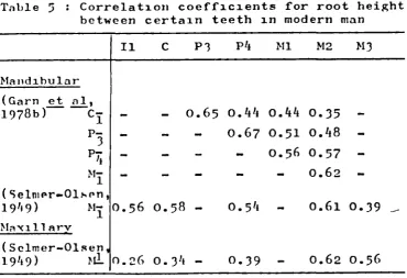
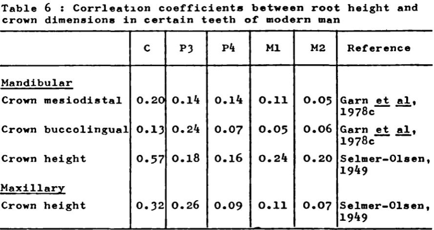
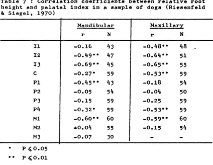
1
A COMPARATIVE STUDY OF TOOTH ROOT MORPHOLOGY IN THE GREAT APES, MODERN MAN AND EARLY HOMINIDS
by
SUSAN ANNE ABBOTT
Thesis submitted for the degree of Doctor of Philosophy
Department of Anatomy
The Middlesex Hospital Medical School University of London
This thesis sets out to document and analyse some aspects of the metric and morphological variation of tootI. roots within
the Jiornirioidea. Saniples of the extant species Gorilla gorilla, Pan troglodytes, Pongo pygmaeus and Homo sapiens formed the basis of this study, but fossil hominid specimens from the East African P1±0/Pleistocene sites of' Koobi Fora, Laetoli, Olduvai and Peninj were also included. The maximum of 182 root, crown and jaw measurements were taken for each specimen included in the comparative samples and included information for all permenent teeth except the maxillary canine. Premolar root number and form were also recorded for each specimen.
The corresponding measurements and observations for the mandib-ular dentition were made, where possible, on the fossil hominid
specimens.
The metrical description of tooth roots in the pongids and modern man was accomplished by computing basic descriptive statistics for each measurement and for indices derived from the measurements. These statistical summaries formed the basis of an assessment of root size in the comparative samples.
The pattern of sexual dimorphism in root dimensions is presented for each comparative sample, and assessment is also made of
the contribution of sexual dimorphism to the variation recorded for root dimensions in the pooled-sex samples. Correlation analysis was used for a preliminary assessment of any inter-relationship between root height and other dimensions.
In the pongid and modern human samples, root number and
3
but in the fossil hominid sample this was possible only for the latter teeth. The assessment of the root and crown dimen-. sions in the fossil hominid sample was conducted by grouping the fossil hominid specimens according to their mandibular premolar root morphology and then examining the dimensions in
I would like to thank Professor Bernard Wood, my supervisor, for his advice, support, help and criticism during the execution of this research. Many others on the staff of the Department of Anatomy and Biology, The Middlesex Hospital Medical School, have helped, in all sorts of ways, during the course of this work and I am grateful to them all.
I would like to thank the staff of the following libraries for their assistance : The Middlesex Hospital Medical School Library, Senate House Library, the Library of the British
Museum (Natural History) and, particularly, the Library of the British Dental Association. Various members of the Linguistic Aid Department of the British Library helped with the translatioi of material published in foreign languages.
My thanks are due to the Director and Trustees of the British Museum (Natural History), the Director and Trustees of the National Museums of Kenya, and the Tanzanian Government for allowing me access
to
collections in their care.I would like
to
thank Mr. D. Piper of the Ferens Institute of Otolaryngology, The Middlesex Hospital Medical School,for the skill, care and patience with which he constructed
the radiographic grid. The staff of the Department of
Computing,
The Middlesexhospital
Medical School, assisted with thecomputing. Mrs. Carol Shoard typed the
manuscript,
with5
The major part of this research was conducted while the author was in receipt of a N.E.R.C. Research Assistantship.
Lastly, I
would
like to thank myfamily
andfriends for
Introduction
(1) Abstract 2
(ii) Acknowledgements 4
(iii) Table of contents 6
(iv) List of tables 8
(v) List of figures 10
(vi) List of appendices 15
(vii) Introduction 17
Part I Literature review
1. The developmental basis of root morphology 20
2. Metrical studies of tooth roots 33
3. Studies on the variation of root form and
number 6
Part II : Samples and methods
The Samples 118
5. Methods 122
Part III : Results
6. Metrical assessment of the root demensions
in the comparative samples 160
7. Sexual dimorphism in the root dimensions
of the comparative samples 223
8. The relationship of root height to other root, crown and jaw dimensions in the
7
9. Premolar root form in the comparative
samples and in the fossil hominid
sample
287
10. The root arid crown dimensions arid
indices in the fossil hominid sample O6
Part IV : Discussion
11. Discussion arid conclusions
Part V References
358
3.. Published mean values of mandibular
root height in modern man 37
2. Published mean values of maxillary
root height iii modern man 38
3. Published male and female mean values
of root height in modern man 41
L1• Correlation coefficients for root
height between adjacent teeth in
modern man 50
5 . Correlation coefficients for root
height between certain teeth in modern
man 50
6. Correlation coefficients between root
height and crown dimensions in certain
teeth of modern man. 52
7. Correlation coefficients between
relative root height and palatal
index in a sample of dogs (Riesenfeld
& Siegel, 1970)
55
8. Correlation coefficients between root
height and jaw dimensions in Papio
anubis (Siegel, 1972)
9. Published mean values of root surface
2
area (mm ) in modern man 61
10. Published values (cm ) of root volume,
and root volume expressed as a
percentage of total tooth volume, in
modern man. 64
11. The percentage incidence of P root
12. The percentage incidence of root
variations in modern man 83
13. Comparison of values obtained for measurements from the angled and lateral radiographs in five Gorilla specimens, illustrating the increased
accuracy resulting from use of the grid 156 1L1. The variability of the root and crowi
dimensions in the comparative samples 213 15. Percentage incidence of mandibular
premolar root number variations in the
comparative samples 289
16. Percentage incidence of maxillary
premolar root number variations in the
(v) List of figures
1. Variation in root form and number between
different teeth 27
2. The developmental basis of molar root
forms 87
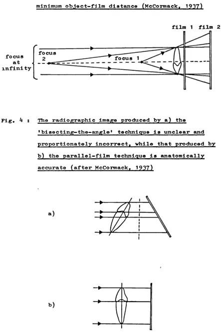
3. Reduced magnification of the radiographic image results from maximum focus-film distance and minimum object-film distance
(McCormack,
1937)
12L4. The radiographic image produced by the 'bisecting-the-angle' technique and by the parallel-film technique (after
McCorrnack,
1937)
12L5. Distal view of the orientation for an angled radiograph of the (right)
mandi-bular premolars 132
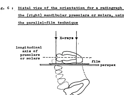
6. Distal view of the orientation for a radiograph of the (right) mandibular
premolars or molars 132
7. Distal view of the orientation for an angled radiograph of the maxillary
cheek-teeth
135
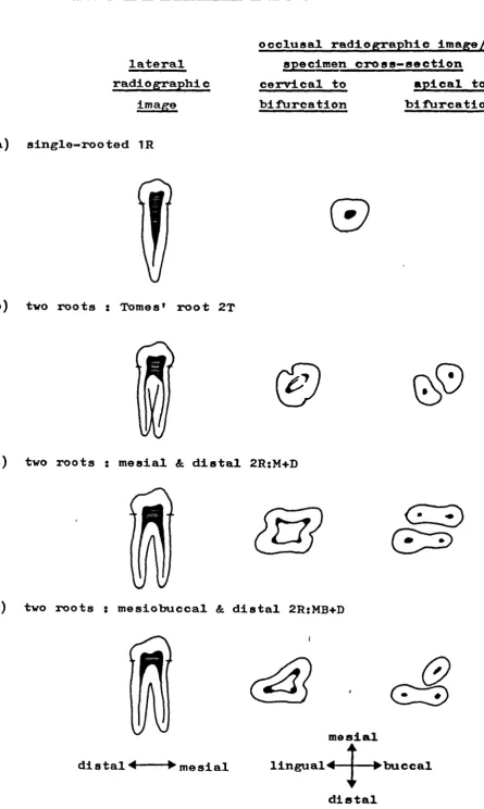
8. Mandibular premolar root form 137
9. Root diagrams of the mandibular teeth 142 10. Root diagrams of the maxillary teeth
143
11. Mean values of neck mesiodistal diameterin the pongids and Homo sapiens 162 12. Mean values of location of bifurcation
11
1 3 .
Mean values of location of bifurcationindex in the mandibular teeth of the
pongids and Homo sapiens 1 69
11! . Mean values of mandibular actual root
height in the pongids and Honio sapiens
176
15. Mean values of maxillary actual root
height in the pongids and Homo sapiens 177
16. Mean values of mandibular root
mesio-distal diameter in the pongids and
Homo sapiens
18Z
17. Mean values of maxillary root mesiodistal
diameter in the pongids and Homo sapiens 185
18. Mean values of the root robusticity index
for the mandibular roots of the pongids
and Homo sapiens
186
19. Mean values of the root robusticity index
for the maxillary roots of the pongids
and Homo sapiens
187
20. Mean values of height of bifurcation in
the pongids and Homo sapiens
196
21. Mean values of the height of bifurcation
index in the pongids and Homo sapiens
197
22. Mean values of mandibular root angulation
in the pongids and Homo sapiens 203
23. Mean values of maxillary root angulation
in the pongids and Homo sapiens 204
24. Mean values of root divergence in the
pongids and Homo sapiens 205
25. Male and female mean values of neck
mesiodistal diameter in the pongids and
26. Male and female mean values for location
of bifurcation in the pongids and Homo
sapiens 230
27. Male and female mean values for the
location of bifurcation index of the
mandibular teeth in the pongids and Homo
sapiens 231
28. Male and female mean values of mandibular
actual root height in the pongids and
Homo sapiens 234
29. Male and female mean values of maxillary
actual root height in the pongids and
Homo sapiens
235
30. Male and female mean values of mandibular
root mesiodistal diameter in the pongids
and Homo sapiens 242
31. Male and female mean values of maxillary
root mesiodistal diameter in the pongids
and Homo sapiens 243
32. Male and female mean values of mandibular
root robusticity index in the pongids
and Homo sapiens 244
33. Male and female mean values of maxillary
root robusticity index in the pongids
and Homo sapiens 245
34. Male and female mean values for height
of bifurcation in the pongids and Homo
13
35. Male and female mean values for the
height of birufcatiori index in the
pongids and Homo sapiens 252
36. Male and female mean values for
mandi-bular root angulation in the pongids
and Homo sapiens 256
37. Male and female mean values of maxillary
root angulation in the pongids and Homo
sapiens 257
38. Male and female mean values of root
divergence in the pongids and Homo sapiens 258
39. Sexual dimorphism in the root and crown
dimensions of Gorilla gorilla 261
110. Sexual dimorphism in the root and crown
dimensions of Pongo pygmaeus 2 6
41. Sexual dimorphism in the root and crown
dimensions of Pan troglodytes 269
42. Sexual dimorphism in the root and crown
dimensions of Homo sapiens 271
113. Correlation coefficients for actual root
height between adjacent teeth in the
pongids and Homo sapiens 277
44. Mandibular premolar root form in the
fossil hominid sample 30 L
45. Mean values of mandibular neck mesiodistal
diameter in the fossil hominid groups 309
46. Mean values of mandibular location of
bifurcation in the fossil hominid groups 311
47. Mean values of location of bifurcation
fossil hominid groups
311 1j8.
Mean valuesof
mandibular actual rootheight in the
fossil
hominid groups 31Z49.
Mean valuesof
mandibular rootmesio-distal diameter in the
fossil
hominidgroups 317
50. Mean values of the root robusticity index for the mandibular roots of the fossil hominid groups
318 51. Mean values of height of bifurcation in
the mandibular teeth of the fossil
hominid groups 320
52. Mean values of the height of bifurcation index in the mandibular teeth of the
fossil
hominid groups 32053. Mean values
of
mandibular rootangula-tion in the fossil hominid groups 322 54. Mean values of root divergence in the
mandibular teeth of the
fossil
hominidgroups 323
55. Mean values of crown mesiodistal diameter 1 n the mandibular teeth
of
the fossilhominid groups 326
56. Mean values of crown buccolingual
diameter in the mandibular teeth of the
15
(vi) List of Appendices
A. The comparative samples 0-1
B. The fossil hominid sample 405
C. The root, crown and jaw variables 07 D. Statistical summaries for the root,
crown and jaw dimensions in the
comparative samples (pooled sexes) 41 E. Statistical summaries for the indices
computed from root, crown and jaw
dimensions in the pongids and Homo
sapiens (pooled sexes) 4 5
F. Statistical tests of significance for the mean values and variances of the root, crown and jaw dimensions and
indices in the comparative samples 463 G. Statistical summaries for the root,
crown and jaw dimensions in the males
and females of each comparative sample 4.2 H. Statistical summaries for the indices
computed from root, crown and jaw
dimensions in the males and females of the porigids and Homo sapiens
5-1p
I. Correlation coefficients computed between root height and other root, crown and jaw variables in the pongids
and Homo sapiens 565
J. Mandibular premolar root form in the
fossil hominid sample 5P9
K. The root and crown dimensions and
crown dimensions and indices in the
If
(vii) Introduction
Aspects of tooth crown morphology, particularly crown size, have long been central to many discussions of hominid taxonomy and phylogeny. In contrast, the roots of the teeth have been almost completely ignored as a source of information
for hominid studies. The subject of this thesis arose as a response to questions concerning tooth root morphology which were prompted by the ' discovery of the fossil hominid sample from the site of Koobi Fora, Kenya, a sample which includes many specimens of the lower jaw in which the tooth crowns are missing. Were there features of the tooth roots which could
contribute to discussions of hominid taxonomy and phylogeny?
This thesis represents an initial foray into the subject of tooth root morphology in the Hominoidea. Its aims are, firstly, to document aspects of the metric and morphological variation of tooth roots in the great apes and modern man so that, secondly, a realistic attempt may be made at evaluating the corresponding aspects of root morphology in a sample of
early fossil hominids. Of particular interest in the comparative samples is the pattern of sexual dimorphism' in root size, and the contribution of sexual dimorphism to variability. The evaluation of root form and number was restricted to the promolar region, it being of particular interest as the site
of reduction in root number during the course of human evolution.
the Hominidae - which is mainly restricted to Homo sapiens and covers root surface area and root volume in addition to
aspects of linear root dimensions, and thirdly, on the variation of root form and number found within the modern human and early fossil hominid dontitions. The second section describes the comparative and early hominid samples used in the study,
and also documents the methodology employed. Included in the latter are outlines of the radiographic method, the assessment of premolar root form, the measurements used and the way in which they were taken, the reliability of the measurement
technique, and the statistical procedures used in the analysis of the data. The third section covers the results of the
investigation, detailing the root dimensions in the comparative samples, the pattern of sexual dimorphism in the root dimensions of the comparative samples, aspects of inter-relationship
19
Part I : Literature review
Chapter 1 : The developmental basis of root morphology
Chapter 2 : Metrical studies of tooth roots
Part I
Chapter 1 : The developmental basis of root morphology
i) Introduction
21
Chapter 1 : The developmental basis of root morphology
j ) Introduction
Tooth formation starts in the region of the cusp tips and proceeds until the root apex or apices are complete; the developing area of the tooth is mapped out in soft tissue, and then calcification starts. The mapping of the crown is determined mainly by growth of the cervical loop of the enamel organ, and when this is complete, the cervical loop becomes modified and is then known as Hertwig's epithelial sheath. It is this structure which is responsible for the mapping of the root and the initiation of its clentine forma-tion (Bhaskar,
1980;
Butler,1956;
Diamond & Applabaum,1942;
Kovacs,1963, 1967; Osborn & Ten Cate, 1976; Schour & Massler,
1940;
Scott & Symons,1974).
Initially, Hertwig's epitbelial sheath bends inwards to form the epithelial diaphragm, this being more or less parallel to the cervical opening of the tooth germ (Bernick &
Levy,
1968;
Bhaskar,1980; Grant & Bernick, 1971; Osborn and
Ten Cate,1976; Sicher,
1942).
Then, the mapping of the root proceeds through cellular proliferation adjacent and coronal to the epithelial diaphragm, and once an area of the root is mapped, the initiation of dentine formation follows shortly (Bhaskar,1980; Grant & Bernick, 1971). It is now
generally accepted that the epithelial diaphragm maintains a relatively fixed position within the jaw during thegreater part of root formation, so that root formation results in movement of the crown towards the alveolar border and
deeper into the jaw (Carleon,
1944; Diab & Stallard,
1965;
Diamond & Applebaum,1942).
These observations contrast with earlier opinion; originally, it was suggested that the developing tooth-edge penetrated into the jaw during crown and root formation (Hertwig,1874,
cited by Diamond & Applebaum,1942;
Prieswerk & Choinpret,1905,
cited by Kovacs,1963),
and later, both the cervical loop and its derivative during root formation were thought to maintain a fixed position within the jaw (Orban, 1928; Orban & Mueller,1929).
The relatively fixed position of the epithelial diaphragi applies only until the crown comes into occlusion; then, the developing root does penetrate into the jaw until the apex is complete (Carison,
1944;
Kovacs,1964, 1967, 1971;
Osborn & Ten Cate,1976).
Kovacs recognises two phases of rootde-velopment : the eruptive phase, which starts with the beginning of root formation and ends when the crown comes into occlusion, and the penetrative phase, which follows the eruptive phase and continues until the root is complete. Each phase results in a distinctive appearance of the root surface; the root
23
Kovacs (1967) considers that during the eruptive phase, the root is protected from environmental influences and is
thus under closer genetic control than it is during the
penetrative phase, when environmental factors may influence the morphology of the developing root. According to Kovacs, this explains differences of morphology such as that noted for the root surface when the parts of the root formed during the two phases of root development are compared. In addition, he claims that when roots of the same tooth type are compared,
they show greater similarity in the parts formed during the eruptive phase than they do in the parts formed during the penetrative phase (Kovacs, 1963, 196k, 1967). hi1e root formation is considered to be under strong genetic control
(Ackerman etal, 1973; Moorrees & Kent, 1981), studies have noted the effect of environmental factors - such as nutrition, masticatory function, growth zones and season of birth - on root formation (Ackerman et al, 1973; Brin & Koyoumdijsky-Kaye,
1981; Korkhaus, 1929; Lowe, 1971; fliesenfeld,
1970; Siegel,1972).
The proportion of the root formed during the eruptive phase of root development appears to vary according to
species and according to tooth-type (Gron, 1962; Korkhaus,
1929; Kovacs, 196k, 1971, 1979).
In human teeth, the proportion of the root generally formed during the eruptive phase hasbeen estimated as wo thirds (Kovacs,
1964), though Gron
herbivore (Kovacs, 1971).
Dutirig the formation of single-rooted teeth, Hertwig's
epithelial sheath maintains a single basal opening - the
primary apical foranien - throughout the period of root formatior
but during the
formation
of multi-rooted teeth, the primary
apical foramen becomes sub-divided into a number of secondary
apical foramina (Bhaskar,
1980;
Butler,
1 95 6 ;Gaunt,
1960;
Jorgensen,
1950;Kovacs, 1963; Liao, 1979; Osborn & Ten Cate,
1976;
Scott
iifldSymons, 197'i). The sub-division of the primary
apical foramen is effected by processes which develop from
llertwig's epithelial sheath, and these have been variously
referred to as the inter-radicular processes, tongues or
projections, or as the horizontal processes.
Before the
iiiter-radicular
processes develop, their
locations are anticipated by the formation of 'bays' in
the basal outline during crown formation arid the early stages
of root formation (I5haskar,
1980;
Butler, 195 6 ; Gaunt, 1960;
Osborn & Ten Cate, 1976; Orban & Mueller, 1929). Thus, the
inter-radicular processes are initially defined by selective
outward expansion of the primary apical foramen. Their
subsequent development proceeds through their own active
inward growth. The
iriter-radicular
processes grow inwards
to the centre of the root, penetrating between the blood
vessels as they go, and fuse with each other to form the
suhpulpal or bifurcal wall (Alexandersen, 1962a, 1963;
Bhaskar,
1980;
Butler, 1956; Campbell, 1925;
Carleen,
1967;
Orban & Mueller, 1929; Osborn & Ten Cate, 1976; Scott &
Symons, 1974). Each of the secondary apical foramina is
formed in relation to blood vessels; prior to the division
of the primary apical foramen, the number and locations of
the secondary apical foramina are anticipated by the number
and locations of the bundles of blood vessels (Aeby, 1878;
Cuvier, 1825; Lepko'wski, 1897, 1901; all cited by Butler,
195 6 ; Gaunt, 1960). Each of the secondary apical foraniina
is bounded by a region of Hertwig's epithelial sheath, and
so the formation of the separate roots proceeds (Bhaskar, 1980
In calcified specimens, the line of fusion between
inter-radicular processes is often identifiable as a ridge
on the subpulpal wall and along the adjacent root surfaces
(Alexandersen, 1962a; Butler, 1956; Carlsen, 1967; Gher &
Vernino, 1980; Kovacs, 19 6
3,
1967, 1971; Lester & Boyde,1970; Osborn & Ten Cate, 1976; Jorgensen, 1950; Von Brunn,
1887, 1891, cited by Butler,
1956).
This ridge has beenreferred to as the inter-radicular ridge (Kovacs, 1971) or
the intermediate bifurcational ridge (Everett et al, 1958)
and it is thought to arise from local growth along the line of
contact of the processes (Jorgensen, 1950). Though it has
a basis in dentine, the ridge is primarily formed of cementum,
and it becomes less defined with age probably as a result of
secondary cementum formation (Carlsen, 1967; Everett
1958; Kovacs, 1971).
In two-rooted teeth with mesial and distal roots, the
inter-.radicular ridge runs from the subpulpal wall down the
distal surface of the mesial root and down the mesial surface
(Lverett etal,
1958;
Jorgensen, 195
0;
Kovacs,1963, 1967).
1verett et al ( 1 95 8 ) classified the inter-radicular ridge
in mandibular first molars by its degree of development, and
noted that it was observable in
73%
of specimens. In teethsuch as maxillary molars, the inter-radicular ridges of the
three roots intersect on the subpulpal wall in the form of
a Y (Jorgensen, 1950; Kovacs, 1967). The inter-radicular
ridge his been identified in animals other than man, an
indication that the process of bifurcation is similar in
wan and other animals (Addison & Appleton, 1923; Butler,
1948;
hovacs, 1971; Lester & I3oyde,
1970).
ii) The developmental basis of root variation and reduction
Variation in root form and number (Fig. 1) between teeth
of different type and between teeth of the same type but from
different specimens or species, is a direct consequence of
the variability shown by several factors operatiig during
root development. As succinctly put by Carlsen,
(1967):
"In order to make the variation in ontogeiletic root
morphology of hurian teeth intelligible, it is very
important that one fully realises that the total
variation is in reality identical with the product
of the variations of a series of individual
These factors, which are really expressions of the growth
process, include: the sites on the primary apical foramen
vhere the inter-radicular processes arise; their number and
their relative degree of development; the timin of the
appearance of t1e inter-radicular processes arid the timing
of their fusion (Carisen,
1967;
kovacs,1963, 1967, 1971;
04.1 Eo '-4 00 I—s r14) p.4,4 (14 I4 Ix .5 4) 0 0 4) 0 0 4.) 4) 0 0 .1-s V 0 0) 0) 0)
27
0
C.)H '.4
S
I-fQjj
rl(U '0,4 llo r-4r-4 ..44) '-lI-S 00 00 00 .1.44.1D 4.1S. U)
•1-4
S. U)
• 41
bDo •T-1 U) 0) E
Iu
S.'-4 r-s ,•-$1U)
A ($it)0
r1 T +)0).4Q
g1I4 1*a'c
'I'he sites where the inter-radicular processes arise on
the periphery of the primary apical foramen determine the
locations of the tooth roots, in a tooth which will exhibit
inesiobuccal, distobuccal arid lingual roots, three
inter-radicular processes arise and are located on the mesial,
distal arid buccal borders of the primary apical foramen
(l3haskar, 1980; Jorgensen, 1
950 ). Thus, on fusion, three
secondary apical foramina are formed in mesiobuccal,
disto-buccal and lingual locations. In a tooth which will exhibit
niesial and distal roots, two inter-radicular processes develop
- one buccally and one lin rually - which form mesial and distal
secondary apical foramina on fusion (I3haskar,
1980;Jorgensen,
1
950 ) . The presence of labial/buccal and lingual roots is
dependant on the formation and fusion of mesial and distal
inter-radicular processes (Alexanderseri, 1962a; Cariseri,
1967).
However, root number is dependant not only on the number
of the inter-radicular processes, but also on the degree to
hich they penetrate towardb the centre of the primary apical
forainen. If all the processes fuse, completely separate roots
result; if the penetration of a process is marked, but fusion
does not take place, the identity of the two adjacent roots
is apparent on the fully-formed specimen sithough their
separation is incomplete; if the penetration is minimal, a
superficial root groove results (Alexandersen, l962a). In
this way, a root groove can be seen as the product of an
abortive inter-radicular process (Carlsen, 1967).
Ifan
inter-radicular process fails to develop there is ciip1ete
29
the absent process. Thus, variation in the develprnent of
the
inter-radicular processes underlies a continuum of
expressaon
of root separation, ranging from complete
separation to Coalescence of the roots, and suppression
of the inter-radicular processes forms a pathway leading to
reduction in root number.
The reasons why inter-radicular processes fail to develop,
or only penetrate inwards for a short distance, have not
apparently been investigated, although it has been suggested
that their growth nay be blocked by growth of the dental papill
or may be inhibited by the proximity of blood vessels
(Alexanderseii, 1962a; Butler,
1956).
while root foriiation
in
molar teeth usually follows its
typical course, one or more of the inter-radicular processes
may be suppressed, particularly
inthe second arid th.rd molars
(Kovacs, 1963, 1967). All the inter-radicular processes
may fail to develop and the resulting root form is referred
to as a pyramidal root; the molar has a single root which
may contain only a single pulp canal and which develops in
the same way as that of a typically single-rooted tooth
(Kovacs, 1963). A longitudinal root groove
or
cleft may bepresent, indicating sonie degree of development of one or
other of' the inter-radi.cular processes.
ormal development of
at least one, but not all, of the inter-radicular processes
leads to the molar root form commonly referred to as fused
roots.
Then this occurs
iii
niandibular molars, there is
continuity between the mesial and distal roots on the side
where the interradicular process has failed to develop
maxillary Molars, root fusion is frequently encountered and
often occurs between the mesiobuccal and lingual roots
-in which -instance the rnesial -inter-ra(Iicular process has
been suppressed to some degree (Jorgensen, 1950). In molars,
variability in the development of the inter-radicular processes
leads to root forms which are considered atypical; but in
preniolars such variability leads to the inter- and
intra-specific variation documented for the roots of these teeth.
In the pongids, maxillary premolars are generally
three-rooted,and mandibular premolars are usually two-rooted,
and so the roots develop in the same way as the corresponding
molars. Human maxillary premolars, particularly the first,
may have one root, to roots or even three roots, and so
variation occurs in the number and degree of development
of the intra-radicular processes (Carisen,
1967).Compared
with the three-rooted maxillary premolar, the formation of
two roots - buccal and lingual - requires only the suppression
of the buccal inter-radicular process. In both maxillary and
mandibular premolars, the presence of a single root indicates
that there is no fusion of inter-radicular processes, though
one or more
may develop to some degree and so produce1onitudinal root grooves.
Under certain circumstances, it is apparent that
inter-radicular piocesses develop which are additional to those
renuired for usual root formation. This is indicated shere
apical bifurcation occurs in anterior roots, or in premolar
31
of the usual inter-radicular processes. Two-rooted mandibular
canines are quite common in some human populations, and
Alexandersen (l96a, 1961) has suggested that these result
from the mesial extension of the morphogenetic field which
governs root formation in the cheek-teeth.
Up to this point, the description of r ,00t development
has ignored another major cause of root variation - the relative
tiriiig of the fusion of the inter-radicular processes 'within
time period of root foriiation. The level at which the
inter-radicular processes fuse determines the height of bifurcation
(Carisen, 1967); this ray be close to the cervical line so
that time root body shows little depth, or it may be delayed
resulting in a deeper root body. Delay in fusion of the
inter-radicular processes partly explains human maxillary
premolar root variation (Cariseri,
1967),
and can also be
seen at work iii the formation of taurodont molars. In
the latter, fusion of the inter-radicular processes may be
so delayed that the separate roots, or even just the subpulpal
wall, are present only in the most apical region (Koyacs,
1963, l96i).
The direction of growth of the inter-radicular processes
determines the contours of the subpulpal wall (Alexandersen,
1962a, 1963;
Cnrlsen, 1967; K ovacs,l97l).
hen this is more
or less horizontal then the subpulpal wall will be approximately
horizontal, but if the inter-radicular processes grow both
inwardly arid apically, so that the level of fusion is apical
to the level at which they arise, then the subpulpal wall
of apical growth relative to the rate of inwards growth.
Vhile the timing of fusion of any two inter-radicular processes is per se the same, the timing of their initial development is not necessarily simultaneous so that the subpulpal wall may show differing slope in its different parts (Carlsen,
1967).
Another feiLture of the inter-radicular processes which underlies variation in root form is their shape. The width of the inter-radicular processes determines whether the roots will he well-separated or closely apposed, and their shape determines the shape of the subpulpal wall and helps determine the contours of the root surfaces adjacent to the subpulpal wall (Alexandersen,
1962a, 1963; Carisen, 1967;
Kovacs,1963,
1971).This survey of root development has identified a variety of factors hich underlie variation .n root form and number. It ha also indicated the probable developmental pathways of root reduction; as recognised by Jorgenseil (1950), root
reduction can he effected through increasing suppression of the inter-radicular processes or through delay in their fusion. Additionally, it can be seen that change in root fortii can be produced by change in the siting of the inter-radicular processes on the primary apical-foramen, and also by change in the contours of the primary apical foramen which itself may reflect changes in crown shape. The
examination of premolar root form in the hominid fossils
33
Part I
Chapter' 2 : Metrical atudies of tooth roote
i) Linear root dimencions
ii) Relative root height
iii) Correlational Btudies on root diaenaiona
Chapter 2 : Metrical studies of tooth roots
While the crowns of the teeth have been the subject
of many and various metrical studies, th. size of tooth
roots ha received little attention - a result, no doubt,
of their usual inaccessibility within the jaws. However,
relative to the paucity of published informationavailable
for the pongid. and fossil hominids, a wealth of information
exists with regard to the human dentition. As far as the
pongid. are concerned, studies concerning the metrical
aspects of tooth roots are virtually non-existent, and
for the hominid., information is mainly restricted to the
reporting of tooth root measurements - the size of tooth
roots has played little part in taxonomic or phylogenetic
discussion. For these reasons, the following survey of
root size is mainly restricted to the modern human dentition.
i) Linear root dimensions
With a few exceptions, measurements of root size in
the fossil hominids have been published with little in the
way of accompanying comment; and so, apart from a few
observations, root size in the hominid. will not be discussed
here, but later where comparison will be made with the
results of this investigation. Published root measurements
for the fossil hominid. have been coll.cted together in
Appendix L.
In a study on the postcanine dentition of the South
35
made where possible of root height, root mesiodistal diameter and root buccolingual diameter. Generally, the mean values for these dimensions in the Swartkrans australopithecines were greater than in the Sterkfontein specimens, being
statistically significantly different for some dimensions : for P! mesiobuccal root height (P = 0.05), for M! mesiobuccal and distobuccal root heights (P = 0.05), for all M 3 root
heights (P = 0.01) and for all dimensions of (P = 0.05) except the mesiodistal diameter of the distal root.
In an experimental study on masticatory function in the Hadar hominids (Ward & Molnar,
1980),
the medal area of a mandibular molar sustained greater force than the distal area, and this was suggested to be compatible with observed mandibular molar morphology. In the Hadar hominids, and in some specimens referred to Australopithecusrobustus and A.africanus, the mesial root of a mandibular molar is buccolingually broader and more robust than the distal root (Ward & Molnar,
1980;
Ward et al,1982).
Similar root morphology has been described for some Laetoli (White, 1977) and, as just noted, South African hominid mandibular molars (Robinson, 1956; Sperber,
1974).
Of the mandibular molars, M has generally the largest roots inthe Hadar hominids (Ward, 1979) and in South African hominid. (Sperber,
1974).
Weidenreich (1937) has made observations onroot
height in Homo erectus pekinensis. In both Jaws the canine
has tho longest root, but while in modern man the root
applied. When compared with Homo erectus', root height in modern man shows much greater reduction anteriorly (Ii to P3) than posteriorly (Pk to M3), and this, Weidenreich
(1937)
suggested, is related to the reduction of the antsrior part of the face in modern man.For the modern human dentition, the measurement of root size has been mainly limited to root height. (Throughout this thesis, the term 'root height' is used in preference to 'root length' as it is compatible with the accepted labelling of crown dimensions). The published mean values of root height in human populations are presented in Table 1 (mandibular dentition) and Table 2 (maxillary dentition). As slightly different methods of measuring root h.ight were used in some studies, not a].1 the mean values are exactly comparable. In addition to root height, projected root height, computed root height and root width have been measured for the poetcanine dentition of American Whites
and Negroes (Moss et al,
1967), and neck mesiodietal diameter
for American Whites (Black, 1902) and Eskimos (Ritchie,1922-23). In an American sample, the root mesiodistal diameter of the apical, middle and cervical thirds was
measured for all roots except those of the second and third molars, and with the exception of the II! lingual root where root buccolingual diameter was measured (Tilk .!.t
i,
1979)C U
F'-4) '.0
$4 a'
4) '4 a'
4) 0% 0%
0%HHHH
H C'.' C
C' --H
4) CC
• .
I) $4 C 4)414)
C: Ii $40
ClQX C a $4 4) 0 B •14 +1 4J 0 0' I-I CI E 0 4) a, I.-' C C a, B a, •1 H H 4) H C IN I1a 'C,.' IH C-) IN H IH H H 4) 4) V H C ".4 4) 41 E 0 H bO p4 4) H C' C'
H-1 0%
I H N
0' It- .0 U.'
.-4f.r -- N ti H N
C' CI C' N C' 0' 0'
NC' H
4) H 0'
C'rl 4) H H
H 4)0 4) CH
+ E I 4)4)4) -$4 H
- $4 C $4 4) 4)4)
, 0 C $4
U -4.0 C B 4) U $44) C
C C $4HV+ Oi-4 E H C $4 4) 4) r4 0 4) 4)
-.1' 0 C In N 0 O '..O 0 H 0 N F- '0 It\ 0 • • • • • • • • • • u . . • • . . U.' N C'.' N - C C N H C'.' N .' H C.' N 0 .'
HHHHHHHHH H H H HH HHH IA LA 0% H 0% N C'.' 0 0' 0 (A C'.' N IA F- C'.' 0
• S • I I S I S S I I I S S S $ • • •
IA C'.' N r' I U.' IA .' N C'.' C'\ - N N C H LA
HHHH HHHHH HH H t-4 Hr4
N it'.
0 -
0
0' LA N N0
-1' N- N (C'. C.' 0 H N• S S I • S • • S I I I I S S S I • • IA C'.' C'.' N U.' - IA - C". C'.' r.' C'.' c N C'.' N
-HHHHHHHHH H H H H,-4H H HH
'.0 N C'.' C'.' - 0 co H u.' N 0 c IA It'.
• • I S I J • I S S S S • I S S I S I
in ir\ 4 LA Ir\ .' LA LA LA LA C'.' C'.' C - N '.0 HHH H H H H H H H H HH H H H H o o 0 H - CO 0 0 C'.' 0 - LA IS.' .' - N C'.'
• . . . I • $ . • • • . • • • • • • I'- LA LA - - - - H - U'. - C c'.' N C". N '.0
HHH H ,-I H H H H H H H H H H H
0' 0 H C'.' C'.' 0 '.0 CO 0 '.0 '.0 C". '.0 H • S • I $ $ I I • . • • • • • I • • • LA N '.0 LA I.' IS.' - '.0 '.0 '.0 %O IA CO
HHH H H H H H H HH H H H
IA N F- - F'- 0 . 0 H '.0 0 .' '.0 F'-• S S S I I I I • • S S I S S S S
C". C C'.' C'.' N - N C". - C'.' H N H
-.-4 H H H H H H H ,-I H H H
N - CO 0 CO LA 0 N N 0' 0% F'
-• . . . I I I I • . a • • • • • • • C'.' N N N H - C'• N N N CO 0% C'.'
HHHH H HHH H H H H
V 0
E I) 4)
B C •.-I
B ".. ,H C '.4
U 4) 4) •,.4 bQ
V
4) 4) 4)0' 4) $4
4) 0 4) L E V H 0
V $4 4- C. C
bO '.4 '.4 Z CC 0
4) 4) , , U .- H H
bO Z
Ii 4) 4)4) 4)
0 CCC 4) ".4
's-i C C C + .1 .4 I. 4) H
0 CU U U 4)4) bOC. 0 •,4 CC
Er4 '.4 'r4r4,4r4 4) B + U) I.
$4 $4 $4+ +-r4 0 4)+)
4) 4)4) 1) 4) .4 4) H $44) , CC) 4)
H
'.0 0'
C' N.I4 H-I4 0'
I H C4
- '.0 C' P N '.0 IA Hj .44 - .44 CI I(\ H C'.I
0' 1 0' C' C'J C' 0' 0"
C4C' H 13HC'r4C'HH 13 H C'
C'H I ri "H
H 130 '.13 13 v-I
"4 0 I 1313 13 -1. H • 14 13 1. 13 ".4 13 13 13 .0 13. 13 14 140.0.0
13 E 13 U 141313
13 i13 14.-lV+ OH E H 13 14 .0 13 13 'v.4 0 13 '.4 13
vp4XZZQ
F,-0'
0' H
0' c\1
'.0 0':
H 13
4.)
13 13 13
13 -13 141313 13 13.014 0: 13 V a 13 bO
S4 a
o 13
W WE o++:
13 a a 131313
,0 13 13 B
.0 H
13 0 13 14 4.) bO -"4 13 z a a 13 13 U U -.4 ".4 $4 14 13 13 B B 13 C.) 13 Ii 13 13 13 E 14 13 0 a -Il 4.) .0 .0 +HI o Z 0 14 14 13 H 13 B o u 13 -13 H H 13 I. 13 H 13 a 13 .0 ".4 H ,0 p4 C'3 13 H 'a 13
N IA '.0 '.0 v-I IA C'\ C') .I 0 IA IA 0 H
r
C') IA C'• S S • S I S S • I I I I • S I S S S S
CH C')C' C') C') H C') C9H H-I4 ,.4C') C'3rH 0 c
H HH Hv-4 HHH Hr.4 HH HHHHH H
C' IA 0 '.0 -I - - H C' IA 0 0 it\ N C' C' .-4 '.0
• S S S S S S S S S I I S I S S I I I • C') 0 C') C\ C C') 44 C'J C') C C'.% C') r C') v-I C') C C" H H HH HHHHH HHHH HH H
C') IA IA H N C' IA '.0 IA C') Q Ir IA N IA IA J C') If'.
• a S • I I S S • I I S • S • S S S S S I
IA {'\ C') H C" C') H - c C') r IA c r C') 0 C') r- c'', c"
HHHH HH HHHH HH
- '.0 '.0 O IA 0 0 - N '.0 C') '.0 Ci O\ 0\
• . I S • 4 • I I . • • • • • • • • • •
'.0 -I4 .I4 .44 IA .44 -I4 IA IA -I4 44 c C\ c c c' '.a
HHHH v-I H HHv-IHHHHr.4HH H
0 0" C' IA IA 0' 0 IA -I 0 t\ H v-f C') C'\ 0 C') C"\
• . S S • S 4 a • 4 • . i • a • • • . .
IA CV\ ,44 .44 .44 C'J .1' .I I4 C" C" C') C'..) '.
HHHP4 HH v-f v-f HHHHP.4HHHHPI v-I
0' C". H C"\ C 0 '.0 C') 0 C"'. - IA C 0 • S S I 4 S I * I • • • • • • • I • • F- N '.0 '.0 N - N '.0 '.0 '.0 IA - 0
HHHH Hr-IH HH HH H C')
N ..0 IA IA 0 N .I4 IA F'. 0 '.0 IA N '.0
• S S S 4 4 I I I I . • . • • I • • • •
F'. C" F'. C') 4"'. .44 Cs.) F'. C') -4' v-f C'.) H If'.
HH rH HHHH HHHH P.4
- IA LA 0 IA 0 F'. v-I H v-I 0' C"'. '.0 • • • • I I I I I I • • • . . I a • • • •
C". F'. Cv-'. C'.) C') I' C"'. C"'. C') C"'. vI C') H
-.0 a B 13 4)
B 13 -P4 a a
'a .0 H . 13 -"4 .0 V C.) 13 VI
a V -"I
13 13 C13 a 14
13 hE H 0
".4 Z 1313 0
4.) Ca, 13 .0
U .-v-I v-I
a .0 a
14 13 1242 13 a 0131313 a 13 -P4
13.0 4J".4.,4 $ 13 H 1313 bOC. O'rI 1313 i.4-vI .pf .'.f 4) 04)13 a 144.0 4".4 W 0 13+) 13-.4 42H $. L. 13 U
B 1414130 1313v-4 13 13
39
size (Nelson,
1938;
Pedereen, 1949; Todd, 1915; Tratman,1 95 0 ). In the Pecos Pueblo Indians, this relative shortness
of the roots was least apparent in the molars (Nelson,
1938).
Pedersen (1949) suggested that the s!iort roots of the East
Greenland Eskimo were the result of apical resorption in
response to heavy occiusal stress and attrition, but Tratman
(195 0 ) has disagreed 'with this opinion on several counts
and has proposed that short roots should be considered
characteristic of the Mongoloid race. For the most part,
the Australian Aborigines (Campbell, 1925) and the Bantu
(Drennan, 1929; Shaw, 1931) show greater root height than
other human groups (Pedersen, 1949), though clearly not
greater than the Teso of Uganda (Barnes,
1969).
Root heightin Caucasian groups tends to be intermediate between that
of the Australian Aborigines/Bantu and that of the Mongoloids
(Tratman,
1950).
However, Selmer-Olsen (1949) has suggestedthat long molar roots are characteristic of the Norwegian
Lapps as, in comparison to crown size, molar root height
is greater in this group than in other human groups. When
the postcanine dentitions of American Whites and Negroes
were compared (Moss et al,
1967),
root height was foundto be similar though the former group did show a greater
mean value for 14! root height
(P(0.05)
and the latter groupfor 143 mesial root height (P( 0.05). Differences between
these two groups were also documented for other root dimensions
The American Negroes showed greater mean values for the
buccolingual root widths of P-i. (P( 0.01), the distal
root (P< 0.05) and the Ml distobuccal root (P< 0.05), while the American Whites did so for the ?4 mesial root (P< 0.05).
roots of the American Negroes were more robust than those
of the American Whites, while for certain mandibular
post-canine roots, notably P, the more robust, roots were displayed
by the American Whites.
Very little attention has been given to the examination
of sexual dimorphism in root dimensions. Male and female
mean values of root height (Table
3)
have been reported forthe Japanese (Miyabara, 1916), the East Greenland Eskimo
(Pedersen, 19119), and the Norwegian Lappe (Selmer-Oleen,
1949). In the East Greenland
Eskimo,
no systematic sexualdifferences were observed in root height, but in many instancea
the sample sizes were particularly small (Pedersen, 1949).
In the Norwegian Lappe, root height was found to exhibit more
sexual dimorphism than either crown buccolingual diameter
or crown mesiodistal diameter, being more marked_in the
mandibular teeth than in the maxillary and more marked in
the anterior teeth than in the molars (Selmer-Olsen, 1949).
Similar observations have been made on an American sample
(Garn et al, 1978b), where sexual dimorphism in root height
for CT to M was found to be greater than sexual dimorphism
in the corresponding crown diameters. These indications of
greater sexual dimorphism in root height led Gem et al
(1979) to investigate the effectiveness of root height in
sexual attribution, and they found that, for CT to M, root
height was generally as effective, or more effective, than
either of the crown diameters. Of the five teeth, the root
height of the canine showed the highest success in determining
4, C.) 4' 14 4, "4 4) 4) E 14 4) ox .4-4 .4-4 4) H z .4) 0-0 14 .44 0 m 4,
H i 4)
U
4
p-C C4
4) H
E 4,
'44
-• H H 4) V-P4 4) E 4) 4) H .0 Ci s-I 4) H C.-' .0 4) •1-4 '-I .0 0 4) 4) x 41 a' 44 a' H a' .44
0" C'
H H 4) 4) - - '-4 4) 0 0 14 4)
4) 4) 14
0 14 4)
4) 4) E
H 4) 4)
(I)
.O44 IA1 C4 N
• . I • I I
H C'H C C4-\C'
H H HHH
444 H NNas
• • I • S •
C'J 0 ( r14
C'-HHHHHH
C'1 04444 H C
• I I I I I
c, c C1-CJ. C1-HHHHHPI
C(\44 00
• • . I I S
r IA
HHHHPH
440H00
• S S S S S
C CI C.444444 P4HHHHH 0 JCrHN
• . S S S S - C.\DOO IA
H H
H C'J NIA
• . I I I
CJ H
H H HH
csJO S I I I I
H HH
4) 4) 4' H H H 4)4)4,4)4)4)
H EH EH E 4)4)4)4)4)4) E'I-4 E'.-d IS-i
4)
Ci Ci
4) 0
E 0 4) .4-I 4) 4) .
4) 4) bO
0 4'
4) •
Ci 5.4
4) • 0 I_) z
0 .44
0
H a'
H 44 -C' C' 0
H H 4)
4) • - H 4) 0 0 14 4) I 4) 4) 54 .0 4) 4) 4' 1 H 4) 4? x Ci Cl)
HH C1
• I I S • S
00 H1 1
HHHHH H
IA H IAHO
• I I • • I
c'J C4-\C1 C34 C
C'4 44
• I I S I I
ci cJ c
HHHHHH
c\I lAO
• I S • iS I
C44 IA44
HHHHH H
C CH H N.O
• I I I I S CJ H44 H44 r' HHHHH
.44
• I I I I I
It\44 N-d NO
HHHHHH
c c
• S I S C'3H CC.J
HH HH
I I I S HO
HH
4) 4' 4' H H H 4)4)4,4)4)4)
H BH EH E 4)4,4)4,4)4) 15-415-485-4 4) Ci p4 4) 0
B P
144' .1-4 4) cm . .1-I HG) 4) bO
H 4)
-1-44) •
84% -
resulted from a total of thirteen measurements which included crown dimensions as well as root heights. Asuccess rate nearly as good as this - 82% - was achieved using the six dimensions of just two teeth - the CI and
The canine has the longest root within each jaw, and the maxillary canine has the longest root of the whole dentition (Black, 1902; Diamond, 1952; Kraus et al, 1969; Scott & Symons, 1974; Tomes, 1923). In the Teso of Uganda
(Barnes, 1969), the mean, minimum and maximum values of root height were greater than the corresponding values of
C1
root height, but these results may well stein fromsmall sample size. Root height in the two maxillary incisors is generally similar (Kraus et al,
1969;
Scott & Symons,1974),
while I typically shows greater root height than'T
(Abrahams,1946-47;
Diamond,1952; Nelson, 1938; Shaw,
1931;
Tomes, 1923). In both jaws, the height of the second premolar root is usually greater than that of the first(Abrahams, 1946-47; Black, 1902; Diamond, 1952;
Salmer-Olsen, 1949; Tomes,
19
23),
and when the is two-rooted, the buccal root is larger in all dimensions than the lingual(Diamond, 1952). In both maxillary and mandibular molars, the third molar typically has the shortest roots (Black, 1902; Kraus et al,
1969;
Scott & Symons,1974; Tomes, 1923).
Within each maxillary molar, lingual root heightis usually greater than mesiobuccal root height, which is usually43
eta]., 1969; Scott & Symons, 1974; Tomes, 1923). In
comparison to root height, for Ii to Ml root inesiodistal
diameter generally shows a similar pattern of size differ
ence between the roots with the notb1e exception that
the root is larger than the c! root (Tilk et a]., 1979).
In multi-rooted teeth, the height of bifurcation - the
distance between the summit of the bifurcation and the
cementoenamel junction - may differ according to the side
on which it is measured, and in teeth with more than two
roots, it may differ between the pairs of roots (Kovac.,
1971). In order to allow better comparison between zoological groups, Kovacs (1971) has suggested the use of the height
of bifurcation index, which relates the height of bifurcation
to total tooth height. However, owing to the effect of
occlusal wear on crown height, it should be considered
preferable to relate height of bifurcation to root height.
In the Taso of Uganda, wide variation has been noted in the
proportion of root height which is occupied by the height
of bifurcation (Barnes, 1969). Quantification of the height of bifurcation is particularly applicable to the examination
of taurodontism, and this will, be discussed later.
In comparison to the crown mesiodietal and buccolingual
diameters, root height tends to show greater variation
(Barnes, 1969; Tratman, 1950); in the Norwegian Lapps, the
average value of the coefficients of variation for root
height was about 12.0, while those for the crown diameters
were about 7.0 (Selmer-Olsen, 1949). The only other study in which coefficients of variation are reported for large
height - about 9.0 - appears lower than in the Norwegian
Lappe.
The roots of certain teeth may be particularly variable
in root height, at least in some human groups. Short root
height for the maxillary central incisor has been noted on
seveil occasions (Barker, 1973; Ketcham, 1927, 1929; Kraus
etal, 1969); in some human groups - the Bantu and Australian
Aborigines, the Norwegian Lapps and the people of Calvinia
and Namaqualand,the Eskimos and American Indians, and in
the people of Jarmo, Iraq - the maxillary central incisor
root has been described as sometimes appearing shorter than
would be expected from crown size (Abrahams, 19 1&6_47; Campbell1
1 9 2 5; Dahlberg, 1960; Hrdlicka, 1910; Pedersen, l9i9; Shaw,
1931). For example, in the East Greenland Eskimos some
sockets were extremely small, measuring only 6 to 7 mm in
height, and ii roots of similar dimensions have been noted
by Kovacs (1971). Selmer-Olsen (19'i9) has proposed that a
relatively short ii root should be regarded as a characteristic
of the Norwegian Lappe.
As characterizing roots as short, or long, involves
comparing the observed root height with what would be
ex-pected from crown size, unusually short or long roots are
more fruitfully examined in the context of relative root
height.
ii) Relative root height
45
or total tooth height has an advantage over examination
of root height alone because it allows more meaningful
comparison between teeth of different species and between
teeth of different type. Kovacs
(1971)
has suggested theuse of the following relative root height index - (root
height/total tooth height) x 100 - and in a preliminary
investigation comparing ?canines and M in some species,
found that man showed greater similarity in relative root
height to a carnivore (lion) than to gorilla andchitnpanzee.
A relative root height index has also been used by
authors interested in short-rooted maxillary central incisors.
In a Swedish sample, Lind
(1972)
considered that some Iiroots were so short that they were anomalous. For this
condition, he proposed the name 'short root anomaly' and
outlined its characteristic features : the relative root
height (root height/crown height) is less than or equal to
1.1; both are affected almost symetrically; the roots
often present a plump onion shape; and if other teeth are
involved they are usually canines and premolars. In this
sample, short-rooted I!s were often associated with external
root resorption of the maxillary anerior dentition, and so
two groups of
affected individuals formed the basis of the
study in conjunction with a control group (C-group) of
individuals with normal-sized i2- roots : one in which
short-rooted ii were associated with root resorption CR-group),
and one in which they were not (S-group).
In the C-group, the mean value of relative root height
all i.Ls was 1.6. This mean value 1. in keeping with the
observation that 11 root height is usually about one and
a half times crown height (Black, 1902; Diamond,
1952).
In both the S-group and the R-group, the mean value of
relative root height was 1.1 and according to Lind (1972),
the difference in relative root height between the C-group
and the R- and S-groups was not contributed to by crown
height. On the contrary, Brook and Bolt (1978) found that
both crown height and root height were significantly shorter
in individuals in 'which relative root height was 1.1 or
less, though the difference in root height was much greater
than that in crown height. The value of relative root
height of 1.1 has been used for assessing an ii root as
short (Brook & Holt, 1978; Jakobsson & Lind, 1973; Lind,
1972), though Ando et a]. (1967) assessed an ii root as
short if its height was less than or equal to crown height.
Using these criteria, the incidence of short-rooted
i2 was 2.4% in a Swedish sample (Jakobseon & Lind, 1973),
2.7% in a British Sample (Brook & Holt, 1978) and 10% in
a Japanese sample (Ando et a]., 1967). Brook & Bolt (1978)
have commented that th. higher incidence in the Japan...
sample may have partly resulted from the differsnt methodology
employed.
In one study, no statistically significant difference
was found between the male and female values of relative
root height for all lie (Jakobbeon & Lind, 1973). When
47
were examined, a sex difference was apparent; the male:female ratios being
1:2.6
and1:2.7
in the Swedish samples (Jakobe-son & Lind, 1973; Lind, 1972), 1:1.5 in the British (Brook & Holt, 1978) and 1:2. 3 in the Japanese (Ando et al,1967).
Where tested, the sex difference was statistically significan(Jakobbeon & Lind, 1973). In the Norwegian Lapps, Selmer-Olsen (1949) observed that a relatively large number of femal exhibited short I- roots.
Opinion varies as to whether or not short-rooted 1.18 represent an anomaly. Lind (1972) and Jakobeson and Lind
(1973)
support this idea, and the former suggests that'short root anomaly' is a constitutional anomaly of genetic origin - where he was able to examine the parents and siblings of affected individuals he established a familial occurrence in most cases. In another study, the hereditary nature of short-rooted I's was established in a family where the father and two daughters were affected (Kovacs,
1971).
Brook & Holt(1978)
refute the idea of a distinct anomaly; in their opinion, short-rooted I!s represent one extreme of the continuous distribution of crown heigh:root height ratios. Moreover, they observed no characteristic root form in short-rooted L&e as Lind (1972) had done. Ando..1(l967)
sought an explanation for short-rootedi1
in physiological factors, and investigated the possibility of an association between hypopituitarism and short roots, but their results were inconclusive. However, in their opinion there was evidence which supported the idea that short roots might result from the crowns being subjected to unusually high occlusal load during the period of root formation.crowding of the teeth (Jakobeson & Lind, 1973); root
resorp-tion and frontal croesbite (Lind, 1972); and an associaresorp-tion
with hypodontia was found to be statistically significant
(Brook & Holt, 1978). Lind (1972) has suggested that an
association with frontal crossbite might indicate a premaxi
liary growth insufficiency in such individuals.
Shortness of the roots in teeth other than I has been
noted, particularly in mandibular canines and premolars, and
especially in P (Ando et al, 1967; Campbell, 1925; Ketcham,
1927; Kraus et al, 1969; Lind, 1972; Nelson, 1938;
S.ler-Olsen, 19k9; Tratman, 1950). Brook & Molt (1978) investigate
the association between short-rooted and short-rooted
I, Ij and Mj, and found that, in individuals with
short-rooted 115 , the mean values of I. crown height and root
height were significantly less (P< 0.001) than in individuals
with normal-rooted 118.
Unusually long roots to certain teeth have also been
commented upon (Barker, 1973; Brabant et al, 1961; Kraus
eta].,
1969; Shaw, 1931). Jakobseon and Lind (1973)
inveati-1
gated the incidence of long-rooted I— in their Swedish sample
an I was assessed as long-rooted if the value of relative
root height was greater than or equal to
2.2.
The incidencewas found to be 2.3% - a similar incidence to the presence
1
of short-rooted I— in the same sample - and a statistically
significant sex difference was demonstrated (male:female
ratio 5:1). They noted an apparent association between
long-rooted Ii and general long-rootedness of the teeth.