A 1:2 crystalline complex of ApA:proflavine: a model for binding to single-stranded reigons in RNA
Stephen Neidle, Garry Taylor and Mark Sanderson
Dep. Biophys.,Kings College, University of London, 26-29 Drury Lane, London WC2B 5RL.UK
Huey-Sheng Shieh and Helen M.Berman
The Institute for Cancer Research, The Fox Chase Cancer Center, Philadelphia, PA 19111, USA
Received 28 July 1978
ABSTRACT
The structure of a 1:2 complex of adenylyl-(3',5')-adenosine phosphate and proflavine hemlsulfate has been determined using the methods of x-ray crystallography. Since the ApA does not form a mini double helix, it may serve as a model for the interaction of planar molecules with single stranded nucleic acids. The dinucleotide adopts an extended conformation with the adenines in adjacent molecules forming base pairs. A most un-usual feature of the molecule is that it does not obey the "rigid nucleo-tide" concept although none of the torsion angles occur in energetically unfavourable regions. This is most probably due to the strong interac-tions between the proflavine and the oligonucleotide.
INTRODUCTION
A large number of drugs, carcinogens and mutagenic agents act biolog-ically by binding to nucleic acids. Interactions with double-stranded nu-1 2 cleic acids, especially DNA have been the subject of many investigations ' , and a number of structural studies on both the natural polymer as well as model systems have attempted to elucidate details of the molecular mecha-nisms of binding and the structures of the complexes formed . It is al-ready apparent from some of these studies that the conformational changes induced in double-stranded nucleic acids by even the simple mutagen profla-vine are complex, and our knowledge of them is far from complete. A recent examination of the model dinucleoside phosphate intercalated complexes has shown that the torsion angle changes induced in them are within the accep-ted ranges of the rigid nucleotide hypothesis , though only just so in some instances. This widely-accepted theory in essence states that the confor-mational flexibility of both nucleic acids and their individual nucleotide constituents, falls within certain well-defined angular ranges, and there-fore that some conformations are distinctly not preferred.
We have now extended our previous structural studies on a
double-heli-© Information Retrieval Limited 1 Falconberg Court London W1V5FG England 4417
at University College London on December 6, 2012
http://nar.oxfordjournals.org/
cal model complex of proflavlne, to one of this drug with a single-stranded dinucleoside phosphate. Single-stranded nucleic acids have long been thought to be of considerable functional importance - examples are looped-out regions of DNA (of importance in unit agenesis), and the non-base-paired regions in tRNAs (which are responsible for much of their functionality). This study reports crystallographic studies on a complex of adenylyl- C3', 5')-adenosine phosphate (ApA), which cannot form an anti-parallel stranded miniature double helix, although in principle a poly A-type double helical
Q
fragment could be formed .
METHODS
Slow evaporation of a solution of proflavine hemisulfate and ApA by vapour diffusion methods yielded deep-red rectangular crystals. Ultravio-let spectroscopic tests indicated that the crystals contained both profla-vine and ApA. A crystal sealed in a glass capillary was used for all sub-sequent X-ray crystallographic measurement.
Crystals were orthorhomic, of space group P.2j2i2 and had unit cell di-mensions a. = 32.157 A, b_ = 21.450 A and c = 10.175 A. A total of 5695 u-nique data up to 26 (CuKa) = 120° (which corresponds to a resolution of 0.9 A ) , were collected on a SYNTEX PI diffractometer.
The structure was solved by direct methods (using a procedure which first obtained the phases of hkO reflections only, then extended these pha-ses to all reflections) and independently by Patterson superposition tech-niques. A blocked full matrix least squares technique was employed for the structure refinement. The current discrepancy factor K is 0.15 based on 3431 observed reflections.
RESULTS AND DISCUSSION
The structure analysis shows that the asymmetric unit contains a 1:2 complex of ApA:proflavine. The molecular structure of ApA is shown in Fig. 1.
The molecule is protonated at Nl at the 5' end and is thus a zwitterion. It adopts an extended conformation, with adenines in adjacent molecules base-pairing together to form infinite chains of ApA units. This feature of opened-out bases is not unlike in general appearance that of
adenylyl-9
(3',5')-uridlne when complexed to 9-aminoacridine . The base pairs, in-volving N6 and N7, have been previously observed in the crystal structures of ApApA and UpA , and is of the type proposed for the structure of acid
at University College London on December 6, 2012
http://nar.oxfordjournals.org/
Figure 1. A single molecule of ApA. The conformational nomenclature for
the torsion angles is shown.
8 °
poly(rA) . Each base pair has stacked (3.4 A apart) on either side of a
proflavine cation, with their long axes both aligned with the base pair.
Furthermore, each independent proflavine is stacked with the other such
that the order of planar groups looking along the stacking direction is
base-pair, proflavine, proflavine, base-pair, proflavine, etc. (Figure 2 ) .
This situation is reminiscent of that in both proflavine CpG and in a 12
platinum-AMP intercalation complex . The rest of the lattice is filled up with water molecules and counter ions which are involved in extensive
hy-drogen bonding and ionic interactions with the proflavines and the
dinu-cleotide.
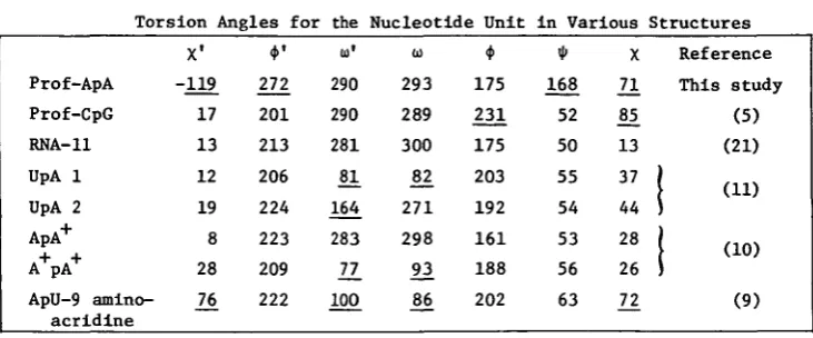
The conformational features of the dinucleoside phosphate are unusual
as shown in Table 1.
In the other oligonucleotide molecules studied to date the nucleoside
por-tions have been shown to obey (at least approximately) the "rigid"
nucleo-tide concept . In rigid nucleonucleo-tides, the glycosidic angles are anti(x •"
0-90°); the torsion angles around C5'-05' and C3'-03' are trans (*M>"VL80°);
the torsion angle around the C4'-C5' bond is gauche (<f> - 60°) and the
ri-bose sugars are either C3' or C2' endo. Only «> and <•>' show large
conforma-tional flexibility. Almost all the exceptions to this principle are found
in this ApA molecule. The two halves of the ApA molecule have very
different conformations. The glycosidic linkage of the 5' adenosine is syn (X^
-4419
at University College London on December 6, 2012
http://nar.oxfordjournals.org/
Figure 2. The arrangement of ApA and proflavine moieties in the crystal structure.
Table 1
Torsion Angles for the Nucleotide Unit in Various Structures
Prof-ApA Prof-CpG RNA-11 UpA 1 UpA 2 ApA+ A+ PA+
ApU-9 amino-acridine X1 -119 17 13 12 19 8 28
21
*'
272 201 213 206 224 223 209 222 to1 290 290 281 81 164 283 _77 100 293 289 300£2
271 298 93 175 231 175 203 192 161 188 202 * 168 52 50 55 54 53 56 63 X2!
85 13 37 44 28 2621
Reference This study (5) (21) (11)1 (10)
(9)The values which deviate significantly from those in RNA-11 are underscored.
119°). Usually, the syn conformation is found only in molecules in which a chemical modification actually excludes an anti-conformation, for example in 8-b romoinos ine13 (X has the values -85° and -76° for the two independent molecules in that structure.) A syn conformation has also been observed for the formycin nucleoside antibiotics - formycin B hydrochloride has X = -140 and oxoformycin B has X = -164° . The other unusual features can be rationalized more easily. The conformation about the C4'-C5' bond is trans,
14
at University College London on December 6, 2012
http://nar.oxfordjournals.org/
a feature which has been observed in some nucleotide structures such as deoxyguanosine 5'-phosphate with X = 175° but in no other oligonucleotides. It has also been implicated in forming kinks in DNA when wound round histone proteins in the nucleosome . Moreover, it appears to be an important
18 feature of the loop regions in tRNA .
The high anti x conformation of the 3' adenosine as well as the mixed sugar puckering has precedent in other dinucleotides with drugs. In the intercalated molecules the C2' endo conformation is found at the 3' rather than the 5' end of the dinucleotide. In the non intercalated ApU-9
amino-9
acridine the sequence of puckering is the same in this structure. The high value of the <t>' angle can be correlated with the C 2 ' endo sugar puc-ker, as observed in pdTpdT and tRNA20. Interestingly, the torsion angles
21 22 $, to and a)' have values normally found in helical ribonucleic acids ' It has always been thought that variation in the latter two angles in par-ticular is responsible for the flexibility in nucleic acids .
The crystal structure of transfer RNA has now been refined indepen-dently by four groups ' . The conformations of some of the loop re-gions are rather ill-defined, and indeed the four analyses do not always agree on interpretation. However, it is likely that at least some of the conformational features observed here in ApA are present in tRNA loop re-gions. Even the rare syn glycosidic conformation may be present in, for
23 example, residue 76 (the end of the amino-acid acceptor stem)
Although the overall conformation that we find for ApA has not been described previously for either a nucleic acid constituent or a polymer, none of the variable angles occur in energetically unfavourable regions. Since w e are not observing in this analysis the structure of a 'naked' nu-cleic acid fragment, but one in a drug-interactive situation (albeit a fro-zen o n e ) , it is tempting to suggest that the binding of the drug is largely responsible for these unexpected conformational features. Furthermore, since the functionality of nucleic acids is often expressed in single-stranded regions, we can expect that protein or drug binding can induce rather more variable and profound conformational changes than have been hitherto suspected, with almost if not all the torsion angles in a
nucleo-tide unit capable of considerable flexibility.
ACKNOWLEDGEMENTS
This work was supported by grants GM 21589, CA 06927, RR 05539 from the National Institutes of Health, by an appropriation from the
at University College London on December 6, 2012
http://nar.oxfordjournals.org/
wealth of Pennsylvania, by the International Cancer Research Data Bank Pro-gramme of the National Cancer Institute with the International Union a-gainst Cancer, and by grants from the Cancer Research Campaign, SRC and Roche Products Ltd.
REFERENCES
1. Lerman, L. S. (1961) J. Molec. Biol. 3, 18-30.
2. Gale, E. F., Cundliffe, E, Reynolds, P. E., Richmond, M. H. and Waring, M. J. (1972) in The Molecular Basis of Antibiotic Action, Wiley, London.
3. Alden, C. J. and Arnott, S. (1975) Nucleic Acids Res. 2, 1701-1717. 4. Tsai, C. C , Jain, S. C. and Sobell, H. M. (1977) J. Molec. Biol. 114,
301-315.
Jain, S. C , Tsai, C. C. and Sobell, H. M. (1977) J. Molec. Biol. 114, 317-331.
5. Neidle, S., Achari, A., Taylor, G. L., Bennan, H. M. , Carrell, H. L., Glusker, J. P. and Stallings, W. C. (1977) Nature 269, 304-307. 6. Bennan, H. M., Neidle, S. and Stodola, R. K. (1978) Proc. Natln. Acad.
Sci. U.S.A. 75, 838-832.
7. Yathindra, N. and Sundaralingam, M. (1973) Biopolymers 12, 297-314. 8. Rich, A., Davies, D. R., Crick, F. H. C. and Watson, J. D. (1961) J.
Molec. Biol. 3, 71-86.
9. Seeman, N. C , Day, R. 0. and Rich, A. (1975) Nature 253, 324-326. 10. Suck, D., Manor, P. C. and Saenger, W. (1976) Acta Crystallogr. B32,
1727-1737.
11. Sussman, J. L., Seeman, N. C , Kim, S. H. and Berman, H. M. J. Molec. Biol. 66, 403-421.
12. Wong, Y. S. and Lippard, S. J. (1977) J. Chem. Soc. Chem. Comm. 824-825. 13. Sternglanz, H., Thomas, J. M. and Bugg, C. E. (1977) Acta. Crystallogr.
B33, 2097-2102.
14. Singh, P. and Hodgson, D. J. (1977) Acta Crystallogr. B33, 3189-3194. 15. Kogama, G., Nakamura, H., Umezana, H. and Iitaka, Y. (1976) Acta
Crys-tallogr. B32, 813-820.
16. Young, D. W., Tollin, P. and Wilson, H. R. (1974) Acta Crystallogr. B30, 2012-2018.
17. Crick, F. H. C. and Klug, A. (1975) Nature 255, 530-533.
18. Jack, A., Klug, A. and Ladner, J. E. (1976) Nature 261, 250-251. 19. Camerman, N., Fawcett, J. K. and Camerman, A. (1976) J. Mol. Biol.
107, 601-621.
20. Jack, A., Ladner, J. E. and Klug, A. (1976) J. Molec. Biol. 108, 619-649. 21. Arnott, S., Chandrasekaran, R. and Seising, E. (1975) in Structure and
Conformation of Nucleic Acids and Protein-Nucleic Acid Interactions, University Park Press, Baltimore.
22. Kallenbach, N. R. and Berman, H. M. (1977) Quart. Rev. Biophy. 10, 138-236.
23. Sussman, J. L. and Kim, S. H. (1976) Biochem. Biophys. Res. Comm. 68, 89-96.
24. Sussman, J. L. and Kim, S. H. (1976) Science 192, 853-858. 25. Quigley, G. J. and Rich, A. (1976) Science 194, 796-805.
26. Quigley, G. J., Seeman, N. C., Wang, A. H. J., Suddath, F. L. and Rich, A. (1975) Nucleic Acids Res. 2, 2329-2339.
27. Stout, C. D., Mizuno, H., Rubin, J., Brennan, T., Rao, S. T. and Sundaralingam, M. (1976) Nucleic Acids Res. 3, 1111-1123.
at University College London on December 6, 2012
http://nar.oxfordjournals.org/