Mechanisms of Bihemispheric Brain Infarctions
in the Anterior Circulation on
Diffusion-Weighted Images
Kozue Saito, Hiroshi Moriwaki, Hiroshi Oe, Kotaro Miyashita, Kazuyuki Nagatsuka, Satoshi Ueno, and Hiroaki Naritomi
BACKGROUND AND PURPOSE: Multiple acute brain infarctions in both cerebral hemi-spheres usually suggest an embolic mechanism, particularly one of aortic or cardiac origin. The purpose of this study was to clarify the etiologic mechanisms and topographic features of bihemispheric infarctions depicted on diffusion-weighted imaging (DWI).
METHODS: Among 411 consecutive patients with ischemic stroke who underwent MR imaging in the acute phase, DWI showed bilateral infarctions in 19 (4.6%). In these patients, we analyzed the presence of carotid, aortic or cardiac embolic sources by using ultrasonography, cerebral angiography, and/or transesophageal echocardiography and evaluated the size and topographic distribution of the lesions. We assessed intracranial cross-flow through the ante-rior communicating artery, mainly on the basis of the anatomic information obatined from angiography or MR angiography.
RESULTS: Bilateral lesions were derived from cardiac and/or aortic embolic sources in 16 (84%) of 19 patients and appeared to originate from unilateral carotid diseases in three (16%). In nine (82%) of 11 patients with cardiac embolic sources, at least one large territorial or subcortical lesion was found in either hemisphere, whereas in all eight patients without a cardiac embolic source, the lesions were small and disseminated bilaterally.
CONCLUSION: Unilateral carotid lesions can cause bihemispheric infarctions through cross-flow in the anterior communicating artery. On DWI, small bihemispheric, disseminated lesions strongly suggest an artery-to-artery embolism. In such cases, aortic and carotid lesions should be assessed as potential embolic sources.
Diffusion-weighted imaging (DWI) has an excellent ability to depict recent small ischemic lesions, and it can depict multiple lesions in patients with acute stroke whose infarction is considered a single event from the clinical aspect (1, 2). According to MR imaging studies, 17–30% of patients with acute stroke have multiple lesions, and 1.4 – 6.1% have bihemi-spheric lesions in the anterior circulation (1– 4). Bi-hemispheric infarctions strongly suggest the presence of embolic sources in the aorta or heart or the asso-ciation of systemic hypercoagulopathy because of
their distributions in multiple arterial territories. Baird et al (2) documented that multiple infarctions may result mainly from multiple emboli or the break up of a single embolus derived from aortic or cardiac disease. However, a single carotid artery lesion can also cause bihemispheric infarctions when the ste-nosed carotid artery supplies the blood flow to the contralateral hemisphere via intracranial cross-flow through the anterior communicating artery (AcoA) (4 – 6). To our knowledge, detailed mechanisms of bihemispheric infarctions, including artery-to-artery embolism of carotid origin, have not previously been investigated. The aim of the present study was to elucidate the etiologic mechanisms and topographic features of bihemispheric infarctions depicted on DWI.
Methods
Patient Population
Between January 1999 and March 2002, 449 patients were consecutively admitted to our department within 3 days after the onset of acute brain infarction. A total of 411 underwent MR imaging, including DWI, during the acute stage. Twenty Received April 22, 2004; accepted after revision August 20.
From the Department of Medicine, Cerebrovascular Division (K.S., H.M., H.O., K.M., K.N., H.N.), National Cardiovascular Center, and the Department of Neurology (S.U.), Nara Medical University, Osaka, Japan.
Supported in part by the Research Grant for Cardiovascular Diseases (14C-1) from the Ministry of Health, Labor and Welfare of Japan.
Address reprint requests to Kozue Saito, M.D., ksaito@ naramed.-u.ac.jp
©American Society of Neuroradiology
(4.9%, 16 men and four women; mean age⫾standard devia-tion, 73.3 ⫾9.9 years) had a bihemispheric infarction in the anterior circulation, as shown on DWI. We excluded patients with systemic disease causing hypercoagulability, such as pro-tein C deficiency or malignancy. Excluding one patient with antiphospholipid antibody syndrome, 19 (4.6%, 15 men and four women; mean age, 73.5⫾10.1 years) underwent further examination. Two patients also had infarctions in the posterior circulation and were included in the study. All patients pro-vided informed consent was obtained before the study.
Evaluation of Risk Factors for Cerebrovascular Diseases All patients underwent systemic examination, including de-termination of their complete blood cell count and blood chem-istry and coagulation tests. Risk factors such as diabetes mel-litus, hypercholesterolemia, habitual cigarette smoking, and cardiac disease were evaluated.
Cardiovascular Evaluation
We evaluated cardiac and aortic disease on the basis of results from several studies. Chest radiography, 12-lead elec-trocardiography, three-lead electrocardiographic monitoring for at least 24 hours after admission, and transthoracic echo-cardiography were performed in all 19 patients. Transesopha-geal echocardiography, including microbubble testing, was per-formed in eight patients. Cardiac diseases that create a risk of potential cardiac embolic sources include intracardiac throm-bus, atrial fibrillation, recent myocardial infarction (⬍4 weeks), sick sinus syndrome, a patent foramen ovale with an interatrial septum aneurysm, mitral valve stenosis, prosthetic valves, and endocarditis (7). The risks of embolus from the thoracic aorta may be due to a mobile plaque or complicated lesion in the arch, which is defined as a lesion thicker than 4.0 mm, a 3.0-mm lesion with wall irregularity, or acoustic shadow in the arch of the thoracic aorta (8 –10).
Cerebrovascular Evaluation
The extracranial carotid and vertebral arteries were evalu-ated with carotid ultrasonography in all 19 patients. Conven-tional cerebral angiography was also performed in nine pa-tients. The intracranial cerebral arteries were evaluated with MR angiography in 10 patients, conventional cerebral angiog-raphy in three, and both studies in the remaining six patients. Stenotic lesions of the extracranial or intracranial arteries were defined as stenosis of at least 50% of the luminal diameter, as shown on Doppler ultrasonography, MR angiography, or con-ventional angiography (7). The degree of intracranial cross-flow through the AcoA was evaluated by using conventional angiography, or if not performed, with MR angiography. When the contralateral side ACA (ACA and MCA in some cases) was detected on conventional angiograms or when the AcoA was detected on MR angiograms, cross-flow via the AcoA was judged to exist. An aplastic A1 segment was defined 1) when the ACA was not detected on the ipsilateral carotid angiograms but was detected on the contralateral carotid angiograms or 2) when no A1 segment was depicted on MR angiograms. A hypoplastic A1 segment was defined when the ACA was de-tected on carotid angiograms and also on the contralateral carotid angiograms or when small A1 segment was present on MR angiograms (11, 12).
MR Imaging Evaluation
All patients underwent conventional MR imaging and DWI with echo-planner imaging (Magnetom Vision, 1.5 T, Siemens Medical Systems, Erlangen, Germany). Conventional imaging consisted of axial MR imaging, including T1-weighted (TR/ TE ⫽ 630/14), T2-weighted (TR/TE ⫽ 5400/99), and
fluid-attenuation inversion recovery (TR/TE/TI ⫽ 9000/105/2400) sequence. DWIs were simultaneously obtained by using a spin-echo planar imaging sequence. Diffusion gradients were ap-plied in each of thex, y,andzdirections withb⫽1000 s/mm2.
Criteria for the diagnosis of acute infarction on DWI were as follows: focal high signal intensities, a location or configuration not representing the normal anisotropy of diffusion, and a location or configuration not thought to represent a magnetic susceptibility artifact (13).
Topographic Distribution and Size of Lesions We assessed the topography of the infarcts by using the mapping templates of Tatu et al (14). The territories of ante-rior circulation were defined as the distribution of the anteante-rior cerebral artery (ACA) and the middle cerebral artery (MCA). When the ipsilateral internal carotid artery (ICA) supplied the posterior cerebral artery (PCA), the distribution of the PCA was also included in the anterior circulation. The watershed area was defined according to the templates of Bogousslavsky and Regli (15) and the atlas of Damasio (16).
Modifying earlier methods (13, 17–19) based on the volume and distribution pattern of the hyperintense area on DWI, we classified infarctions into three types: 1) territorial infarction more than 15 mm in diameter, including more than one infarct with a continuous lesion from the cortex to the subcortex (Fig 1A, right side); 2) subcortical infarction more than 15 mm in diameter in the territory of deep perforating branches originat-ing from the distal ICA or the MCA trunk (Fig 1A); and 3) disseminated small infarction less than 15 mm in diameter (Fig 1B). On the basis of the total volume of hyperintense areas on DWI, we defined the hemisphere with larger volume of the infarcts as the predominantly affected hemisphere, and that with the smaller volume was the subordinately affected hemi-sphere (Fig 1). The lesions were classified in blinded fashion with regard to the results of source identification, and vice versa.
Classification According to Arterial and Cardiac Risks On the basis of arterial and cardiac risks, we divided the patients into three groups, as follows: Group C included pa-tients with cardiac disease; group CA, papa-tients with carotid disease without cardiac disease; and group AO; patients with aortic arch lesions without cardiac or carotid artery disease.
Results
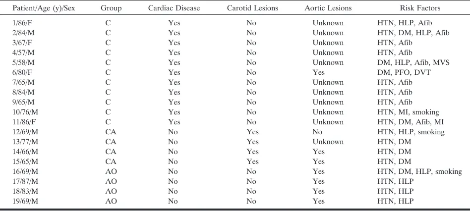
Seventeen patients had hypertension, eight had di-abetes mellitus, eight had hypercholesterolemia, and three were habitual smokers. As shown in Table 1, 11 patients had cardiac disease, namely, chronic atrial fibrillation (n⫽9), recent myocardial infarction (n⫽ 2), mitral valve stenosis (n⫽1), or a patent foramen ovale with deep venous thrombosis (n⫽1), and they were classified into group C. At least one patient also had aortic lesions, though none had both cardiac and carotid artery lesions. Four patients had carotid ar-tery lesions without cardiac disease and were classi-fied into group CA. In this group, three patients underwent an evaluation of the aortic arch, and two were found to have aortic lesions. The remaining four had aortic lesions without cardiac disease or carotid disease and were classified into group AO. In all 19 patients, the manifestation of ischemic symptoms was abrupt, suggesting an embolic mechanism.
FIG 1. Classification of infarction.
A, Right-sided infarction was defined as territorial; left sided, subcortical. Total vol-ume of the infarcts was smaller on the left than right, which was defined as the pre-dominantly affected hemisphere.
[image:3.585.53.370.55.474.2]B, DWI shows small disseminated le-sions. The predominantly affected hemi-sphere is the left side, and infarcts on the contralateral side are localized in the ACA area.
TABLE 1: Patient characteristics
Patient/Age (y)/Sex Group Cardiac Disease Carotid Lesions Aortic Lesions Risk Factors
1/86/F C Yes No Unknown HTN, HLP, Afib 2/84/M C Yes No Unknown HTN, DM, HLP, Afib 3/67/F C Yes No Unknown HTN, Afib
4/57/M C Yes No Unknown HTN, Afib
5/58/M C Yes No Unknown DM, HLP, Afib, MVS
6/80/F C Yes No Yes DM, PFO, DVT
7/65/M C Yes No Unknown HTN, Afib 8/84/M C Yes No Unknown HTN, Afib 9/65/M C Yes No Unknown HTN, Afib 10/76/M C Yes No Unknown HTN, MI, smoking 11/86/F C Yes No Unknown HTN, DM, Afib, MI 12/69/M CA No Yes No HTN, HLP, smoking 13/77/M CA No Yes Unknown HTN, DM
14/66/M CA No Yes Yes HTN, DM
15/65/M CA No Yes Yes HTN, DM
16/69/M AO No No Yes HTN, DM, HLP, smoking
17/87/M AO No No Yes HTN, HLP
18/83/M AO No No Yes HTN, HLP
19/69/M AO No No Yes HTN, HLP
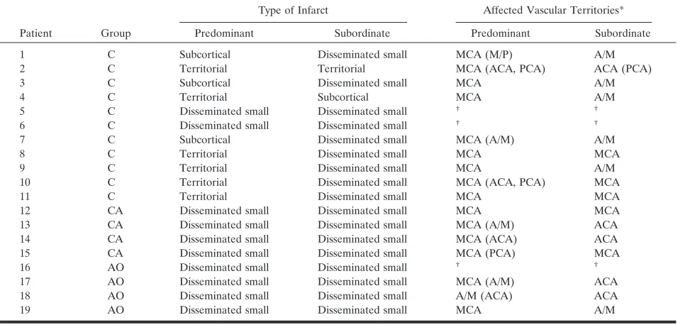
[image:3.585.54.528.501.714.2]least one large territorial infarction (n ⫽ 6) or sub-cortical infarction (n⫽3) in either hemisphere. Only two (19%) patients had small disseminated lesions alone in both hemispheres; in one (patient 6), aortic lesions were also found. In group AO and CA, all eight patients had only small disseminated lesions, which were bilateral.
In all four patients in group CA, ICA stenosis was found on the side ipsilateral to the predominantly affected hemisphere. Figure 2 shows the angiographic and DWI findings in group CA. In patient 12, the contralateral carotid artery was occluded, and pa-tients 13 and 14, the contralateral A1 segment was aplastic or hypoplastic; in these three patients, the stenotic ICA supplied both ACA areas via AcoA cross-flow. In patients 13 and 14, lesions in the con-tralateral hemisphere were located in the ACA area. In patient 15, the stenotic ICA supplied the blood flow to only the ipsilateral hemisphere because of the absence of AcoA cross-flow. This patient also had aortic lesions as a possible embolic source; therefore, bilateral infarcts were derived from the aortic lesions instead of the ICA lesion. Thus, bilateral lesions are considered to have derived from cardiac and/or aortic embolic sources in 16 patients (all 15 patients in group C and group AO plus one of four patients in group CA) and appeared to have originated from unilateral carotid diseases in three patients (three of four patients in group CA).
Discussion
We detected acute infarctions in both cerebral hemispheres in the anterior circulation in 20 (4.9%)
of 411 patients with stroke. Our prevalence of DWI-depicted bihemispheric infarctions was similar to that previously reported (1.4 – 6.1%) (1– 4). Multiple in-farctions are generally thought to be caused by an embolic mechanism (2). In cases of bihemispheric infarctions, cardiac disease or an aortic lesion is usu-ally suspected as a possible embolic source. However, embolic sources of bihemispheric infarctions are sel-dom evaluated. Bogousslavsky et al (3) found a car-diac embolic source on one-half of their patients with bihemispheric infarctions.
Roh et al (4) suggested that unilateral carotid ar-tery lesions may cause bihemispheric infarctions. In their study, five of 20 patients with bihemispheric infarctions had stenotic lesions in the unilateral ICA, which supplied the contralateral ACA through intra-cranial cross-flow via the AcoA. In these five patients, the bihemispheric infarctions were all located in the territories supplied by the stenotic ICA. Unfortu-nately, the association of cardiac disease or aortic lesions was not evaluated, and hence, the importance of ICA lesions in the etiologic mechanisms of bihemi-spheric infarctions remains unclear. In our study, three of four patients with unilateral ICA lesion had AcoA cross-flow from the stenotic ICA to the con-tralateral ACA (Fig 2). In these patients, the stenotic ICA supplied blood flow to the territories including the infarcts. (Fig 2). The development of bihemispheric in-farctions was presumably attributed to an embolism originating from the unilateral carotid lesion.
[image:4.585.57.535.72.301.2]We found cardiac disease in 58% of patients with bihemispheric infarctions, similar Bogousslavsky et al (3). At least one large territorial infarction or subcor-tical infarction was found in nine of 11 patients with TABLE 2: Distribution pattern of infarcts
Patient Group
Type of Infarct Affected Vascular Territories*
Predominant Subordinate Predominant Subordinate
1 C Subcortical Disseminated small MCA (M/P) A/M 2 C Territorial Territorial MCA (ACA, PCA) ACA (PCA) 3 C Subcortical Disseminated small MCA A/M 4 C Territorial Subcortical MCA A/M 5 C Disseminated small Disseminated small † †
6 C Disseminated small Disseminated small † †
7 C Subcortical Disseminated small MCA (A/M) A/M 8 C Territorial Disseminated small MCA MCA 9 C Territorial Disseminated small MCA A/M 10 C Territorial Disseminated small MCA (ACA, PCA) MCA 11 C Territorial Disseminated small MCA MCA 12 CA Disseminated small Disseminated small MCA MCA 13 CA Disseminated small Disseminated small MCA (A/M) ACA 14 CA Disseminated small Disseminated small MCA (ACA) ACA 15 CA Disseminated small Disseminated small MCA (PCA) MCA 16 AO Disseminated small Disseminated small † †
17 AO Disseminated small Disseminated small MCA (A/M) ACA 18 AO Disseminated small Disseminated small A/M (ACA) ACA 19 AO Disseminated small Disseminated small MCA A/M
* Data in parentheses indicates the vascular territory in which a small number of infarcts were localized. A/M⫽watershed area between the ACA and MCA, M/P⫽watershed area between the MCA and PCA.
†
cardiac disease, whereas such territorial or subcortical infarction was not found in patients without cardiac disease. In our eight patients with carotid or aortic lesions unaccompanied by cardiac disease, all lesions were small and scattered in both cerebral hemi-spheres in a disseminated manner. Such a pattern of small bihemispheric, disseminated lesions was found in two of 11 patients with cardiac disease; one of them also had aortic lesions other than cardiac disease. Kang et al (6) evaluated the lesion pattern of infarc-tions in 35 patients with acute stroke and ICA steno-sis or occlusion by using DWI and reported that large territorial infarction was rare. Matsumoto et al (20) studied the size of infarctions in acute stroke using DWI and reported that lesions in patients with ca-rotid lesions or aortic lesions were generally small and disseminated. Therefore, the pattern of small bihemi-spheric, disseminated lesions appears to strongly sug-gest the existence of carotid lesions or aortic lesions as the possible embolic sources rather than cardiac disease. The role of aortic lesions in mechanism of
bihemispheric infarctions is seldom evaluated, most likely because aortic lesions are often occur concom-itantly with cardiac disease or carotid lesions. How-ever, in four of our 19 patients, aortic lesions were found solely and not associated with other embolic sources. If we extrapolate the results from our rela-tively small samples to the general population, this finding implies that aortic lesions cause at least 20% of all bihemispheric infarctions.
The present study had some limitations. First, the apparent diffusion coefficient on DWI remains low for 2–10 days after the onset of acute stroke (21, 22). Provided that stroke occurs repeatedly within a short interval, the first and subsequent infarctions are likely demonstrated as lesions that develop simultaneously on DWI. The possibility that such recurrent infarc-tions were included as bihemispheric infarcinfarc-tions in the present study could not be excluded completely. Second, the patients were classified into three groups according to the type of embolic source found. How-ever, the classification was incomplete: In group C,
the presence or absence of aortic lesions was un-known in 10 of 11 patients, though none had carotid lesions. In group CA, aortic lesions coexisted in two of three patients and were not evaluated in the other patient, though none of them had cardiac disease. Only in group AO was the association of other em-bolic sources completely excluded in all patients. This partial lack of information and the coexistence of two embolic sources in some patients should be taken into account when the present results are interpreted.
Conclusion
Despite the general belief that bihemispheric in-farctions are caused mainly by cardiogenic embolism, such infarctions may occasionally result from an ar-tery-to-artery embolism originating from aortic le-sions or unilateral carotid artery lele-sions. Small bilat-eral, spotty lesions strongly suggest an artery-to-artery embolism originating from an aortic or unilateral ca-rotid artery lesion. Cardiac disease, aortic lesions, and/or carotid lesions may concomitantly occur in a patient with bihemispheric infarctions. In such pa-tients, the possible embolic source must be deter-mined speculatively to select either anticoagulation or antiplatelet therapy to prevent recurrent attacks. Our results provide information useful in selecting such preventive therapies.
References
1. Altieri M, Metz RJ, Muller C, Maeder P, Meuli R, Bogousslavsky J. Multiple brain infarcts: Clinical and neuroimaging patterns using diffusion-weighted magnetic resonance.Eur Neurol1999;42:76 – 82 2. Baird AE, Lovblad KO, Schlaug G, Edelman RR, Warach S.
Multiple acute stroke syndrome: marker of embolic disease? Neu-rology2000;54:674 – 678
3. Bogousslavsky J, Bernasconi A, Kumral E.Acute multiple infarc-tion involving the anterior circulainfarc-tion.Arch Neurol1996;53:50 –57 4. Roh JK, Kang DW, Lee SH, Yoon BW, Chang KH.Significance of acute multiple brain infarction on diffusion-weighted imaging.
Stroke2000;31:688 – 694
5. Siebler M, Sitzer M, Rose G, Bendfeldt D, Steinmetz H.Silent cerebral embolism caused by neurologically symptomatic
high-grade carotid stenosis: event rates before and after carotid endar-terectomy.Brain1993;116:1005–1015
6. Kang DW, Chu K, Ko SB, Kwon SJ, Yoon BW, Roh JK.Lesion patterns and mechanism of ischemia in internal carotid artery dis-ease: a diffusion-weighted imaging study. Arch Neurol 2002;59: 1577–1582
7. Adams HP, Bendixen BH, Kappelle LJ, et al.Classification of subtype of acute ischemic stroke: definitions for use in a multi-center clinical trial.Stroke1993;24:35– 41
8. Amarenco P, Duyckaerts C, Tzourio C, Henin D, Bousser MG, Hauw JJ.The prevalence of ulcerated plaques in the aortic arch in patients with stroke.N Engl J Med1992;326:221–225
9. Toyoda K, Yasaka M, Nagata S, Yamaguchi T.Aortogenic embolic stroke: a transesophageal echocardiographic approach. Stroke
1992;23:1056 –1061
10. Amarenco P, Cohen A, Tzourio C, et al.Atherosclerotic disease of the aortic arch and the risk of ischemic stroke.N Engl J Med
1994;331:1474 –1479
11. Stock KW, Wetzel S, Kirsch E, Bongartz G, Steinbrich W, Radue EW.Anatomic evaluation of the circle of Willis: MR angiography versus intraarterial digital subtraction angiography.AJNR Am J Neuroradiol1996;17:1495–1499
12. Osborn AG. The circle of Willis. In: Osborn AG, ed.Cerebral Angiography.2nd ed. Philadelphia: Lippincott, Williams & Wilkins; 1999:105–116
13. Singer MB, Chong J, Lu D, Schonewille WJ, Tuhrim S, Atlas SW. Diffusion-weighted MRI in acute subcortical infarction. Stroke
1998;29:133–136
14. Tatu L, Moulin T, Bogousslavsky J, Duvernoy H.Arterial territories of the human brain: cerebral hemispheres.Neurology1998;50:1699 –1708 15. Bogousslavsky J, Regli F.Unilateral watershed cerebral infarcts.
Neurology1986;36:373–377
16. Damasio H.A computed tomographic guide to the identification of cerebral vascular territories.Arch Neurol1983;40:138 –142 17. Szabo K, Kern R, Gass A, Hirsch J, Hennerici M.Acute stroke
patterns in patients with internal carotid artery disease: a diffu-sion-weighted magnetic resonance imaging study.Stroke2001;32: 1323–1329
18. Rodda RA.The arterial patterns associated with internal carotid disease and cerebral infarcts.Stroke1986;17:69 –75
19. Ringelstein EB, Schneider R, Koschorke S.Analysis of patterns of hemispheric brain infarctions on CT: embolic stroke mechanism, territorial infarction and lacunae.Psychiatry Res1989;29:273–276 20. Matsumoto N, Yokota T, Hasegawa Y, Yamaguchi T, Minematsu K. Analysis of acute multiple brain infarction using diffusion-weighted MR imaging.Jpn J Stroke2001;23:248 –254
21. Warach S, Gaa J, Siewert B, Wielopolski P, Edelman RR. Acute human stroke studied by whole brain echo planar diffusion-weighted magnetic resonance imaging.Ann Neurol1995;37:231–241 22. Yamada N, Imakita S, Sakuma T. Value of diffusion-weighted