research communications
Acta Cryst.(2017). E73, 1747–1750 https://doi.org/10.1107/S2056989017015171
1747
Received 9 September 2017 Accepted 17 October 2017
Edited by A. J. Lough, University of Toronto, Canada
Keywords:crystal structure; hydrogen bonding; zwitterions; Hirshfield analysis.
CCDC reference:1580559
Supporting information:this article has supporting information at journals.iucr.org/e
Crystal structure and Hirshfield analysis of the
4-(dimethylamino)pyridine adduct of
4-methoxy-phenylborane
Jesse Shooter,aCaleb J. Allen,aColby W. K. Tinsley,aLev N. Zakharovband Eric R. Abbeya*
aDepartment of Chemistry, Biochemistry, and Physics, Eastern Washington, University, Cheney, WA 99004, USA, and bDepartment of Chemistry and Biochemistry, CAMCOR, University of Oregon, Eugene, OR 97403, USA.
*Correspondence e-mail: [email protected]
The title compound [systematic name: 4-(dimethylamino)pyridine–4-methoxy-phenylborane (1/1)], C14H19BN2O, contains two independent molecules in the
asymmetric unit. Both molecules exhibit coplanar, mostly sp2-hybridized methoxy and dimethylamino substituents on their respective aromatic rings, consistent with-donation into the aromatic systems. The B—H groups exhibit an intramolecular close contact with a C—H group of the pyridine ring, which may be evidence of electrostatic attraction between the hydridic B—H and the electropositive aromatic C—H. There appears to be weak C—H (arene) interactions between two of the H atoms of an aminomethyl group and the methoxy-substituted benzene ring of the other independent molecule, and another C—H (arene) interaction between one of the pyridine ring H atoms and the same benzene ring.
1. Chemical context
Monoorganoboranes (RBH2) have been the focus of chemical
research for over fifty years, most notably for their use in the indispensable hydroboration reaction, which permits reduc-tion of olefins, carbonyl compounds and others (Brown & Krishnamurthy, 1979; Crudden & Edwards, 2003.) Such boranes are often isolated as their Lewis base adducts, in which the base donates a lone pair into the vacantporbital of thesp2borane. Among the most common class of Lewis bases
for the formation of borane adducts are amines. Amine boranes are widely used as hydroboration reagents (Clay & Vedejs, 2005), precursors for borenium cation synthesis (De Vrieset al., 2012), frustrated Lewis pairs (Stephan, 2015), and have been investigated as hydrogen-storage materials (Campbellet al., 2010). We have synthesized the zwitterionic title compound by hydride removal from sodium 4-methoxy-phenylborohydride with chlorotrimethylsilane in the presence of 4-dimethylaminopyridine. This compound is slightly unusual, as examples of monoorganoboranes with hetero-atoms on the organic substitituent are limited.
2. Structural commentary
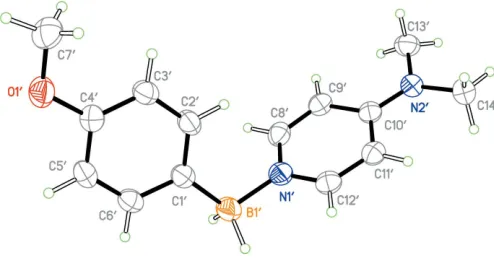
The asymmetric unit contains two independent molecules (Figs. 1 and 2) with only slightly different geometric features (Fig. 3). In both molecules, the boron atom appears to besp3
hybridized [C1—B1—N1 = 110.8 (1) and C10—B10—N10 =
1.611 (2) A˚ , respectively] are consistent with a formal C—B single bond. The oxygen atom of both methoxy groups appears to be mostlysp2hybridized, [C7—O1—C4 = 117.3 (1) and C70—O10—C40= 117.4 (1)] and is close to coplanar with
the phenyl ring [torsion angles C7—O1—C4—C3 =7.4 (2) and C70—O10—C40—C30 = 7.1 (2)], consistent with
-donation into the phenyl ring.
The geometries of the 4-(dimethylamino)pyridine (DMAP) fragment of both molecules is similar to other structures of DMAP–borane adducts. The nitrogen atom of the dimethyl-amino fragment appears to besp2hybridized [torsion angles C13—N2—C10 = 121.0 (1) and C130—N20—C100 =
122.2 (1)] and is close to coplanar [torsion angles C13—N2—
C10—C11 = 2.4 (2) and C130—N20—C100—C110 = 3.4 (1)]
consistent with-donation into the pyridine ring.
The B1—N1 and B10—N10 distances [1.597 (2) and
1.595 (2) A˚ , respectively] are consistent with formal N—B single bonds, and are within the range observed for other DMAP–borane adducts (seeDatabase survey). Interestingly, the B—H atoms exhibit intramolecular close contacts with the C—H atoms of the pyridine ring [H12 H2B= 2.26 (3) and H120 H2B0= 2.27 (3) A˚ ] and are close to coplanar [torsion angles H2B—B1—N1—C12 = 4(1) and H2B—B1—N1—C12 = 16 (1)], which may be evidence of electrostatic interactions
between the hydridic B—H atoms and electropositive aromatic C—H atoms, and is observed in other DMAP– borane adducts (see Database Survey). The planes of the pyridine rings and the benzene rings are almost normal to one another [the dihedral angle between the C1–C6 and C8–C12/ N1 rings is 73.14 (7)and that between the C10–C60and C80–
C120/N10 rings is 74.15 (7)]. Perhaps the most significant
difference between the two molecules is the 9.0difference in
the torsion angle about the B—N bond [C1—B1—N1—C8 = 63.9 (2) while C10—B10—N10—C80=72.9 (2)] (Fig. 3).
3. Supramolecular features
The molecules within the asymmetric unit exhibit weak C— H (arene) interactions between two of the hydrogen atoms of the aminomethyl group and the methoxyphenyl group of a neighboring molecule (see Table 1) as well as a C— H (arene) interaction between one of the pyridine hydrogen atoms and the same methoxyphenyl ring (Fig. 4).
1748
Shooteret al. C14H19BN2O Acta Cryst.(2017). E73, 1747–1750
[image:2.610.313.566.107.157.2]research communications
Figure 2
[image:2.610.62.279.170.256.2]The molecular structure of the other independent molecule of the title compound with displacement ellipsoids drawn at the 50% probability level.
Figure 3
An overlay of the two independent molecules. Table 1
Hydrogen-bond geometry (A˚ ,).
Cgis the centroid of the C10–C60ring.
D—H A D—H H A D A D—H A
[image:2.610.48.298.358.509.2]C9—H9A Cg 0.95 3.12 4.069 (2) 178 C13—H13A Cg 0.97 3.12 3.662 (2) 112 C13—H13C Cg 0.97 3.23 3.662 (2) 109
Figure 1
[image:2.610.46.293.576.705.2] [image:2.610.316.564.597.725.2]4. Hirshfield analysis
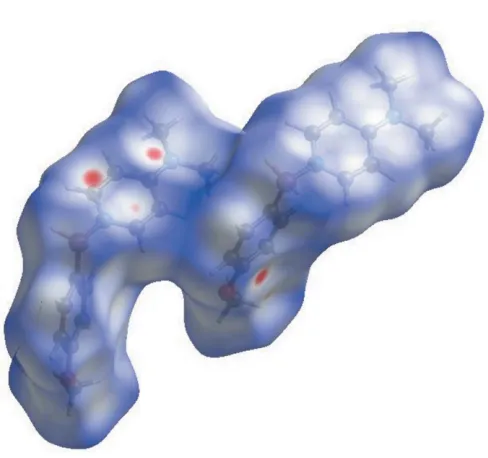
The weak intermolecular interactions of the title compound were explored by Hirshfield analysis. Hirshfield surfaces were generated usingCrystal Explorer 3.1(McKinnonet al., 2007; Spackman & Jayatilaka, 2009). The space within a crystal is partitioned so that the ratio of promolecule to procrystal is equal to 0.5, generating continuous surfaces that permit the visualization of weak interactions. The dnormvalues illustrate
whether the intermolecular contact is shorter or longer than the van der Waals radii. Red areas of the Hirshfield surface indicate negativednormvalues contacts closer than the van der
Waals radii. This analysis lends further support to the weak C—H (arene) interactions described in the previous section (Fig. 5.)
5. Database survey
A search of the Cambridge Structural Database (Version 5.37, update February 2017; Groomet al., 2016) for DMAP–borane adducts yielded only two structures: VOGJEI (Chu, et al., 2014) and JUDQAA (Lesleyet al., 1998). A search for phenyl-based monoorganoborane–amine adducts (Ph–BH2–NR3)
yielded four structures: UTOZEJ (Hubner et al., 2012), BEXQOM (Me´nard & Stephan, 2013), EPOYAK (Franzet al., 2011), and GEBNAE (Jacobset al., 2012). In all four of these structures, the B—N bonds are approximately perpendicular to the plane of the arene rings. In all six cases, the boron atom is tetrahedral and displays structural features consistent with
sp3hybridization. Additionally, the C—B and B—N bonds are all within the range for formal C—B and C—N single bonds.
6. Synthesis and crystallization
In a nitrogen-filled glove box, sodium 4-methoxyphenyl-borohydride (97mg, 0.67 mmol) and 4-dimethylaminopyridine (82 mg, 0.67 mmol) were combined in a 20 mL vial containing a stir bar and dissolved in anhydrous THF (4 mL). The solu-tion was cooled to 247 K in the freezer and chlorotrimethyl-silane (73 mg, 0.67 mmol) was added dropwiseviasyringe. The reaction was allowed to come to 295 K and was stirred for 1 h. The solvent was then removed in vacuoand the residue was
washed with anhydrous diethyl ether (4 mL), followed by extraction with anhydrous dichloromethane (4 mL). The extract was filtered through a 0.45mm PTFE syringe filter. The
research communications
Acta Cryst.(2017). E73, 1747–1750 Shooteret al. C
[image:3.610.46.296.71.180.2]14H19BN2O
1749
Figure 5 [image:3.610.319.563.76.305.2]Hirshfield surface mapped overdnorm. Red areas highlight intermolecular contacts shorter than the sum of the van der Waals radii.
Figure 4
Weak C—H (arene) interactions between the two independent molecules in the unit cell shown as dashed lines.Cgis the centroid of the C10–C60 benzene ring. Only H atoms involved in the interactions are shown.
Table 2
Experimental details.
Crystal data
Chemical formula C14H19BN2O
Mr 242.12
Crystal system, space group Orthorhombic,Pbca
Temperature (K) 173
a,b,c(A˚ ) 12.3538 (6), 18.7727 (10), 23.4056 (12)
V(A˚3) 5428.1 (5)
Z 16
Radiation type CuK
(mm1) 0.58
Crystal size (mm) 0.140.090.07
Data collection
Diffractometer Bruker APEXII CCD Absorption correction Multi-scan (SADABS; Bruker,
2008) Tmin,Tmax 0.695, 0.753
No. of measured, independent and observed [I> 2(I)] reflections
46022, 4800, 3948
Rint 0.063
(sin/)max(A˚
1) 0.595
Refinement
R[F2> 2(F2)],wR(F2),S 0.047, 0.144, 1.08 No. of reflections 4800
No. of parameters 353
H-atom treatment H atoms treated by a mixture of independent and constrained refinement
max,min(e A˚
3) 0.22,0.22
[image:3.610.313.560.445.726.2]solvent was again removed in vacuo to afford a white solid (51 mg, 37%). Crystals suitable for X-ray diffraction were grown by diffusion of pentane into a concentrated solution of the title compound in anhydrous dichloromethane.
1
H NMR (500 MHz, CDCl3)(ppm): 8.12 (d, 2H,J =7 Hz),
7.23 (d, 2 H,J= 8 Hz), 6.80 (d, 2H,J= 8.5 Hz), 6.52 (d, 2H,J= 8 Hz), 3.78 (s, 3H), 3.11 (s, 6H).13C NMR (126 MHz, CDCl3)
(ppm): 157.3, 154.9, 146.7, 145.0 (br s), 134.5, 122.9, 106.5, 55.0, 39.5.11B NMR (160 MHz, CDCl3)(ppm):5.0 (br,s). FTIR
(ATR, cm1): 3012, 2952, 2923, 2853, 2610, 2346, 2288, 2227, 1634, 1548, 1442, 1418, 1392, 1237, 1223, 1161, 1076, 1031, 811, 797, 548, 515.
7. Refinement
Crystal data, data collection and structure refinement details are summarized in Table 2. H atoms were refined in calculated positions (C—H = 0.95 A˚ withUiso(H) = 1.5Ueq(C-methyl) and
1.2eq(C) for other H atoms. The B-bound H atoms were located in a difference-Fourier map and freely refined. Methyl H atoms were refined without restrictions on rotation around the C—C bonds, HFIX 138 inSHELXL(Sheldrick, 2015).
Funding information
Funding for this research was provided by: Eastern Washington University Faculty Grants for Creative Works.
References
Brown, H. C. & Krishnamurthy, S. (1979).Tetrahedron,35, 567–607. Bruker (2008). APEX2, SAINT and SADABS. Bruker AXS Inc.,
Madison, Wisconsin, USA.
Campbell, P. G., Zakharov, L. N., Grant, D. J., Dixon, D. A. & Liu, S.-Y. (2010).J. Am. Chem. Soc.132, 3289–3291.
Chu, J., Han, X., Kefalidis, C. E., Zhou, J., Maron, L., Leng, X. & Chen, Y. (2014).J. Am. Chem. Soc.136, 10894–10897.
Clay, J. M. & Vedejs, E. (2005).J. Am. Chem. Soc.127, 5766–5767. Crudden, C. M. & Edwards, D. (2003).Eur. J. Org. Chem.pp. 4695–
4712.
De Vries, T. S., Prokofjevs, A. & Vedejs, E. (2012).Chem. Rev.112, 4246–4282.
Franz, D., Bolte, M., Lerner, H.-W. & Wagner, M. (2011). Dalton Trans.40, 2433–2440.
Groom, C. R., Bruno, I. J., Lightfoot, M. P. & Ward, S. C. (2016).Acta Cryst.B72, 171–179.
Jacobs, E. A., Fuller, A., Coles, S. J., Jones, G. A., Tizzard, G. J., Wright, J. A. & Lancaster, S. J. (2012).Chem. Eur. J.18, 8647–8658. Lesley, M. J. G., Woodward, A., Taylor, N. J., Marder, T. B., Cazenobe, I., Ledoux, I., Zyss, J., Thornton, A., Bruce, D. W. & Kakkar, A. K. (1998).Chem. Mater.10, 1355–1365.
Macrae, C. F., Bruno, I. J., Chisholm, J. A., Edgington, P. R., McCabe, P., Pidcock, E., Rodriguez-Monge, L., Taylor, R., van de Streek, J. & Wood, P. A. (2008).J. Appl. Cryst.41, 466–470.
McKinnon, J. J., Jayatilaka, D. & Spackman, M. A. (2007).Chem. Commun.pp. 3814–3816.
Me´nard, G. & Stephan, D. W. (2013).Dalton Trans.42, 5447–5453. Sheldrick, G. M. (2008).Acta Cryst.A64, 112–122.
Sheldrick, G. M. (2015).Acta Cryst.C71, 3–8.
Spackman, M. A. & Jayatilaka, D. (2009).CrystEngComm,11, 19–32. Stephan, D. L. (2015).J. Am. Chem. Soc.137, 10018–10032.
1750
Shooteret al. C14H19BN2O Acta Cryst.(2017). E73, 1747–1750
supporting information
sup-1
Acta Cryst. (2017). E73, 1747-1750
supporting information
Acta Cryst. (2017). E73, 1747-1750 [https://doi.org/10.1107/S2056989017015171]
Crystal structure and Hirshfield analysis of the 4-(dimethylamino)pyridine
adduct of 4-methoxyphenylborane
Jesse Shooter, Caleb J. Allen, Colby W. K. Tinsley, Lev N. Zakharov and Eric R. Abbey
Computing details
Data collection: APEX2 (Bruker, 2008); cell refinement: SAINT (Bruker, 2008); data reduction: SAINT (Bruker, 2008);
program(s) used to solve structure: SHELXS97 (Sheldrick 2008); program(s) used to refine structure: SHELXL2014
(Sheldrick, 2015); molecular graphics: SHELXTL (Sheldrick, 2008) and Mercury (Macrae et al., 2006); software used to
prepare material for publication: SHELXTL (Sheldrick, 2008).
4-(Dimethylamino)pyridine–4-methoxyphenylborane (1/1)
Crystal data
C14H19BN2O Mr = 242.12
Orthorhombic, Pbca a = 12.3538 (6) Å
b = 18.7727 (10) Å
c = 23.4056 (12) Å
V = 5428.1 (5) Å3 Z = 16
F(000) = 2080
Dx = 1.185 Mg m−3
Cu Kα radiation, λ = 1.54178 Å Cell parameters from 6122 reflections
θ = 3.8–66.5°
µ = 0.58 mm−1 T = 173 K
Cut-block, colorless 0.14 × 0.09 × 0.07 mm
Data collection
Bruker APEXII CCD diffractometer
Radiation source: Incoatec IµS
φ and ω scans
Absorption correction: multi-scan (SADABS; Bruker, 2008)
Tmin = 0.695, Tmax = 0.753
46022 measured reflections
4800 independent reflections 3948 reflections with I > 2σ(I)
Rint = 0.063
θmax = 66.6°, θmin = 3.8° h = −14→14
k = −22→21
l = −27→27
Refinement
Refinement on F2
Least-squares matrix: full
R[F2 > 2σ(F2)] = 0.047 wR(F2) = 0.144 S = 1.08 4800 reflections 353 parameters 0 restraints
Hydrogen site location: mixed
H atoms treated by a mixture of independent and constrained refinement
w = 1/[σ2(F
o2) + (0.0934P)2 + 0.2472P]
where P = (Fo2 + 2Fc2)/3
(Δ/σ)max = 0.001
Δρmax = 0.22 e Å−3
supporting information
sup-2
Acta Cryst. (2017). E73, 1747-1750 Special details
Geometry. All esds (except the esd in the dihedral angle between two l.s. planes) are estimated using the full covariance matrix. The cell esds are taken into account individually in the estimation of esds in distances, angles and torsion angles; correlations between esds in cell parameters are only used when they are defined by crystal symmetry. An approximate (isotropic) treatment of cell esds is used for estimating esds involving l.s. planes.
Fractional atomic coordinates and isotropic or equivalent isotropic displacement parameters (Å2)
x y z Uiso*/Ueq
supporting information
sup-3
Acta Cryst. (2017). E73, 1747-1750
C1′ 0.66395 (12) 0.06221 (8) 0.22354 (7) 0.0353 (3) C2′ 0.55974 (13) 0.05774 (9) 0.24570 (7) 0.0390 (4) H2′A 0.5010 0.0533 0.2197 0.047* C3′ 0.53692 (13) 0.05950 (9) 0.30416 (7) 0.0391 (4) H3′A 0.4642 0.0567 0.3172 0.047* C4′ 0.62082 (13) 0.06528 (8) 0.34280 (7) 0.0355 (3) C5′ 0.72647 (12) 0.07031 (8) 0.32248 (7) 0.0375 (4) H5′A 0.7850 0.0747 0.3486 0.045* C6′ 0.74621 (12) 0.06892 (8) 0.26434 (7) 0.0365 (4) H6′A 0.8189 0.0727 0.2515 0.044* C7′ 0.50222 (15) 0.06648 (10) 0.42327 (8) 0.0505 (4) H7′1 0.4633 (7) 0.1089 (7) 0.4080 (6) 0.076* H7′2 0.50509 (15) 0.0693 (8) 0.4658 (6) 0.076* H7′3 0.4632 (7) 0.0223 (7) 0.4117 (6) 0.076* C8′ 0.54070 (13) −0.01494 (9) 0.11568 (7) 0.0389 (4) H8′A 0.5701 −0.0528 0.1376 0.047* C9′ 0.45100 (13) −0.02840 (8) 0.08343 (7) 0.0370 (3) H9′A 0.4193 −0.0745 0.0839 0.044* C10′ 0.40477 (12) 0.02603 (8) 0.04920 (6) 0.0335 (3) C11′ 0.45656 (13) 0.09326 (8) 0.05317 (6) 0.0360 (3) H11B 0.4290 0.1326 0.0322 0.043* C12′ 0.54561 (13) 0.10182 (8) 0.08691 (7) 0.0372 (3) H12B 0.5782 0.1476 0.0885 0.045* C13′ 0.26225 (14) −0.05283 (9) 0.01282 (8) 0.0450 (4) H13D 0.3125 (7) −0.0908 (5) 0.0187 (6) 0.068* H13E 0.2285 (10) −0.0583 (4) −0.0236 (5) 0.068* H13F 0.2083 (10) −0.0539 (3) 0.0421 (5) 0.068* C14′ 0.26866 (14) 0.07399 (10) −0.01557 (8) 0.0459 (4) H14D 0.2354 (11) 0.1068 (6) 0.0122 (4) 0.069* H14E 0.2125 (11) 0.0554 (2) −0.0414 (5) 0.069* H14F 0.3238 (7) 0.0996 (6) −0.0380 (5) 0.069* H1′B 0.7533 (16) 0.0147 (10) 0.1468 (8) 0.048 (5)* H1B 1.0840 (16) 0.2841 (11) 0.3930 (8) 0.051 (5)* H2′B 0.7314 (16) 0.1141 (10) 0.1398 (9) 0.052 (5)* H2B 1.0475 (17) 0.3752 (11) 0.4179 (9) 0.056 (6)*
Atomic displacement parameters (Å2)
U11 U22 U33 U12 U13 U23
supporting information
sup-4
Acta Cryst. (2017). E73, 1747-1750
C6 0.0273 (7) 0.0431 (8) 0.0387 (8) −0.0005 (6) 0.0005 (6) −0.0017 (6) C7 0.0574 (12) 0.0904 (15) 0.0391 (10) 0.0067 (10) 0.0129 (8) 0.0114 (10) C8 0.0417 (8) 0.0327 (8) 0.0407 (8) −0.0009 (6) −0.0021 (7) 0.0070 (6) C9 0.0418 (8) 0.0314 (7) 0.0385 (8) −0.0039 (6) −0.0003 (7) 0.0005 (6) C10 0.0337 (7) 0.0379 (8) 0.0277 (7) 0.0013 (6) 0.0039 (6) −0.0001 (6) C11 0.0435 (8) 0.0324 (8) 0.0336 (8) 0.0030 (6) 0.0000 (6) −0.0006 (6) C12 0.0434 (8) 0.0334 (8) 0.0328 (8) −0.0032 (6) 0.0018 (6) 0.0000 (6) C13 0.0459 (9) 0.0540 (10) 0.0507 (10) −0.0061 (8) −0.0117 (8) −0.0059 (8) C14 0.0568 (12) 0.0560 (11) 0.0618 (12) 0.0110 (9) −0.0197 (9) 0.0004 (9) B1′ 0.0333 (9) 0.0635 (12) 0.0392 (10) −0.0031 (8) 0.0021 (8) 0.0000 (8) O1′ 0.0467 (7) 0.0583 (7) 0.0345 (6) 0.0036 (5) −0.0030 (5) −0.0021 (5) N1′ 0.0349 (7) 0.0465 (8) 0.0315 (7) −0.0003 (5) 0.0042 (5) −0.0018 (5) N2′ 0.0345 (7) 0.0418 (7) 0.0378 (7) −0.0006 (5) −0.0013 (5) 0.0025 (5) C1′ 0.0328 (8) 0.0339 (7) 0.0393 (8) −0.0006 (6) −0.0021 (6) 0.0000 (6) C2′ 0.0308 (8) 0.0505 (9) 0.0358 (8) −0.0013 (6) −0.0052 (6) −0.0034 (7) C3′ 0.0300 (8) 0.0469 (9) 0.0403 (9) −0.0005 (6) 0.0006 (6) −0.0014 (7) C4′ 0.0398 (8) 0.0323 (7) 0.0345 (8) 0.0018 (6) −0.0025 (6) −0.0024 (6) C5′ 0.0351 (8) 0.0343 (8) 0.0431 (9) 0.0009 (6) −0.0108 (7) −0.0014 (6) C6′ 0.0291 (7) 0.0354 (8) 0.0449 (9) 0.0008 (6) −0.0013 (6) −0.0001 (6) C7′ 0.0538 (11) 0.0577 (11) 0.0399 (9) 0.0058 (8) 0.0071 (8) −0.0059 (8) C8′ 0.0408 (8) 0.0427 (8) 0.0333 (8) 0.0051 (7) 0.0034 (6) 0.0034 (6) C9′ 0.0402 (8) 0.0365 (8) 0.0345 (8) −0.0002 (6) 0.0049 (6) 0.0004 (6) C10′ 0.0329 (7) 0.0383 (8) 0.0292 (7) 0.0024 (6) 0.0064 (6) −0.0022 (6) C11′ 0.0375 (8) 0.0358 (8) 0.0346 (8) 0.0037 (6) 0.0041 (6) 0.0007 (6) C12′ 0.0386 (8) 0.0375 (8) 0.0354 (8) −0.0011 (6) 0.0067 (6) −0.0026 (6) C13′ 0.0406 (9) 0.0491 (9) 0.0454 (9) −0.0092 (7) 0.0018 (7) −0.0001 (7) C14′ 0.0403 (9) 0.0520 (10) 0.0455 (10) 0.0028 (7) −0.0049 (7) 0.0069 (7)
Geometric parameters (Å, º)
B1—N1 1.597 (2) B1′—N1′ 1.595 (2)
B1—C1 1.608 (2) B1′—C1′ 1.611 (2)
B1—H1B 1.13 (2) B1′—H1′B 1.150 (19) B1—H2B 1.14 (2) B1′—H2′B 1.166 (19) O1—C4 1.3752 (19) O1′—C4′ 1.370 (2)
O1—C7 1.413 (2) O1′—C7′ 1.423 (2)
N1—C12 1.342 (2) N1′—C12′ 1.347 (2)
N1—C8 1.350 (2) N1′—C8′ 1.350 (2)
N2—C10 1.338 (2) N2′—C10′ 1.344 (2) N2—C14 1.455 (2) N2′—C14′ 1.456 (2) N2—C13 1.459 (2) N2′—C13′ 1.458 (2)
C1—C2 1.396 (2) C1′—C2′ 1.390 (2)
C1—C6 1.402 (2) C1′—C6′ 1.400 (2)
C2—C3 1.389 (2) C2′—C3′ 1.397 (2)
C2—H2A 0.9500 C2′—H2′A 0.9500
C3—C4 1.387 (2) C3′—C4′ 1.380 (2)
C3—H3A 0.9500 C3′—H3′A 0.9500
supporting information
sup-5
Acta Cryst. (2017). E73, 1747-1750
C5—C6 1.381 (2) C5′—C6′ 1.383 (2)
C5—H5A 0.9500 C5′—H5′A 0.9500
C6—H6A 0.9500 C6′—H6′A 0.9500
C7—H7A 0.996 (14) C7′—H7′1 0.996 (14) C7—H7B 0.996 (14) C7′—H7′2 0.996 (14) C7—H7C 0.996 (14) C7′—H7′3 0.996 (14)
C8—C9 1.361 (2) C8′—C9′ 1.364 (2)
C8—H8A 0.9500 C8′—H8′A 0.9500
C9—C10 1.419 (2) C9′—C10′ 1.419 (2)
C9—H9A 0.9500 C9′—H9′A 0.9500
C10—C11 1.413 (2) C10′—C11′ 1.418 (2) C11—C12 1.366 (2) C11′—C12′ 1.364 (2)
C11—H11A 0.9500 C11′—H11B 0.9500
C12—H12A 0.9500 C12′—H12B 0.9500
C13—H13A 0.967 (13) C13′—H13D 0.955 (12) C13—H13B 0.967 (13) C13′—H13E 0.955 (12) C13—H13C 0.967 (13) C13′—H13F 0.955 (12) C14—H14A 0.973 (15) C14′—H14D 0.985 (12) C14—H14B 0.973 (15) C14′—H14E 0.985 (12) C14—H14C 0.973 (15) C14′—H14F 0.985 (12)
supporting information
sup-6
Acta Cryst. (2017). E73, 1747-1750
C4—C5—H5A 120.0 C4′—C5′—H5′A 120.0 C5—C6—C1 122.85 (14) C5′—C6′—C1′ 123.02 (14) C5—C6—H6A 118.6 C5′—C6′—H6′A 118.5 C1—C6—H6A 118.6 C1′—C6′—H6′A 118.5 O1—C7—H7A 109.5 O1′—C7′—H7′1 109.5 O1—C7—H7B 109.5 O1′—C7′—H7′2 109.5 H7A—C7—H7B 109.5 H7′1—C7′—H7′2 109.5 O1—C7—H7C 109.5 O1′—C7′—H7′3 109.5 H7A—C7—H7C 109.5 H7′1—C7′—H7′3 109.5 H7B—C7—H7C 109.5 H7′2—C7′—H7′3 109.5 N1—C8—C9 123.92 (14) N1′—C8′—C9′ 123.76 (14) N1—C8—H8A 118.0 N1′—C8′—H8′A 118.1 C9—C8—H8A 118.0 C9′—C8′—H8′A 118.1 C8—C9—C10 120.23 (14) C8′—C9′—C10′ 120.40 (14) C8—C9—H9A 119.9 C8′—C9′—H9′A 119.8 C10—C9—H9A 119.9 C10′—C9′—H9′A 119.8 N2—C10—C11 122.89 (14) N2′—C10′—C9′ 123.00 (14) N2—C10—C9 121.99 (14) N2′—C10′—C11′ 122.00 (14) C11—C10—C9 115.11 (14) C9′—C10′—C11′ 114.99 (14) C12—C11—C10 120.41 (14) C12′—C11′—C10′ 120.44 (14) C12—C11—H11A 119.8 C12′—C11′—H11B 119.8 C10—C11—H11A 119.8 C10′—C11′—H11B 119.8 N1—C12—C11 123.83 (14) N1′—C12′—C11′ 123.83 (14) N1—C12—H12A 118.1 N1′—C12′—H12B 118.1 C11—C12—H12A 118.1 C11′—C12′—H12B 118.1 N2—C13—H13A 109.5 N2′—C13′—H13D 109.5 N2—C13—H13B 109.5 N2′—C13′—H13E 109.5 H13A—C13—H13B 109.5 H13D—C13′—H13E 109.5 N2—C13—H13C 109.5 N2′—C13′—H13F 109.5 H13A—C13—H13C 109.5 H13D—C13′—H13F 109.5 H13B—C13—H13C 109.5 H13E—C13′—H13F 109.5 N2—C14—H14A 109.5 N2′—C14′—H14D 109.5 N2—C14—H14B 109.5 N2′—C14′—H14E 109.5 H14A—C14—H14B 109.5 H14D—C14′—H14E 109.5 N2—C14—H14C 109.5 N2′—C14′—H14F 109.5 H14A—C14—H14C 109.5 H14D—C14′—H14F 109.5 H14B—C14—H14C 109.5 H14E—C14′—H14F 109.5
supporting information
sup-7
Acta Cryst. (2017). E73, 1747-1750
C2—C3—C4—C5 1.1 (2) C2′—C3′—C4′—C5′ 1.0 (2) O1—C4—C5—C6 177.77 (14) O1′—C4′—C5′—C6′ 178.10 (13) C3—C4—C5—C6 −0.9 (2) C3′—C4′—C5′—C6′ −0.5 (2) C4—C5—C6—C1 0.1 (2) C4′—C5′—C6′—C1′ −0.3 (2) C2—C1—C6—C5 0.6 (2) C2′—C1′—C6′—C5′ 0.6 (2) B1—C1—C6—C5 178.75 (15) B1′—C1′—C6′—C5′ −179.77 (15) C12—N1—C8—C9 0.4 (2) C12′—N1′—C8′—C9′ 0.6 (2) B1—N1—C8—C9 −178.28 (15) B1′—N1′—C8′—C9′ −179.78 (14) N1—C8—C9—C10 0.2 (2) N1′—C8′—C9′—C10′ 0.9 (2) C14—N2—C10—C11 −1.7 (2) C14′—N2′—C10′—C9′ 174.39 (14) C13—N2—C10—C11 −177.60 (15) C13′—N2′—C10′—C9′ 4.4 (2) C14—N2—C10—C9 179.35 (16) C14′—N2′—C10′—C11′ −6.6 (2) C13—N2—C10—C9 3.4 (2) C13′—N2′—C10′—C11′ −176.65 (14) C8—C9—C10—N2 178.37 (14) C8′—C9′—C10′—N2′ 177.12 (14) C8—C9—C10—C11 −0.7 (2) C8′—C9′—C10′—C11′ −1.9 (2) N2—C10—C11—C12 −178.48 (14) N2′—C10′—C11′—C12′ −177.51 (14) C9—C10—C11—C12 0.6 (2) C9′—C10′—C11′—C12′ 1.5 (2) C8—N1—C12—C11 −0.5 (2) C8′—N1′—C12′—C11′ −1.0 (2) B1—N1—C12—C11 178.13 (15) B1′—N1′—C12′—C11′ 179.36 (14) C10—C11—C12—N1 0.1 (2) C10′—C11′—C12′—N1′ −0.1 (2)
Hydrogen-bond geometry (Å, º)
Cg is the centroid of the C1′–C6′ ring.
D—H···A D—H H···A D···A D—H···A