The 1:1 charge-transfer complex
dibenzotetrathiafulvalene–pyromellitic
dianhydride (DBTTF–PMDA)
Margaret E. Payne,aKatelyn P. Goetz,aCynthia S. Dayb* and Oana D. Jurchescua*
aDepartment of Physics, Wake Forest University, Winston-Salem, NC 27109, USA,
andbDepartment of Chemistry, Wake Forest University, Winston-Salem, NC 27109,
USA
Correspondence e-mail: [email protected], [email protected]
Received 21 May 2014; accepted 7 June 2014
Key indicators: single-crystal X-ray study;T= 213 K; mean(C–C) = 0.002 A˚;
Rfactor = 0.032;wRfactor = 0.091; data-to-parameter ratio = 19.6.
The title charge-transfer (CT) complex, C10H2O6C14H8S4,
composed of donor dibenzotetrathiafulvalene (DBTTF) and acceptor pyromellitic dianhydride (PMDA), forms a mixed stacking pattern along the [110] direction. The constituent molecules occupy crystallographic inversion centers. They are nearly parallel and lieca.3.41 A˚ from each other. The crystals exhibit a high degree of donor/acceptor overlap [88.20 (4)%] in the long direction of the DBTTF and PMDA molecules as compared with 51.27 (5)% in the shortest direction of the molecules.
Related literature
General properties and potential applications of charge-transfer complexes in electronic devices are outlined by Goetz
et al. (2014); Horiuchi et al. (2006); Tsutsumi et al. (2012); Kobayashi et al. (2012); Kagawa et al. (2010); Herbstein (2005); Ferraris et al. (1973); Kistenmacher et al. (1981); Takahashi et al. (2006); Wu et al. (2013). Related CT struc-tures, containing the acceptor pyromellitic dianhydride
(PMDA) include anthracene–PMDA (Robertson &
Stezowski, 1978), phenanthrene–PMDA (Evans & Robinson, 1977), pyrene–PMDA (Herbstein & Snyman, 1969) and two polymorphs of biphenylene–PMDA (Stezowski et al., 1986). Structure–property relationships in molecular crystals have been described theoretically by Coropceanuet al.(2007) and experimentally by Meiet al.(2013), among others.
Experimental
Crystal data
C10H2O6C14H8S4
Mr= 522.56 Triclinic,P1
a= 7.2292 (4) A˚
b= 8.9572 (5) A˚
c= 9.5224 (5) A˚
= 70.051 (1) = 68.712 (1)
= 70.136 (1)
V= 523.39 (5) A˚3
Z= 1
MoKradiation
= 0.50 mm 1
T= 213 K
0.200.200.02 mm
Data collection
Bruker APEX CCD diffractometer Absorption correction: multi-scan
(SADABS; Sheldrick, 2012)
Tmin= 0.703,Tmax= 0.746
10004 measured reflections 3023 independent reflections 2668 reflections withI> 2(I)
Rint= 0.021
Refinement
R[F2> 2(F2)] = 0.032
wR(F2) = 0.091
S= 1.07 3023 reflections
154 parameters
H-atom parameters constrained
max= 0.30 e A˚ 3
min= 0.33 e A˚ 3
Data collection:SMART(Bruker, 2002); cell refinement:SAINT
(Bruker, 2011); data reduction:SAINT; program(s) used to solve structure: SHELXLS2013 (Sheldrick, 2008); program(s) used to refine structure:SHELXL2013(Sheldrick, 2008); molecular graphics:
SHELXTL(Sheldrick, 2008); software used to prepare material for publication:SHELXTL.
The WFU X-ray Facility thanks the National Science Foundation (grant CHE-0234489) for funds to purchase the X-ray instrument and computers. This work has been partly supported by the National Science Foundation grant DMR-1105147. KPG acknowledges the NSF Graduate Research Fellowship Program under grant DGE-0907738.
Supporting information for this paper is available from the IUCr electronic archives (Reference: PK2526).
References
Bruker (2002).SMART. Bruker AXS Inc., Madison, Wisconsin, USA. Bruker (2011).SAINT. Bruker AXS Inc., Madison, Wisconsin, USA. Coropceanu, V., Cornil, J., da Silva Filho, D. A., Olivier, Y., Silbey, R. &
Bredas, J.-L. (2007).Chem. Rev.107, 926–952.
Evans, D. L. & Robinson, W. T. (1977).Acta Cryst.B33, 2891–2893. Ferraris, J., Cowan, D. O., Walatka, V. & Perlstein, J. H. (1973).JACS,95, 948–
949.
Goetz, K. P., Vermeulen, D., Payne, M. E., Kloc, C., McNeil, L. E. & Jurchescu, O. D. (2014).J. Mater. Chem. C,2, 3065–3076.
Herbstein, F. H. (2005).Crystalline Molecular Complexes and Compounds: Structures and Principles. New York: Oxford University Press Inc. Herbstein, F. H. & Snyman, J. A. (1969).Phil. Trans. Roy. Soc. (London) A,
264, 635–662.
Horiuchi, S., Hasegawa, T. & Tokura, Y. (2006).J. Phys. Soc. Jpn,75, 051016.
organic compounds
o844
Payneet al. doi:10.1107/S1600536814013324 Acta Cryst.(2014). E70, o844–o845 Acta Crystallographica Section EStructure Reports
Online
Phys.6, 169–172.
Kistenmacher, T. J., Emge, T. J., Wiygul, F. M., Bryden, W. A., Chappell, J. S., Stokes, J. P., Chiang, L. Y., Cowan, D. O. & Bloch, A. N. (1981).Solid State Commun.39, 415–417.
Kobayashi, K., Horiuchi, S., Kumai, R., Kagawa, F., Murakami, Y. & Tokura, Y. (2012).Phys. Rev. Lett.108, 237601.
Mei, Y., Loth, M. A., Payne, M., Zhang, W., Smith, J., Day, C. S., Parkin, S. R., Heeney, M., McCulloch, I., Anthopoulos, T. D., Anthony, J. E. & Jurchescu, O. D. (2013).Adv. Mater.25, 4352–4357.
Sheldrick, G. M. (2008).Acta Cryst.A64, 112–122.
Sheldrick, G. M. (2012).SADABS. University of Go¨ttingen, Germany. Stezowski, J. J., Stigler, R. D. & Karl, N. (1986).J. Chem. Phys.84, 5162–5170. Takahashi, Y., Hasegawa, T., Abe, Y., Tokura, Y. & Saito, G. (2006).Appl.
Phys. Lett.88, 073504.
Tsutsumi, J., Matsui, H., Yamada, T., Kumai, R. & Hasegawa, T. (2012).J. Phys. Chem.C116, 23957–23964.
supporting information
sup-1
Acta Cryst. (2014). E70, o844–o845supporting information
Acta Cryst. (2014). E70, o844–o845 [doi:10.1107/S1600536814013324]
The 1:1 charge-transfer complex dibenzotetrathiafulvalene
–
pyromellitic
dianhydride (DBTTF
–
PMDA)
Margaret E. Payne, Katelyn P. Goetz, Cynthia S. Day and Oana D. Jurchescu
S1. Comment
Organic charge-transfer (CT) complexes are combinations of electron donor (D) and electron acceptor (A) materials.
They have been studied for decades, but have attracted significant interest recently due to their intriguing properties such
as photoconductivity, tunable semiconductivity, metallicity, ferroelectricity, etc., which make them viable candidates for versatile electronic devices (Goetz et al., 2014; Horiuchi et al., 2006; Tsutsumi et al., 2012; Kobayashi et al., 2012; Kagawa et al., 2010). In the 1:1 D:A stoichiometry, they can exhibit either mixed stacking, where the repeating motif in the π-stacking direction is ···D—A—D—A···, or segregated stacking, where the donor and acceptor π-stack separately, as ···A—A—A—A··· and ···D—D—D—D··· (Herbstein, 2005). CT complexes of the acceptor
7,7,8,8-tetracyanoquinodi-methane (TCNQ) have been widely explored. Examples include the organic metal with donor tetrathiafulvalene (TTF)
(Ferraris et al., 1973) or the ambipolar semiconductor with dibenzotetrathiafulvalene (DBTTF) (Kistenmacher et al., 1981; Takahashi et al., 2006; Wu et al., 2013). With the exception of a few reports, CT complexes containing pyromellitic dianhydride (PMDA) as an acceptor have received little attention. Examples of CT complexes of PMDA include
anthracene-PMDA (Robertson & Stezowski, 1978), phenanthrene-PMDA (Evans & Robinson, 1977), pyrene-PMDA
(Herbstein & Snyman, 1969), and two polymorphs of biphenylene-PMDA (Stezowski et al., 1986). Here we report for the first time on the growth and crystal structure of the 1:1 CT complex containing the donor DBTTF and acceptor
PMDA.
Single crystals of DBTTF-PMDA are triclinic, space group P1, with Z=1, where the DBTTF and PMDA molecules
occupy crystallographic inversion centers at (0,0,0) and (1/2,1/2,0), respectively. The crystals are platelets, with their
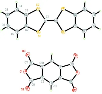
largest face corresponding to the (001) plane. The molecular structure is shown with thermal ellipsoids in Figure 1, where
only the contents of the asymmetric unit are labeled. The DBTTF and PMDA molecules pack in a mixed-stack pattern,
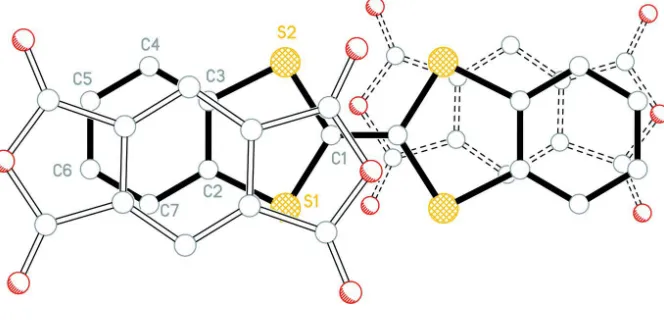
previously observed in other PMDA-based CT complexes. As shown in Figure 2, the DBTTF molecules lie on the
corners of the unit cell, while the PMDA molecules lie in the center of the ab crystal faces. The mixed DA stacks build along the [-1 1 0] direction and are tilted by 45.43 (6)° (DBTTF) and 46.40 (6)° (PMDA) with respect to ab face. This tilt leads to a molecular overlap between the donor and acceptor wherein the fused 5-and 6-membered rings of each half
DBTTF overlap ("straddle") the 3 fused rings of the PMDA molecules in the stack (Figure 3). The centroid of the central
PMDA 6-membered ring to centroid of the 5- and 6-membered rings of the adjacent DBTTF molecules in the stack are
3.648 (1)Å and 3.585 (1)Å, respectively. The shortest centroid-centroid contact involving 5-membered rings of the
DBTTF and PMDA molecules is 3.611 (1)Å while the shortest contacts involving the DBTTF 6-membered ring centroid
are 3.527 (1)Å and 3.538 (1)Å to the centroids of the 5-membered PMDA and 6-membered DBTTF rings, respectively.
The planes of the D/A molecules are nearly parallel with an interplanar angle of 1.31 (5)° and the long axes of the
alternates between 3.32Å and 3.4Å (Robertson & Stezowski, 1978). There is a high degree of overlap between donor and
acceptor molecules: the DBTTF molecule overlaps with 88.20 (4)% of the PMDA molecule in the longest direction of the
molecule, and with 51.27 (5)% of the PMDA molecule in the shortest direction of the molecule. Measurements are in
progress to evaluate the degree of charge transfer between the two moieties; however, this high degree of overlap
suggests that a high value is to be expected. The large molecular overlap is a signature of good electrical properties, as
suggested by theoretical (Coropceanu et al., 2007) and experimental studies (Mei et al., 2013).
S2. Experimental
Dibenzotetrathiafulvalene (DBTTF) and pyromellitic dianhydride (PMDA), both obtained from Sigma Aldrich, were
separately dissolved in xylenes and acetonitrile, respectively. The solid weights of the compounds were measured in the
molar ratio 1:1. The solution concentrations were saturated, such that all of parent compound dissolved in as little solvent
as possible. The solutions were mixed, and the complex was then crystallized by slow evaporation under ambient
conditions. After about two days of evaporation, crystals were obtained as green-gold plates with approximate
dimensions of 0.20 mm x 0.20 mm x 0.02 mm.
S3. Refinement
The hydrogen atoms were included in the structural model as fixed atoms (using idealized sp2-hybridized geometry and C
—H bond lengths of 0.94 Å) "riding" on their respective carbon atoms. The isotropic thermal parameter of each hydrogen
atom was fixed at a value 1.2 times the equivalent isotropic thermal parameter of the carbon atom to which it is
supporting information
[image:5.610.136.477.70.382.2]sup-3
Acta Cryst. (2014). E70, o844–o845Figure 1
The atom numbering scheme for DBTTF-PMDA with non-hydrogen atoms represented by 50% probability ellipsoids.
Each molecule occupies an inversion center in the unit cell and only contents of the asymmetric unit are labeled.
Figure 2
[image:5.610.151.465.446.588.2]Figure 3
Projection perpendicular to plane of DBTTF with PMDA (above - open bonds) (below - dashed open bonds) showing
overlap of rings.
Dibenzotetrathiafulvalene–pyromellitic dianhydride (1/1)
Crystal data
C10H2O6·C14H8S4
Mr = 522.56
Triclinic, P1 Hall symbol: -P 1
a = 7.2292 (4) Å
b = 8.9572 (5) Å
c = 9.5224 (5) Å
α = 70.051 (1)°
β = 68.712 (1)°
γ = 70.136 (1)°
V = 523.39 (5) Å3
Z = 1
F(000) = 266
Dx = 1.658 Mg m−3
Mo Kα radiation, λ = 0.71073 Å Cell parameters from 5268 reflections
θ = 3.5–31.3°
µ = 0.50 mm−1
T = 213 K Plate, green-gold 0.20 × 0.20 × 0.02 mm
Data collection
Bruker APEX CCD diffractometer
Radiation source: sealed x-ray tube Graphite monochromator
φ and ω scans
Absorption correction: multi-scan (SADABS; Sheldrick, 2012)
Tmin = 0.703, Tmax = 0.746
10004 measured reflections 3023 independent reflections 2668 reflections with I > 2σ(I)
Rint = 0.021
θmax = 30.0°, θmin = 3.5°
h = −10→10
k = −12→12
l = −13→13
Refinement
Refinement on F2
Least-squares matrix: full
R[F2 > 2σ(F2)] = 0.032
wR(F2) = 0.091
S = 1.07 3023 reflections 154 parameters 0 restraints
Primary atom site location: structure-invariant direct methods
Hydrogen site location: inferred from neighbouring sites
H-atom parameters constrained
w = 1/[σ2(F
o2) + (0.0529P)2 + 0.115P]
where P = (Fo2 + 2Fc2)/3
(Δ/σ)max < 0.001
Δρmax = 0.30 e Å−3
supporting information
sup-5
Acta Cryst. (2014). E70, o844–o845Special details
Experimental. Absorption correction: data were corrected for scaling and absorption effects using the multi-scan
technique [SADABS (Sheldrick, 2012)]. The ratio of minimum to maximum apparent transmission was 0.942.
Geometry. All e.s.d.'s (except the e.s.d. in the dihedral angle between two l.s. planes) are estimated using the full
covariance matrix. The cell e.s.d.'s are taken into account individually in the estimation of e.s.d.'s in distances, angles and torsion angles; correlations between e.s.d.'s in cell parameters are only used when they are defined by crystal symmetry. An approximate (isotropic) treatment of cell e.s.d.'s is used for estimating e.s.d.'s involving l.s. planes.
Fractional atomic coordinates and isotropic or equivalent isotropic displacement parameters (Å2)
x y z Uiso*/Ueq
S1 −0.06704 (4) 0.15387 (4) 0.15691 (4) 0.02936 (10)
S2 −0.31329 (5) −0.02880 (4) 0.11851 (4) 0.03025 (10)
C1 −0.08003 (17) 0.02644 (15) 0.05769 (14) 0.0250 (2)
C2 −0.32321 (18) 0.17849 (14) 0.27098 (14) 0.0245 (2)
C3 −0.43823 (18) 0.09185 (14) 0.25298 (14) 0.0246 (2)
C4 −0.64247 (19) 0.10259 (16) 0.34069 (16) 0.0291 (3)
H4 −0.7200 0.0443 0.3290 0.035*
C5 −0.7294 (2) 0.20070 (17) 0.44543 (16) 0.0333 (3)
H5 −0.8672 0.2096 0.5042 0.040*
C6 −0.6146 (2) 0.28587 (17) 0.46427 (15) 0.0332 (3)
H6 −0.6754 0.3511 0.5362 0.040*
C7 −0.4108 (2) 0.27550 (16) 0.37773 (15) 0.0292 (3)
H7 −0.3334 0.3329 0.3909 0.035*
O1 0.08852 (14) 0.63648 (12) 0.27821 (12) 0.0338 (2)
O2 0.30802 (18) 0.75698 (14) 0.29107 (13) 0.0441 (3)
O3 −0.05194 (14) 0.48790 (13) 0.21480 (13) 0.0395 (2)
C8 0.2784 (2) 0.67350 (16) 0.23163 (15) 0.0301 (3)
C9 0.41355 (18) 0.59058 (14) 0.10654 (14) 0.0237 (2)
C10 0.30252 (17) 0.50435 (14) 0.08434 (14) 0.0237 (2)
C11 0.09415 (18) 0.53456 (16) 0.19353 (15) 0.0282 (2)
C12 0.61565 (17) 0.58997 (14) 0.02281 (14) 0.0252 (2)
H12 0.6905 0.6484 0.0375 0.030*
Atomic displacement parameters (Å2)
U11 U22 U33 U12 U13 U23
S1 0.02281 (15) 0.03348 (17) 0.03676 (18) −0.00701 (12) −0.00525 (12) −0.01796 (14)
S2 0.02316 (15) 0.03528 (18) 0.03745 (19) −0.00885 (12) −0.00331 (12) −0.01913 (14)
C1 0.0214 (5) 0.0260 (5) 0.0281 (6) −0.0048 (4) −0.0051 (4) −0.0102 (5)
C2 0.0249 (5) 0.0227 (5) 0.0237 (5) −0.0048 (4) −0.0055 (4) −0.0057 (4)
C3 0.0234 (5) 0.0239 (5) 0.0246 (5) −0.0043 (4) −0.0059 (4) −0.0062 (4)
C4 0.0249 (5) 0.0291 (6) 0.0303 (6) −0.0072 (5) −0.0050 (5) −0.0060 (5)
C5 0.0282 (6) 0.0341 (7) 0.0274 (6) −0.0061 (5) 0.0008 (5) −0.0063 (5)
C6 0.0378 (7) 0.0298 (6) 0.0255 (6) −0.0067 (5) −0.0007 (5) −0.0094 (5)
C7 0.0347 (6) 0.0266 (6) 0.0261 (6) −0.0084 (5) −0.0059 (5) −0.0083 (5)
O1 0.0265 (4) 0.0378 (5) 0.0343 (5) −0.0065 (4) 0.0015 (4) −0.0179 (4)
C8 0.0295 (6) 0.0318 (6) 0.0280 (6) −0.0075 (5) −0.0028 (5) −0.0119 (5)
C9 0.0242 (5) 0.0249 (5) 0.0228 (5) −0.0061 (4) −0.0051 (4) −0.0082 (4)
C10 0.0204 (5) 0.0253 (5) 0.0239 (5) −0.0065 (4) −0.0048 (4) −0.0050 (4)
C11 0.0234 (5) 0.0288 (6) 0.0287 (6) −0.0054 (4) −0.0031 (4) −0.0084 (5)
C12 0.0239 (5) 0.0270 (6) 0.0274 (6) −0.0082 (4) −0.0065 (4) −0.0086 (5)
Geometric parameters (Å, º)
S1—C1 1.7543 (13) C6—H6 0.9400
S1—C2 1.7560 (12) C7—H7 0.9400
S2—C3 1.7505 (13) O1—C11 1.3923 (16)
S2—C1 1.7567 (12) O1—C8 1.3993 (16)
C1—C1i 1.353 (2) O2—C8 1.1878 (17)
C2—C7 1.3967 (17) O3—C11 1.1907 (16)
C2—C3 1.3985 (17) C8—C9 1.4800 (17)
C3—C4 1.3963 (16) C9—C12 1.3864 (16)
C4—C5 1.3892 (19) C9—C10 1.3933 (16)
C4—H4 0.9400 C10—C12ii 1.3839 (16)
C5—C6 1.391 (2) C10—C11 1.4836 (16)
C5—H5 0.9400 C12—C10ii 1.3838 (16)
C6—C7 1.3913 (19) C12—H12 0.9400
C1—S1—C2 95.38 (6) C6—C7—C2 118.93 (13)
C3—S2—C1 95.34 (6) C6—C7—H7 120.5
C1i—C1—S1 122.00 (13) C2—C7—H7 120.5
C1i—C1—S2 122.26 (13) C11—O1—C8 110.17 (10)
S1—C1—S2 115.74 (6) O2—C8—O1 121.25 (13)
C7—C2—C3 120.37 (11) O2—C8—C9 131.52 (13)
C7—C2—S1 123.12 (10) O1—C8—C9 107.23 (11)
C3—C2—S1 116.50 (9) C12—C9—C10 122.97 (11)
C4—C3—C2 120.30 (12) C12—C9—C8 129.32 (11)
C4—C3—S2 122.83 (10) C10—C9—C8 107.70 (10)
C2—C3—S2 116.88 (9) C12ii—C10—C9 123.01 (10)
C5—C4—C3 119.04 (12) C12ii—C10—C11 129.51 (11)
C5—C4—H4 120.5 C9—C10—C11 107.48 (11)
C3—C4—H4 120.5 O3—C11—O1 121.29 (12)
C4—C5—C6 120.69 (12) O3—C11—C10 131.31 (13)
C4—C5—H5 119.7 O1—C11—C10 107.40 (10)
C6—C5—H5 119.7 C10ii—C12—C9 114.03 (10)
C5—C6—C7 120.66 (13) C10ii—C12—H12 123.0
C5—C6—H6 119.7 C9—C12—H12 123.0
C7—C6—H6 119.7
C2—S1—C1—C1i 176.20 (15) C11—O1—C8—O2 −178.78 (13)
C2—S1—C1—S2 −4.14 (8) C11—O1—C8—C9 0.82 (14)
C3—S2—C1—C1i −176.37 (15) O2—C8—C9—C12 −0.6 (2)
supporting information
sup-7
Acta Cryst. (2014). E70, o844–o845C1—S1—C2—C7 −178.74 (11) O2—C8—C9—C10 178.37 (15)
C1—S1—C2—C3 2.71 (10) O1—C8—C9—C10 −1.17 (14)
C7—C2—C3—C4 0.54 (18) C12—C9—C10—C12ii 0.2 (2)
S1—C2—C3—C4 179.13 (9) C8—C9—C10—C12ii −178.82 (11)
C7—C2—C3—S2 −179.00 (9) C12—C9—C10—C11 −179.90 (11)
S1—C2—C3—S2 −0.41 (13) C8—C9—C10—C11 1.05 (13)
C1—S2—C3—C4 178.35 (11) C8—O1—C11—O3 179.96 (12)
C1—S2—C3—C2 −2.12 (10) C8—O1—C11—C10 −0.18 (14)
C2—C3—C4—C5 0.16 (18) C12ii—C10—C11—O3 −0.9 (2)
S2—C3—C4—C5 179.68 (10) C9—C10—C11—O3 179.27 (14)
C3—C4—C5—C6 −0.7 (2) C12ii—C10—C11—O1 179.29 (12)
C4—C5—C6—C7 0.5 (2) C9—C10—C11—O1 −0.57 (13)
C5—C6—C7—C2 0.2 (2) C10—C9—C12—C10ii −0.21 (19)
C3—C2—C7—C6 −0.73 (18) C8—C9—C12—C10ii 178.62 (12)
S1—C2—C7—C6 −179.23 (10)