DEVELOPMENT OF NEW NMR TECHNIQUES FOR
CONFORMATIONAL ANALYSIS OF ¹³C-ENRICHED
OLIGOSACCHARIDES
Richard Harris
A Thesis Submitted for the Degree of PhD
at the
University of St Andrews
1998
Full metadata for this item is available in
St Andrews Research Repository
at:
http://research-repository.st-andrews.ac.uk/
Please use this identifier to cite or link to this item:
http://hdl.handle.net/10023/14226
Development of NMR methods for conformational
analysis of^^C-enriched oligosaccharides.
Richard Hanis
A thesis submitted for the degree of Doctor of Philosophy.
School of Biomedical Sciences University of St. Andrews
ProQuest Number: 10166231
All rights reserved
INFORMATION TO ALL USERS
The quality of this reproduction is d ep en d en t upon the quality of the copy submitted.
In the unlikely ev en t that the author did not send a co m p lete manuscript
and there are missing p a g es, these will be noted. Also, if material had to be rem oved, a n o te will indicate the deletion.
uest
ProQuest 10166231
Published by ProQuest LLO (2017). Copyright of the Dissertation is held by the Author.
All rights reserved.
This work is protected against unauthorized copying under Title 17, United States C o d e Microform Edition © ProQuest LLO.
ProQuest LLO.
789 East Eisenhower Parkway P.Q. Box 1346
Table of Contents
Acknowledgements
Declaiations vii
Abstiact X
List of Figures xi
List of Tables xvii
List of Abbreviations xix
List of Symbols Used xx
Chapter 1 - Introduction 1
1.1 Biological Function of Carbohydrates 2
1.1.1 Carbohydrate Structure 2
1.1.2 Blood Group Antigens 6
1.1.2.1 ABH and Lewis Antigens 6
1.1.3 Caibohydrates in Disease 9
1.1.3.1 Oncology 9
1.1.3.2 Bacterial Infection 9
1.1.4 Inflammation 10
1.2 Selectins 13
1.2.1 Stucture 13
1.2.2 Expression 15
1.2.2.1 E-selectin 15
1.2.2.2 P-selectin 16
1.2.2.3 L-selectin 16
1.2.3 Additional Selectin Functions 17
1.2.4 Cai'bohydi'ate Ligands 18
1.2.5 Glycoprotein Ligands 19
1.3 Carbohydrate Therapeutics 21
1.3.1 Rational Drug Design 23
1.4 NMR Spectroscopy 24
1.4.1 Conformational Analysis 24
Ill
1.4.3 Full Relaxation Matiix Analysis 29
1.4.4 Spin-Spin Coupling Constants 30
1.4.5 Two and Thi'ee Dimensional NMR Spectroscopy 31
1.4.6 Transferred NOE experiments 31
1.5 Cai'bohdiate Conformation 34
1.5.1 Theoretical Determination of Conformation 35
1.6 Outline of Investigation 39
Chapter 2 - Synthesis of ^^C-labelled Carbohydrates 40
2.1 Introduction 42
2.1.1 NMR Investigation of ^^C enriched Oligosacchaiides 42
2.1.2 Synthesis of sialyl Lewis’" 43
2.2 Materials and Methods 46
2.2.1 Synthesis of A^-acetyl [U-‘^C] neuraminic acid 46
2.2.2 Multi-enzymatic synthesis of N-acetyl [U-^^C] lactosamine, 46 Gaipi-4GlcNAc
2.2.3 Multi-enzymatic synthesis of lactose, Galpl-4[U-‘^C, 50%-^H]Glc 47 2.2.4 Enzymatic synthesis of [U-^^C] sialyla2,3-A^-acetyllactosamine 47 2.2.5 Enzymatic synthesis of [U-^^C] Lewis’" and [U-^^C] sialyl Lewis’" 51
2.3 Results and Discussion 54
2.3.1 Chemical synthesis of 7/-acetyl [U-^^C] mannosamine 54
2.3.2 Enzymatic synthesis of iV-acetyl [U-‘^C] neuraminic acid 55
2.3.3 Enzymatic synthesis of 7/-acetyl [U-^^C] lactosamine 56
2.3.4 Enzymatic synthesis of [U-^^C] sialyla2,3-iV-acetyllactosamine 58
2.3.5 Enzymatic fucosylation 60
IV
Chapter 3 - Theoretical and Practical Aspects of Gaining Additional 63 NMR Derived Restraints for Modelling of Oligosaccharides
3.1 Introduction 64
3.2 Distance Resti'aints 67
3.2.1 "C-edited ‘H-‘H NOEs 68
3.2.2 "C -'H NOEs 72
3.2.3 NOEs to exchangeable protons 79
3.2.3.1 Sample Preparation 80
3.2.3.2 Vaiiable temperature study of chemical shifts 80
3.2.3.3 Optimisation of pH 82
3.2.3.4 Water suppression 83
3.2.3.5 Pulse sequences for the observation and measurement of NOEs to 85 hydroxyls protons
3.2.3.5 Quantitation of NOEs 89
3.3 Angular Restraints 93
3.3.1 One bond Coupling Constants 93
3.3.2 Three bond ^H-‘^C Coupling Constants 94
3.3.3 ^^C-^^C Coupling Constants 99
3.3.3.1 Kai'plus Parametiisation 102
Chapter 4 - Three Dimensional Structure and Dynamics of the 105 disaccharide W-acetyllactosamine
Introduction 107
Materials and Methods 107
4.2.1 Sample Preparation 107
4.2.2 NMR Experiments 108
4.2.3 Moleculai' Modelling 110
Results and Discussion 111
4.3.1 Spectral Assignments 111
4.3.1.1 Non-exchangeable protons and caibons 111
4.3.1.2 Exchangeable protons 113
4.3.2 Inter-glycosidic NOEs 116
4.3.3 Exchangeable Protons
4.3.3.1 ROEs involving exchangeable protons
4.3.3.2 Quantitation of ROE data involving exhangeable protons 4.3.3.3 Evidence of hydrogen bonds
4.3.4 Trans-glycosidic Coupling Constants 4.3.4.1 ^^C-'^C Coupling Constants
4.3.4.2 Coupling Constants
4.3.5 Modelling
4.3.5.1 Structural implictions of ROE data for exchanegable protons 4.3.5.2 Assessment of model
4.4 Conclusions
119 119 121 124 126 126 128 129 129 131 133
Chapter 5 » Heteronuclear NMR Investigation of the Solution Structure 134 and Dynamics of the carbohydrate moiety
sialyIa2,3-A"acetyllactosamine
5.1 Introduction 136
5.2 Materials and Methods 137
5.2.1 Sample Preparation 137
5.2.2 NMR Experiments 137
5.2.3 Molecular Modelling 139
5.3 Results and Discussion 140
5.3.1 Specti'al Assignments 140
5.3.2 ^H-‘H Homonuclear NOEs 144
5.3.3 Exchangeable Protons 146
5.3.3.1 Evidence of hydi'ogen bonds 146
5.3.3.2 NOEs to hydoxyl protons 148
5.3.4 Trans-glycosidic Coupling Constants 151
5.3.5 Modelling 153
5.3.5.1 Time-averaged vs. Conventional restraints 153
5.3.5.2 Conventionally restrained moleculai* dynamics simulations 156 5.3.5.3 Time-averaged restrained moleculai* dynamics simulations 158
VI
Chapter 6 - Heteronuclear NMR Investigation of the three dimensional 167 conformation of sialyl Lewis’" in free solution and in
complex with E-selectin
6.1 Introduction 169
6.2 Materials and Methods 172
6.2.1 Sample Preparation 172
6.2.2 NMR Experiments 172
6.2.2.1 Free Solution Studies 172
Ô.2.2.2 Bound State Conformational Studies 174
6.2.3 Conventions 174
6.2.4 Moleculai' Modelling 175
6.3 Results and Discussion 176
6.3.1 Spectral Assignments 176
6.3.2 Homonuclear' NOEs 181
6.3.3 Exchangeable Protons 183
6.3.3.1 Evidence of hydiogen bonds 183
6.3.3.2 NOEs to hydoxyl protons 185
6.3.4 Trans-glycosidic Coupling Constants 188
6.3.5 ModeUing 193
6.3.5.1 Conventionally restrained molecular' dynamics simulations 196 6.3.5.2 Time-averaged restrained molecular' dynamics simulations 200
6.3.6 Bound Conformation of sialyl Lewis’" 206
6.4 Conclusions 211
vil
Acicnowledgements
I would like to thank my supervisor, Steve Homans, for all of his help and support throughout my Ph.D. project.
I would also like to thank Julia Richardson, Trevor Rutherford, and Charles Weller for all of their help with the NMR experiments and theory. I would like to thank Mark Milton, and Mark Probert, without whom the fully ^^C labelled sialyl Lewis’" would never have been made, to Martek Biosciences for the gift of labelled star ting materials, and to Monica Palcic for the gift of fucosyltransferase. I would also like to acknowledge the Idnd gift of E- selectin by Beat Ernst, and the advice from Thomas Weimar on the transferred NOE study. In addition I would like to thank Mark Milton, again, for all of his help and advice with the computer modelling, especially the time-averaged restrained dynamics.
Finally I have to thank Graham Kiddle and Grant Fuller for keeping me entertained throughout the three years, and to my family for all their continued support during my life as a student.
Thank you all.
Vlll
Declarations
I, Richard Haiiis, hereby certify that this thesis, which is approximately 50 000 words in length, has been written by me, that it is a record of work caiiied out by me and that it has not been submitted in any previous application for a higher degree.
14" ’ November 1997 Richard Harris
I was admitted as a research student in October 1994 and as a candidate for the degree of Doctor of Philosophy in October 1994; the higher study for which this is a record was carried out in the University of St. Andrews between 1994 and 1997.
IX
I hereby certify that the candidate has fulfilled the conditions of the Resolution and Regulations appropriate for the degree of Doctor of Philosophy in the University of St. Andrews and that the candidate is qualified to submit this thesis in application for that degree.
14* November 1997 Steve William Homans
In submitting this thesis to the University of St. Andrews I understand that I am giving permission for it to be made available for use in accordance with the regulations of the University Library for the time being in force, subject to copyright vested in the work not being affected thereby. I also understand that the title and abstract will be published, and that a copy of the work may be made and supplied to any bona fide library or research worker.
Abstract
The conformation and dynamics of sialyl Lewis’" and related oligosacchaiides were investigated using high resolution nuclear magnetic resonance measurements, and molecular* dynamics calculations.
In order to increase the number of structural parameters for inclusion in the molecular* modelling simulations, the oligosaccharide sialyl Lewis’" was chemo-enzymatically synthesised to a high degree of carbon-13 enrichment (<99%). The incorporation of labelling allows editing of standard homonuclear* NMR experiments by the chemical shift, thus overcoming the spectral overlap which plagues carbohydrate NMR.
Three dimensional heteronuclear* NOESY/ROESY-HSQC experiments have allowed the unambiguous assignment and quantitation of NOEs/ROEs in a number of oligosaccharides including evidence that the anti-conformer* is populated in aqueous solution for* the Gaipi- 4GlcNAc linkage in sialyla2,3-AF-acetyllactosamine trisaccharide. Additional structural information was gained from the measurement of trans-glycosidic thr*ee bond carbon- carbon coupling constants. A Karplus relationship was derived for* the C-O-C-C fragment allowing the back-calculation of ^Jcocc values from molecular* dynamics simulation for comparison to experimental data.
XI
Table of Figures
Chapter 1
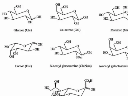
1.1 Schematic representation of the most commonly found constituent monosacchiide units of glycporteins and glycosphingolipids
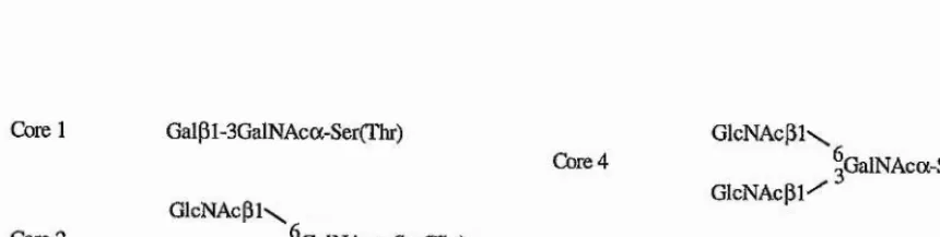
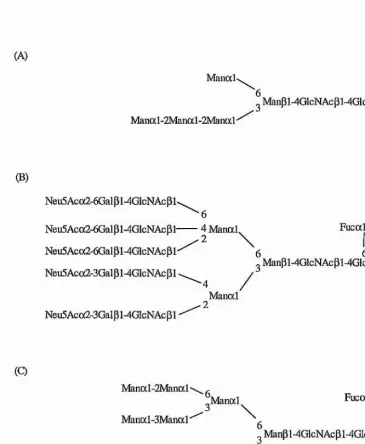
1.2 Examples of the core structures of animal glycosphingolipids 1.3 Examples of the three major sub-groups of N-linked sugar* chains 1.4 Core structures found in 0-linked glycans
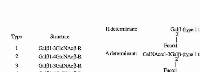
1.5 Acceptors for* the synthesis of, and structures of the ABH determinants
1.6 Determinants of the Lewis'", Lewis'", Lewis"", Lewis"*, Lewis’", and Lewis^ antigens 1.7 A car toon representation of diapedesis
1.8 (A) Ribbon representation of the lectin and EGF domains of E-selectin (B) Schematic representation of the overall structure of the selectins 1.9 Schematic representation of the sialyl Lewis’" antigen
1.10 Structures and percentages of the O-glycans in PSGL-1
1.11 Energy level digram for* two non-coupled spins, I and S, in close spatial proximity 1.12 Chair conformations of pyranose rings.
Chapter 2
2.1 (A) 'H spectrum of [U-'^C] Neu5Ac
(B) ID 'H-'^C HSQC spectrum of [U-‘^C] Neu5Ac ('^C-decoupled 'H spectrum) (C) *H spectrum of standard unlabelled Neu5Ac (a-glycoside)
2.2 (A) 'H spectrum of 7/-acetyl [U-'^C] lactosamine
(B) '^C-decoupled 'H spectrum of N-acetyl [U-'^C] lactosamine (C) 'H spectr um of standard unlabelled 77-acetyllactosamine 2.3 (A) 'H spectrum of [U-'^C] sialyla2,3-A-acetyllactosamine
(B) '^C-decoupled 'H spectrum of [U-'^C] sialyla2,3-7/-acetyllactosamine (C) 'H spectrum of standard unlabelled sialyla2,3-N-acetyllactosamine 2.4 (A) 'H spectrum of [U-'^C] Lewis’"
X II
(B) '^C-decoupled spectrum of [U-'^C] sialyl Lewis’" (C) 'h spectrum of standard unlabelled sialyl Lewis’"
Chapter 3
3.1 Definition of (j) and ij/ for a generalised oligosaccharide linkage
3.2 Schematic representation of the NOEs observed in solution for sialyl Lewis’" between (A) non-exchangeable protons, and (B) exchangeable - non-exchangeable protons 3.3 3D ROESY-HSQC pulse sequence
3.4 '^C-'H {F2F3) HSQC plane at the *H chemical frequency of Gal-Hl from a three dimensional NOESY-HSQC spectrum on [U-‘^C] Galpl-4Glc
3.5 Region of the two dimensional proton detected HOBS Y of unlabelled Gaipi-4Glc 3.6 Full relaxation matrix simulation of the Gal-Hl - Glc-H4 inter-glycosidic heteronuclear
NOE in Gaipi-4[U-'^C, ^H]Glc
3.7 Full relaxation matrix simulations of trans-glycosidic heteronuclear* NOEs in Gaipi- 4[50%^H,100%'^C]Glc
3.8 (A) '^C spectrum of Gaip 1-4[50%^H, 100%*^C]Glc
(B) difference spectrum upon saturation of Gal-Hl in Gaipi-4[50%^H, 100%'^C]Glc without ^H decoupling
(C) '^C difference spectrum upon saturation of Gal-Hl in Gaipi-4[50%^H, 100%'^C]Glc with ^H decoupling
3.9 *^C Chemical shift dependence upon temperature of Gaipi-4GlcNAc in supercooled H2O
3.10 Exchange rates vs. pH for selected hydroxyl protons in Galpl-4Glc measured at 268K 3.11 Pulse sequences for the assignment and measurement of NOE/ROEs to exchangeable
protons; (A) ES-COSY, (B) ES-NOESY, (C) ES-TOCSY, and (D) ES-ROESY 3.12 Pulse sequences for (A) gd-NOESY-HSQC, (B) gd-HOHAHA-HSQC, (C) gd-
ROESY-HSQC techniques
3.13 (A) Plot of ln(peak intensity) vs. spin lock time for hydroxyl protons GlcNAca 0H3 and Gal 0H4
X lll
3.14 Pulse sequence for the constant time heteronuclear* COSY experiment (CT-LRCH) for* measuring long range carbon-proton couplings
3.15 LRCH spectrum of Galpl-4Glc
3.16 Sum projection rule for ^Jcoc values about oligosaccharide linkages 3.17 Karplus Curve for ^Jcocc
Chapter 4
4.1 Schematic representation of 7V-acetyllactosamine
4.2 (A) 2D COSY spectrum of iV-acetyllactosamine
(B) 2D HCCH-HOHAHA spectrum of A/-acetyllactosamine
4.3 (A) 2D HSQC spectrum of A-acetyllactosamine
(B) 2D *H-'H BS-HOHAHA spectrum of A-acetyllactosamine at 258K 4.4 2D 'H-'H n o b s Y spectrum of unlabelled N-acetyllactosamine
4.5 Planes from the three dimensional NOESY-HSQC spectrum of [U-'^C]
N-acetyllactosamine at 303K at the chemical frequencies of (A) Gal-C5, and (B) GlcNAca/p-C4
4.6 (A) Region of the two dimensional 'H-'H ES-ROESY spectrum of unlabelled iV-acetyllactosamine at 256K
(B) Plane from the three dimensional gd-ROESY-HSQC spectra at the chemical frequencies of GlcNAc(x/p-C6 of iV-acetyl [U-'^Cj lactosamine at 256K
4.7 Plot of recovery of water* magnetisation during the acquisition period and delay of the gd-ROESY-HSQC pulse sequence
4.8 Simulations of the rate of recover*y of exchangeable proton magnetisation during the acquisition and relaxation delay of the pulse sequences in figure 3.12
4.9 ID ES-‘H spectra of (A) Galpl-OMe, (B) Gaipi-4GlcNAc, and (C) GlcNAc in H20/acetone-d6 (85:15) at 258K
4.10 FI strips of the 2D 'H-'^C LRCC spectrum at the chemical frequencies of (A) Gal-Hl, and (B) Gal-H2
4.11 (A) Instantaneous values of the glycosidic torsion angles <j) and \|/ over the 500ps restrained MD simulation in vacuo for Gaipi-4GlcNAc
XIV
(C) Instantaneous values of the glycosidic torsion angles \}/ vs. time over the 500ps restr ained MD simulation
Chapter 5
5.1 Schematic representation of sialyloc2,3-N-acetyllactosamine
5.2 (A) 2D *H-'^C HSQC spectrum of sialyla2,3-7/-acetyllactosamine (B) 2D *H-'H ES-COSY spectrum of sialyla2,3-iV-acetyllactosamine
5.3 (A) Section of the 2D ROESY spectrum of sialyla2,3-N-acetyllactosamine centred at Gal-Hl (FI)
(B) An F2F3 plane from the 3D ROESY-HSQC spectrum at the proton chemical frequency of Gal-Hl
5.4 ID ES-'H spectra of (A) sialyla2,3-//-acetyllactosamine and (B) A-acetyllactosamine in H20/acetone-d6 at 258K
5.5 Region of the 2D *H-‘H ES-NOESY spectrum of sialyla2,3-77-acetyllactosamine in H20/acetone-d6 at 258K
5.6 (A) Section of the 2D LRCC spectrum of sialyla2,3-A-acetyllactosamine
(B) F1F3 strips from the 3D LRCC spectr um of sialyla2,3-A-acetyllactosamine 5.7 Potential energy surface of the Neu5Aca2-3Gal linkage
5.8 Superposition of nine geometries derived from the restrained dynamical simulated annealing calculations on sialyla2,3-//-acetyllactosamine. Instantaneous values for (j) vs. \|/ for the Neu5Aca2-3Gal and Galpl-4GlcNAc linkages, and the (j) and \|/ vs. time plots for the Neu5Aca2-3Gal hnkage from the 5ns conventionally restrained MD simulation
5.9 Instantaneous values for (j) vs. \j/, (j) vs. time, and \\f vs. time plots for the Neu5Ac-Gal linkage from Ins time-averaged MD simulations
5.10 Instantaneous vBîlues for (j) vs. i}/, ({) vs. time, and \[/ vs. time plots for the Gal-GlcNAc linkage from Ins time-averaged MD simulations
XV
Chapter 6
6.1 Schematic representation of sialyl Lewis’"
6.2 (A) 2D 'H-'^C HCCH-COSY spectrum of sialyl Lewis’"
(B) 2D 'H-'H ES-COSY spectmm of sialyl Lewis’" in H20/acetone-d6 at 258K 6.3 (A) 2D *H-'^C HSQC spectrum of sialyl Lewis’" in free solution
(B) 2D *H-'^C HSQC spectr um of sialyl Lewis’" in complex with E-selectin
6.4 F2F3 planes from the 3D ROESY-HSQC spectrum of sialyl Lewis’" at the *H chemical frequencies of (A) Gal-Hl, and (B) Gal-H3
6.5 ID ES-'H spectra of sialyl Lewis’", Lewis’", and sialyla2,3-7V-acetyllactosamine at 258K 6.6 (A) Region of the 'H-'H ES-NOESY spectrum of sialyl Lewis’" at 258K, showing the
through space connectivities from the non-exchangeable (F2) to exchangeable protons {FI)
(B) Region of the 'H-'H ES-ROESY spectrum of sialyl Lewis’" at 258K, showing the through space connectivities from the non-exchangeable {F2) to exchangeable protons {FI)
6.7 (A) FI strip from the 2D LRCC spectrum of sialyl Lewis’" centred at the proton chemical frequency of Gal-Hl
(B) A region of the F2F3 plane of the thr ee dimensional LRCC spectrum at the carbon chemical frequencies of Gal-C3/C5 and GlcNAcP-C3/C5
(C) A region of the F2F3 plane of the thr ee dimensional LRCC spectrum at the carbon chemical frequency of Gal-C2
6.8 F2F3 strips from the three dimensional LRCC spectrum of sialyl Lewis’" showing the long range trans-glycosidic correlations for the Fuc-GlcNAc linkage, and the three- bond corxelations along the sialic acid glycerol side chain
6.9 Superposition of seven geometries derived from the restrained dynamical simulated annealing calculations on sialyl Lewis’". Instantaneous values for glycosidic torsion angles (j) vs. \\f for the Neu5Aca2-3Gal, Fucal-3GlcNAc, and Galpl-4GlcNAc linkages from the 5ns conventionally restrained MD simulation
XVI
6.11 Regions of the F2F3 planes from the three dimensional TRNOES Y spectrum of sialyl Lewis’" in complex with E-selectin at the proton chemical frequencies of (A) Gal-Hl, and (B) Gal-H3
X V ll
List of Tables
Chapters
3.1 Thi'ee-bond *^C-'^C coupling constants measured in '^C model compounds and utilised for the parametrisation of a Kaiplus curve for the C-O-C-C fragment.
Chapter 4
4.1 *H and *^C chemical shift assignments for A-acetyllactosamine at 303K.
4.2 NMR data for the exchangeable protons of N-acetyllactosamine in HzO/acetone-cf^ (85:15)at258K.
4.3 Values of the apparent 'Jcc from the LRCC experiment compared with 'Jcc values obtained from a ID '^C spectrum.
4.4 Experimental ROEs involving exchangeable protons compared with theoretical values derived from two 500ps restr ained MD simulations in vacuo for Galpl-4GlcNAc. 4.5 Comparison of experimental trans-glycosidic coupling constants vs. theoretically
computed values derived from a 51 Ops in vacuo molecular* dynamics simulation.
Chapter 5
5.1 'H and '^C Chemical Shift Assignments for* sialyla2,3-V-acetyllactosamine in D2O at 303K.
5.2 NMR data for* the exchangeable protons from sialyla2,3-iV-acetyllactosamine in H20/acetone-(7g (85:15) at 258K.
5.3 Experimental ROEs (non-exchangeables) and NOEs (involving exchangeables).
5.4 Energies and torsion angles of structures from the simulated annealing process which satisfied the distance restraints obtained from ROESY data at 303K and NOESY data at 258K.
5.5 Experimental trans-glycosidic scalar* coupling constants in sialyla2,3-V- acetyllactosamine vs. theoretical values computed from the Ins MD simulations using ‘conventional’ and time-averaged restraints.
XVlll
Chapter 6
6.1 *H and chemical shift assignments for sialyl-Lewis’" in D2O at 303K.
6.2 NMR data for the exchangeable protons from sialyl Lewis’" in H20/acetone-<i6 (85:15) at 258K.
6.3 Experimental ROEs (non-exchangeables) and NOEs (involving exchangeables). 6.4 Experimental values for the inter-glycosidic ^Jcocc and ' Jch couplings in sialyl Lewis’". 6.5 Energies and torsion angles of structures from the simulated annealing process which
satisfied the distance restraints obtained from ROESY data at 303K and NOESY data at 258K.
6.6 Back-calculated inter-glycosidic (R)NOEs for sialyl Lewis’" from the “conventionally” restrained MD simulations using DISCOVER.
6.7 Experimental vs. Back-calculated caibon-carbon and carbon-proton coupling constants from the Ins MD simulation using DISCOVER.
6.8 Back-calculated inter-glycosidic (R)NOEs for sialyl Lewis’" from the time-averaged restrained MD simulations using XPLOR.
6.9 Experimental trans-glycosidic scalar coupling constants in sialyl Lewis’" vs. theoretical values computed from the Ins MD simulations using time-averaged restraints.
XIX
List of Abbreviations
COSY Correlated Spectroscopy
ES Excitation Sculpting
HMQC Heteronucleai* Multiple Quantum Coherence Spectroscopy
HOHAH A Homonucleai* Haitmann Hahn
HSQC Heteronucleai’ Single Quantum Coherence Spectroscopy
LRCC Long Range Carbon-Carbon Coupling Spectroscopy
LRCH Long Range Carbon-Proton Coupling Spectroscopy
NMR Nuclear Magnetic Resonance
NOE Nuclear Overhauser Effect
NOESY Nuclear Overhauser Effect Spectroscopy
ns nanosecond
ROE Rotating Frame Nuclear Overhauser Effect
ROESY Rotating Frame Nucleai* Overhauser Effect Spectroscopy
ps picosecond
p-NP pam-nitrophenol
TOCS Y Total Correlated Spectroscopy
TRNOE Transferred Nuclear Overhauser Effect
TRNOESY Transfened Nuclear Overhauser Effect Spectroscopy TSP 3-(ti*imethylsilyl) proprionic-2,2,3,3,-<i4-acid, sodium salt.
XX
Symbols
*H Hydrogen atom (proton)
Cai‘bon-13 atom
"JcH n-bond carbon-proton coupling constant "Jcc n-bond caiton-caiton coupling constant "Jhh n-bond proton-proton coupling constant
Ô Chemical Shift
(|) Dihedial angle Hi-Ci-Oi-Cx in a glycosidic linkage (|)i Dihedral angle He-Ce-Cv-H? in the glycerol side chain
(])2 Dihedial angle H7-C7-C8-H8 in the glycerol side chain (j)3 Dihedial angle Hs-Cs-Cg-Oç in the glycerol side chain
y Gyromagnetic ratio
J(co) Spectral Density
koff Exchange off-rate
kon Exchange on-rate
Kd Equilibrium dissociation constant
Tc Conelation time for moleculai* reorientation
Tm NOES Y/ROES Y mixing time
Acquisition time (ID NMR experiment); first evolution period (nD NMR experiment)
t2 Acquisition time (2D NMR experiment); second evolution period (nD NMR
experiment)
ts Acquisition time (3D NMR experiment)
Chapter 1
Chapter 1: Introduction
1.1 Biological Function of Carbohydrates
Until the late 1960’s the biological role of carbohydiates was generally believed to be hmited to energy storage and production, and as a structural material. That carbohydrates may play a role in cellular recognition and differentiation was first suggested by the discovery of the ABH blood group antigens, and the first evidence that caibohydrates can function as cellular receptors was the discovery that iV-acetyl neuraminic acid {see figure I.I) on the surface of erythi'ocytes is essential for the binding and entiy of the influenza virus. Research over the last 30 years has expanded the range of biological processes in which caibohydrates play a pivotal role, for example, cell-cell recognition, toxin adhesion and invasion, immune evasion and activation, leukocyte extravasation, and the clearance of glycoproteins from the circulation by hepatic or reticuloendothelial cells (reviewed by Vaiki, 1993).
i . i . l Carbohydrate Structure
Chapterl: Introduction
.OH
OH
Glucose (Glc)
Fucose (Fuc)
OH OH
HO ,OH
OH Galactose (Gal)
NAc
A-acetyl glucosamine (GlcNAc)
OH OH
Mannose (Man)
OH
^OH O
HO OH
NAc
A-acetyl galactosamine (GalNAc)
COjH
A-acetyl neiuaminic acid (Neu5Ac)
Figure 1.1 - Schematic representation of the most commonly found constituent monosaccharide units of glycoproteins and glycosphingolipids.
[image:26.613.82.490.82.390.2]Chapter'1: Introduction
Ganglia
Galp l-3GalNAcp l-4Galp l-4Glc-Cer
Globa
GalNAcP l-3Galal-4Galp l-4Glc-Cer
Isaglaba
GalNAcp l-3Galal-3Galp l-4Glc-Cer
Lacta
Galp l-3GlcNAcp l-3Galp l-4Glc-Cer
Nea-lacta
Galp 1 -4GlcNAcP 1 -3Galp 1 -4Glc~Cer lacto-A/'-neotetraose
Figure 1.2 - Examples of the core structures of animal glycosphingolipids.
asialo-GMi
globoside, Gb4
isogloboside
lacto-A-tetraose
0-linked glycans aie classified as belonging to one of six groups according to different core structures (figure 1.3). These cores can be elongated to form the backbone region by addition of galactose in P1-3 and p i-4 linkages, and A-acetylglucosamine in P1-3 and pi-6 linkages. Although the glycans aie often linked to a serine or threonine residue via GalNAc, they may link through other residues such as fucose.
Core 1
Cbre2
Core 3
Galpl-3GalNAca-Ser(rhr)
G lcN A cpl\
Gaipi/ aGalNAca-Ser(Thr)
Cbre4
Core 5
G lcN A cpi\
gGalNAca-Ser(Thr)
GIcNAcpi/
GlcNAcPF6GalNAca-Ser(Thi-)
[image:27.612.91.521.492.601.2]Chapter 1: Introduction 5
A^-linked glycoprotein glycans fall into three broad families (figure 1.4): high mannose, complex, and hybrid type oligosaccharides. All A-linked oligosaccharides contain the pentasaccharide Manal-6(Manal-3)Manpi-4GlcNAcpl-4GlcNAc as a common core (the “trimannosyl core”). High mannose type sugars contain only a-mannosyl residues in addition to the trimannosyl core, complex type oligosaccharides contain no mannose residues other than those in the core, and the chains normally terminate with variations of the Neu5Aca2-3/6Galpl-4GlcNAcpi-R (where R = trimannosyl core), and hybrid type oligosaccharides have a combination of the complex and high mannose type characteristics.
(A)
Manod-Manal-2Nfanal-2Manal *
Manpi-4GlcNAcpi-4GlcnAc-Asn
(B)
Neu5Ac(x2-6Gaipi-4GlcNAcPl'
Neu5Acoc2-6Gaipi-4GlcNAcpi-Neu5Aca2-6GaipWGlcNAcpl'
Neu5Aca2-3Galpl4GlcNAcpl
Neu5Ac(x2-3Gal pNGIcNAc pi •
■6
4 Maiial
\
6 Fucal6o Manpl-4GIcNAcpl-4GlcNAc-Asn
4 /
Manal
■ 2
(Q
Manal-2Manal'
Manocl-3Manal'
Neu5Aca2-3Galpl-4GlcNAcPF
Manal Fucal
6 6
„ Manpi-4GlcNAcpl-4GlcNAc-Asn
[image:28.612.114.479.246.690.2].Manal GlcNAcPl
Chapter 1: Introduction 6
1.1.2 Blood Group Antigens
Human blood group antigens are expressed on red blood cells, and at present over 200 antigens have been identified and assigned to 22 blood group systems (Daniels et a l, 1993). Of these the most well studied and characterised are the carbohydrate based blood group antigens, namely ABH, Lewis (Le), li, P-related, Pi, T and Tn (for recent reviews: Oriol et a l, 1986; Clausen and Hakomori, 1989; King, 1994; Greenwell, 1997), which may occur as either glycolipids or glycoproteins. For the purposes of this introduction only the structural char acteristics of the ABH and Lewis blood group antigens will be discussed.
L I.3.1 ABH and Lewis antigens
The ABO blood system, first to be identified, was discovered by Landsteiner in 1900 and remains the most important class of antigens for blood transfusion. By mixing the blood of different people, Landsteiner made the discovery that some combinations resulted in agglutination of the blood, whilst others did not. This subsequently lead to the suggestion that there were three blood group antigens. A, B, and O. The simplest way to demonstrate the presence of an antigen on erythrocytes is by testing whether they will be agglutinated by specific antibodies. Individuals who lack an antigen have, in their sera, natural antibodies that agglutinate cells carrying that antigen. So in the case of the ABO blood system, individuals of blood group A have anti-B antibodies, and those of blood group B have anti-A antibodies. Hence on mixing blood from two individuals from different blood groups wiU result in agglutination. In the 1940’s it was first demonstrated that these antigens were not confined to the erythrocytes but were present in other cell type and in secretions (Hartmann,
1941), and so they are more appropriately termed ‘histo-blood group antigens’.
Apart from antibodies, lectins may also be used to identify the antigens present as some are also blood group specific (Sharon and Lis, 1972). For example, the lectins of Phaseolus limensis (lime bean) and Dolichos biflorus are blood group A specific, whilst Lotus tetragonolobus and Ulex europeus are blood group O specific. Some of these lectins are still used in blood transfusion centres today.
Chapterl: InWoduction 1
antigen on O cells, is a product of a different gene system, Hh (Watkins and Morgan, 1959). This conclusion is based upon the fact that individuals from blood groups A, B, and O often secrete a glycoprotein that inhibits the agglutination of O erythrocytes by anti-0 reagents. Hence this glycoprotein is now called an H substance.
The nature of the relationship between the A, B, and H antigens became apparent when the str uctures were determined (figure 1.5), and found to be of carbohydrate structure (reviewed by Sharon and Lis, 1972). The H antigen is the precursor to both A and B antigens. The only difference between these is the terminal sugai* residue, where GalNAc and Gal are the immunodominant sugars for A and B antigens, respectively. The H disacchaiide may be synthesised on four main acceptor sequences, depending upon the source of the blood group substances (Clausen and Hakomori, 1989). Type 2 based ABH oligosaccharides are the major species in erythrocytes, glycoproteins, and glycolipids (reviewed by Hakomori, 1982), whilst type 1 and 3 glycolipids have been isolated as minor components (Breimer et a l,
1988; Clausen et a l, 1985). Type 1 is found mostly in mille oligosaccharides, and secreted glycoproteins. Type 3 is found in glycoconjugates of epithelial cells in the gut and lungs. Type 4 is associated with glycolipids paiticulaiiy those found in the human kidney.
Type Stmcture
1 Gaipi-3GlcNAcp-R 2 Galpl-4GlcNAcP-R 3 Galpl-3GalNAcp-R 4 Gaipi-4GaINAcP-R
H determi nant: Gal (T(type 1 to 4 chain)
f
Fucal
A determinant: GalNAcal-3Gaip-(type 1 to 4 chain)
f
Fucal
B determinant: Galal-3Gaip-(type 1 to 4 chain)
2
Fucal
Figure 1.5 - Acceptors for the synthesis of, and structures of the ABH determinants.
[image:30.612.97.489.470.612.2]Chapter !: Introduction 8
The Lewis® antigen is formed from the precursor of the H antigen. The Lewis*' antigen is a hybrid structure of both H and Lewis® antigens and requires the glycosyltransferases involved in the production of both H and Lewis® antigens to be present.
The structures of the Lewis® and Lewis** antigens are based upon the type 1 chain, whilst Lewis'" and Lewis^ antigens are based upon the type 2 chain and are therefore regarded as their structural isomers.
Le“ determinant: Gaip l-3GIcNAc P-R 4
Fucal
Le^ determinant: Gal P1 -4GlcNAc P-R
I
Fucal
Le'’ determinant: Fucal-2Gaipi-3GlcNAcP-R
I
Fucal
Ley detenninant: Fucal-2Galpl-4Glc^AcP-R
Fucal
Le'’ glycolipid: Gaipi3GlcNAcpi
-GalpMGlcNAcpF
f
Fucal
Le‘> glycolipid: Galpl-3GlcNAcpi-3Galpl-4Glc-Cer
Fucal
[image:31.614.75.504.236.566.2]Gal pl-3GlcNacpi-3Galpl-4Glc-Cer
Chapterl: Introduction 9
1.1.3 Carbohydrates in disease 1.1.3.1 Oncology
Increased cellular- glycosylation is a common change in malignancy, with altered glycosylation a unifymg feature of cancer cells (Kim and Vai'ki, 1997). De-differentiation of normal cells leads to cancerous growth and certain malignant cells exhibit glycolipid surface antigens resembling those occuiTing during early embryonysis, and are often termed oncodevelopmental (or oncofoetal) associated antigens (Halcomori, 1985). For example in foetal colon, blood group ABH and Lewis** antigens are expressed during carcinomas of the colon, but are not usually expressed in the adult colon (Lloyd, 1987). Therefore, antibodies to these antigens can be used as markers in tumour diagnosis (reviewed by King, 1994).
The sialylated form of Lewis'" (sialyl Lewis'") has been identified on carcinoma cells and a correlation between the metastatic potential of colon carcinoma cell lines in mice and the expression of this antigen has been demonstrated. The recognition of sialyl Lewis'" by a group of proteins, which allows leukocytes to migrate to areas of inflamed tissue (for discussion see section 1.1.4), is used by the carcinoma cells to facilitate evasion of the host’s immune system.
1.1.3.2 Bacteriallnfection
Chapter!: Introduction 10
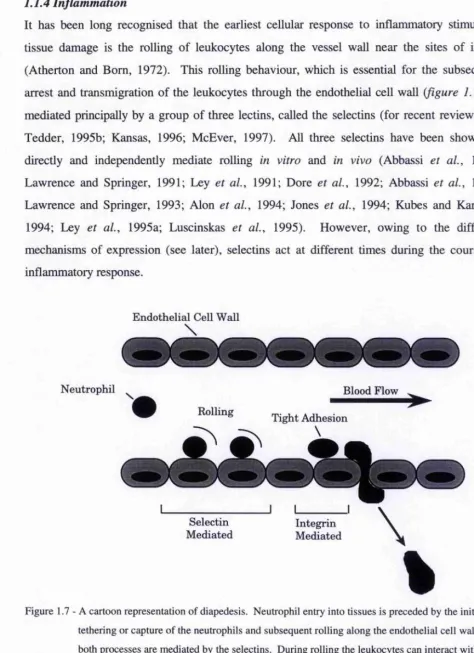
1.1.4 Inflammation
It has been long recognised that the earliest cellular response to inflammatory stimuli or tissue damage is the rolling of leukocytes along the vessel wall near the sites of injury (Atherton and Born, 1972). This rolling behaviour, which is essential for the subsequent arrest and transmigration of the leukocytes through the endothelial cell wall (figure 1.7), is mediated principally by a group of three lectins, called the selectins (for recent reviews see Tedder, 1995b; Kansas, 1996; McEver, 1997). All three selectins have been shown to directly and independently mediate rolling in vitro and in vivo (Abbassi et at., 1991; Lawrence and Springer, 1991; Ley et a l, 1991; Dore et a l, 1992; Abbassi et a l, 1993; Lawrence and Springer, 1993; Alon et a l, 1994; Jones et a l, 1994; Kubes and Kanwar, 1994; Ley et a l, 1995a; Luscinskas et a l, 1995). However, owing to the different mechanisms of expression (see later), selectins act at different times during the course of inflammatory response.
Endothelial Cell Wall
\
Neutrophil ^ Blood Flow ^
Bolling Tight Adhesion
Selectin Mediated
[image:33.612.63.537.73.726.2]Chapterl: Introduction 11
The earliest response to inflamniatoiy stimuli, within 20 minutes after induction, is predominately mediated by P-selectin (Ley et al., 1993; Ley et al., 1995a; Ley et al., 1995b; Dore et a l, 1992; Nolte et al., 1994; Mayadas et al., 1993). Subsequently, the levels of P- selectin activity decrease, whilst rolling after 20 minutes is principally mediated by L-selectin. It is only after two hours that E-selectin plays a part in the rolling phenomena, although both L-selectin and P-selectin also appeal* to have a role in the later periods in inflammatory response.
One important aspect of the selectin function is the ability of cooperativity, especially on populations of leukocytes that can potentially use more than one selectin simultaneously (Ley and Tedder, 1995). For example, comparison of spontaneous rolling flux in normal (wild type) mice against mice deficient in L- or P-selectin show that there is a higher flux of rolling at neaily all times for the wild type as compared to the combination of both deficient mice (Ley et al., 1995a). Therefore, interaction of neutrophils with endothelium is more efficient when more than one selectin is present. This can be explained in terms of the differences in the chaiacteristic rolling velocities that each selectin can sustain. L-selectin supports the greatest rolling velocities, between 50 to 150 |ims'% whilst P-selectin mediates slower rolling, between 20 to 50 |ims‘*, and E-selectin the slowest of all, between 3 to 10 jxms'* (Ley et a i, 1993; Kunkel et a i, 1995; Jung et a l, 1996; Kunkel et a l, 1996). This therefore suggests that L-selectin is involved in the initial capture or tethering of the leukocytes which is likely to then facilitate and enhance slower rolling mediated by E- or P-selectin. Consistent with this proposal is evidence where spontaneous rolling occurs at the lower rolling velocity of P-selectin in normal mice even though both L- and P-selectin are present (Ley et a l,
1995a).
Another factor important in the rolling phenomenon, is that of “tensile strength” (Alon et a l,
1995), which is the resistance of the selectin interaction to brealc down on increasing the applied force. Without tensile strength the rolling phenomenon would not occur because the selectin-ligand interaction would have vanishingly short lifetimes, lower than the minimum required to maintain rolling. This factor may be important in distinguishing the selectins from other molecules which have similar rates of interaction but cannot mediate rolling (Alon
Chapterl : Introduction 12
The generation of mice lacking one or more of the selectins (Mayadas et al., 1993; Arbones
et al., 1994; Labow et a l, 1994) has provided further insight into the recruitment of leukocytes and the roles of the selectins. In mice deficient of both E- and P-selectin (Bullard
et a l, 1996), the recruitment of neutiophils is completely absent during eaily time points, which suggests that whilst L-selectin can capture the neutrophils it is incapable of efficiently airesting the neuti'ophils and allowing transmigration. However, recruitment of neutrophils to the peritoneum after 24 hours is at normal levels, and leukocytes can be also be found in the dermis, suggesting that additional L-selectin dependent pathways are active. To summarise the findings on knockout mice, L-selectin deficient mice show defects in leukocyte recruitment up to 48 hours after initiation (Tedder et a l, 1995a), whilst P-selectin knockout mice show partial defects at early stages in the inflammatory response but not during late stage (Mayadas et a l, 1993), and no significant defects can be found in E-selectin knockout mice (Labow et a l, 1994). In the E- and P-selectin knockout mice there are near- total defects in the early stages but recruitment appears to be normal after 24 hours. Hence cooperativity is important for effective leukocyte recruitment during inflammation, but functions of individual selectins are distinguishable and not completely essential, in all settings of chronic inflammation.
Chapterl: Introduction 13
1.2 Selectins
1.2.1 Structure
The selectins are a group of three proteins which enable adhesion between the endothelial cell wall and leukocytes, and although they have had a wide selection of names, are now referred to as E(endothelial)-, P(platelet)-, and L(leukocyte)-seiectin. They are multi-domain proteins, with a unique overall structure (figure 1.8A). The primary sequence shows highly conserved regions in the lectin and EGF domains (~65%), and very high conservation between different mammalian species. The lectin domain is essential for binding carbohydrates, and is homologous to others found in mammalian proteins which require Ca^^ to bind carbohydrates. The EGF domain influences the lectin’s ability to mediate cell binding, possibly through stabilising the protein. Although, the crystal structure of the lectin and EGF domains of E-selectin (Graves et a l, 1994) showed that these were distinct domains with little apparent association (figure l.SB).
The complement regulatory domains (CRDs) although not essential for binding, have been reported to facilitate lectin activity, possibly by contributing to the conformational stabilisation or to the orientation of the protein within the membrane. The number of CRDs is dependent upon the selectin and mammalian system; for humans E-selectin has six CRDs, P-selectin has nine; and L-selectin has two. The CRDs also may also have a function in mediating receptor oligomerisation. The number of CRDs is thought to extend the lectin and EGF domains the appropriate distance away from the membrane for optimal ligand binding.
Chapter 1 : Introduction 14
(A)
I I I h i | g
L-selectin
E-selectin
I I I I I
I
P-selectin□ Signal Peptide H Transmembrane E Lectin Domain O Cytoplasmic Tail
H Epidermal Growth Factor (EGF) Q Complementary Regulatory Domains
(B)
Lectin Domain
EGF Domain
Carbohydrate Binding Site
Figure 1.8 - (A) A schematic representation of the overall structure of the selectins, and (B) a ribbon representation of the lectin and EGF domains derived from the x-ray crystal structure (Graves
[image:37.612.26.522.26.578.2]Chapterl: Introduction 15
Although the selectins have similarities with C-type lectins (such as the requirement for a calcium ion for binding, the high conservation of amino acids in the CRDs between selectins and lectins, and the possibility of mutating a small number of amino acids in the mannose binding protein (MBP) lectin domain to confer E-selectin binding activity) they also exhibit significant differences. The selectins only require a single Ca^"*" ion for binding whereas MBP is known to require two ions for binding. Selectins have only a single carbohydrate binding domain per whole protein, whilst other known C-type lectins, except proteoglycan C-type lectin, have multiple carbohydrate binding domains per protein. The transmembrane orientation of the selectins is type I which is similar to the macrophage mannose receptor, but other C-type lectins exhibit type II. Finally, the selectins are unique in that they have all thi'ee lectin, EGF, and CRD domains together, unlike all other lectins, and hence the spatial aiTangement of these domains may be important in the receptor function.
1.2.2 Expression 1.2.2.1 E-Selectin
Chapterl: Introduction 16
1.2.23 P-Selectin
P-Selectin or CD62P (previously Icnown as PADGEM, or GMP-140), is a 140 IdDa glycoprotein and unlike E-selectin is preformed and stored in secretory granules of platelets and endothelial cells (Hsu-Lin et a l, 1984; McEver et a l, 1989). Its rapid expression at the cell surface by the process of degranulation, occurs within minutes after stimulation of the endothelial cells by either histamine or thiombin, and is also expressed later on in the course of inflammatoiy response by gene induction by TNF-a, lipopolysaccharides (LPS), or IL-1, leading to expression between 2 and 4 hours after induction.. The cytoplasmic tail has multiple motifs (though these are not completely defined) and this dynamic pattern may be important in the regulation of the expression of P-selectin at the surface (Kansas, 1996). The exact role of acylation and phosphoiylation of certain amino acids in the tail is still uncleai* (Fujimoto and McEver, 1993), however, the cytoplasmic tail is imphcated in a number of important regulatoiy functions. First, the sorting of P-selectin into granules is controlled by sequences in the cytoplasmic tail which interacts with the sorting machineiy in cells that have the regulated sorting pathway (Disdier et al., 1992). Second, the transient expression on the surface of endothelial cells is due to the rapid internalisation (McEver et a l, 1989) and followed by lysosome degradation which is regulated by amino acids within the cytoplasmic tail (Green et a l, 1994).
1.2.2.3 L-Selectin
Chapterl: Introduction 11
1.2.3 Additional Selectin Functions
Apart from the function of leukocyte rolling, the selectins have also been implicated in a number of other biologically important functions. For a normal immune response the circulation of mature T and B lymphocytes throughout the secondaiy lymphoid organs is required as it ensures that a full complement of antigen receptors aie exposed to the full range of antigens present in the system. Entiy to these secondary lymphoid organs by lymphocytes occurs across specialised endothelial cells, known as high endothelial venules (HEV) (Picker and Butcher, 1992), and L-selectin is the principal if not sole HEV receptor for lymphocyte traffic to the lymph nodes (Kansas, 1996). Additionally L-selectin may play a role in interactions between leukocytes, and in paiticulai- neutrophils, which may be important in the amplification of the inflammatoiy response. It has been noted that neutrophils can roll on other neutrophils and that this is blocked by monoclonal antibodies to L-selectin (Bargatze et a l, 1994). Neutiophil aggregation can also be blocked by a monoclonal antibody to L-selectin or carbohydrates that inhibit L-selectin activity (reviewed by Kansas, 1996).
The fact that P-selectin is found on platelets suggests a role in platelet-leukocyte interactions during wound healing and hemostasis, which could amplify the recruitment to sites of vascular injuiy (Larsen et a l, 1989). P-selectin is known to mediate platelet binding to monocytes which in turn induces tissue factor, thereby activating the blood coaggulation cascade (Cell et a l, 1994). E-selectin has been implicated as a tissue specific homing receptor for leukocyte recruitment specifically to the skin, particularly in the case of memory T cells (Picker <2/., 1991).
Chapterl: Introduction 18
L2.4 Carbohydrate Ligands
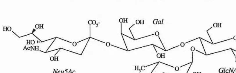
The identification of the physiological ligands for the selectins is complicated because, like most lectins, they bind to a wide variety of carbohydrate epitopes in vivo. The dependence of the selectins upon a caibohydrate ligand was illustrated in two patients with leukocyte adhesion deficiency type II disease, which is an inherited inability to recruit neutrophils to the sites of inflammation (Harlan, 1993). This inability to generate the ligands required for selectin mediated rolling is attributed to a defect in the fucose metabolism, and they are, therefore, unable to synthesise fucosylated caibohydiates. The observation that cell adhesion is sensitive to neuraminidase, indicates that fucose and sialic acid residues are essential for selectin binding, and a prototype caibohydrate ligand, sialyl Lewis^, Neu5Aca2-3Gaipi- 4[Fucal-3]GlcNAc, was proposed as the minimal required structure (figure 1.9).
OH OH
HO
OH OH
OH NHAc
GlcNAc
NeuSAc OH
OH OH
[image:41.614.84.491.311.437.2]Fuc
Figure 1.9 - Schematic representation of the sialyl Lewis'" antigen, the minimal ligand proposed for selectin binding.
Chapter!: Introduction 19
1.2,5 Glycoprotein Ligands
Purified forms of the selectins or IgG fusion proteins exhibit a higher level of specificity in compaiison to monoclonal antibodies against their putative ligands. This suggests that the selectins recognise not just a simple oligosaccharide, but a three dimensional surface composed of several carbohydrate moieties contributed by several molecular species attached to a specific glycoprotein.
Mouse L-selectin binds to at least three heavily glycosylated mucin-like proteins in vivo, namely GLYCAM-1, CD34, and MAdCAM-1. Each glycoprotein bears sulfated, sialylated, and fucosylated 0-lrnked oligosaccharide chains that appear essential for L-selectin binding (reviewed by Kansas, 1996). Although first reported to be the ligand for L-selectin, and thought to be constitutively expressed on peripheral lymph node HEV, GLYCAM-1 contains no apparent transmembrane region, and it appears that it is predominately released into circulation where it competitively inhibits, rather than promotes, L-selectin mediated attachment (Tedder e ta l, 1995b).
The second glycoprotein, a glycoform of CD34, is constitutively expressed on most endothelial and hematopoietic stem cells, and therefore has been proposed to regulate tissue specific selectin binding because of the tissue-specific glycosylation of CD34. In mice it is universally expressed on the endothelium both during mouse development and in the mature animal, so it is perfectly placed as a ligand for L-selectin. In CD34 knockout mice normal levels of rolling and lymphocyte homing is observed, and in puiified human tonsil, CD34 accounts for approximately half of the L-selectin activity. Therefore, the third ligand, MAdCAM-1, may constitute the principal adhesive ligand(s) for L-selectin, although it has yet to be identified at the moleculai* level. Interestingly, in knockout mice of both CD34 and GLYCAM-1, an up-regulation of this third ligand is seen, indicating the evolution of dynamic mechanisms to maintain adequate levels of lymphocyte traffic and recirculation.
The O-linked carbohydrates of GLYCAM-1 have been characterised (Hemmerich et a l,
O-Chapter!: Introduction 20
linked glycans, le., Gaipi-3[GlcNAcpl-6]GalNAc. These ligands provide all the requirements for L-selectins ligands, but whether these are the actual recognition motifs, or whether CD34 or MAdCAM-1 contain these or different oligosaccharides structures is as yet unknown.
The P Selectin Glycoprotein Ligand -1 (PSGL-1) is a disulfide linked homo-dimer of two identical ~120kDa type I mucin-like proteins which have a unique stmcture (Moore et al,
1992). This glycoprotein is expressed by all blood neutrophils, monocytes, and lymphocytes but requires specific glycosylation for its function. Studies on the P-selectin - PSGL-1 binding have provided the first direct evidence that leukocyte rolling under physiological shear forces requires the specific interaction of a selectin with a single cell-surface glycoprotein. A specific anti-PSGL-1 monoclonal antibody completely inhibits P-selectin mediated rolling of leukocytes (Moore et a l, 1995). PSGL-1 is mainly O-linlced glycosylated, although it has two or three A-linked sugars which do not appear* to be necessary for binding, with extensive branched chain polylactosamine glycans, which contain fucose and sialic acid and many terminate in the sialyl Lewis* motif (Moore et al., 1994). The structure of these glycans has been characterised (Wilkins et a l, 1996) and contain
Chapterl: Introduction 21
Gaipi-3GalNAc 1 4% Gaipi-4GlcNAcPK .
oGalNAc 52% G aipK ^
{
Galpl^GlcNAcpK _ Neua2-3Gaipi-4GlcNAcpl\ ^qGalNAc 6% gGalNAc 14%
Gaipi"^^ Neua2-3Gaipr
Fucal
I
3
Neua2-3Galpl-4GlcNAcpl\
gGaINAc 2% Neua2-3Gaipi"^
Fucal Fucal Fucal
1
I
I
3 3 3
[image:44.612.90.502.64.397.2]
Neua2-3Galpl-4GlcNAcpi-3Gaipi-4GlcNAcPl-Gaipi-4GlcNAcpl-^GalNAc 12% G aip i^^
Figure 1.10 - Structures and percentages of the 0-glycans in PSGL-1 (from Wilkins et al., 1996)
1.3 Carbohydrate Therapeutics
Chapterl: Introduction 22
reducing the likelihood of resistance. The mode of action of these caibohydrate drugs is varied:
L Inhibition o f protein attachment
The most common approach is to design a drug based upon the natural carbohydrate receptor which would bind iixeversibly and competitively with the natural substrate, thus preventing cellular* adhesion. This method is the current approach for* mimics to the sialyl Lewis* - selectin interaction for* the treatment of reperfusion injury, where the accumulation of leukocytes in tissues after heart attack and surgery can lead to tissue damage. The clearance of bacteria and their toxins from the gastrointestinal tract with oligosaccharides is used in nature. The high levels of oligosaccharides present in mammalian milk, often found on the surface of the gut, is believed to protect infants from infection. Mimics of this process include the development of a Gbg conjugate linked to an insoluble powder* to prevent verotoxin adhesion.
2. Alteration of carbohydrate biosynthesis
Many diseases are associated with defects in oligosaccharide metabolism, such as diabetes. Azasugars (nitrogen analogous of carbohydrates) are believed to be a possible treatment by inhibiting sucrase and amylase, responsible for the breakdown of polysaccharides in the gut, and therefore prevent the adsorption of glucose into the bloodstream. Inhibition of the catabolism of Neu5Ac ternrinating oligosaccharides has been successfully used in the treatment of influenza (von Itzstein et a l, 1993).
3. Eliciting an immune response
Chapter!: Introduction 23
1.3.1 Rational Drug Design
A prerequisite for the rational design of drugs is a detailed understanding of the interaction of a carbohydrate with its receptor. A high resolution three dimensional structure of the complex is required, and several crystallographic studies have been described recently which illustrate in detail the precise nature of certain carbohydrate-protein interactions. The most celebrated example of model based drug design was the work performed in von Itzstein’s laboratory on the development of an influenza sialidase inhibitor (von Itzstein et a l, 1993). Sialidase is critical in the release of newly synthesised influenza virions from infected cells by the cleavage of terminal NeuSAc residues from host cell-surface glycans. Inhibition of this enzyme limits the establishment and progression of infection. Computer-aided molecular modelling was used to analyse the active site and predict the functional group changes which would increase the binding affinity of the sialic acid residue. Modelling studies predicted that the substitution of the 4-OH group with an amino or guanidynl group would produce an increase in overall binding. Results show that the 4-guanidino derivative has an inhibition constant, for various strains of influenza, in the 10'^° range. Additionally the affinity of this compound against human sialidases is a million times lower, therefore offering excellent selectivity for vii-us sialidases.
Detailed x-ray crystal structures of biologically important oligosaccharide complexes are rare, as the relatively flexible oligosaccharide often results in poor electron densities for the carbohydrate residues, and when observed they are often stabilised by crystal packing forces which may orient the sugar in an unnatural conformation. Many lectins only weakly bind isolated oligosaccharide analogues of their receptor glycans, and so may not be co crystallised (Stein et ah, 1992). An additional problem is that carbohydrate binding sites in the well ordered solid state may not be active in the less well ordered solution state (c./. Wright, 1992, with Wright and Kellog, 1996, and reviewed by Rossi, 1992).
Chapterl: Introduction 24
1.4 NMR Spectroscopy
The nucleai” magnetic resonance phenomenon, first observed in 1946, has become a widespread tool for the non-desti'uctive analysis of both organic and inorganic compounds. NMR techniques can provide information on structure, conformation, and internal mobility. Comprehensive introductions include Sanders and Hunter (1987), and Derome (1987).
The fundamental basis of the NMR experiment is perturbation by a radio-frequency pulse of the equilibrium bulk magnetisation from an axis parallel to the external static magnetic field.
Bo, into a vector perpendicular* to this axis. The individual spins precess around this plane at a characteristic (Larmor) frequency (co), which is dependent upon the shielding of the nucleus from the external field by nearby electron-inductive groups:
co=yBo [1.1]
Once perturbed from its equilibrium position the magnetisation can be detected as an oscillating current in a coil orthogonal to the Bq field. The detected signal, known as the Free Induction Decay (FID), is a function of time, which upon Fourier transformation is converted into a domain of the frequencies at which the nuclei ar e precessing.
1.4.1 Conformational Analysis
NMR can be used to study the conformation of oligosaccharides in free solution by the measurement of three parameters:
(a) Relaxation Properties (NOE, Ti, T%) (b) Spin-spin coupling constants (J)
(c) Conformation and str ucture dependent chemical shifts
and the conformation of oligosaccharides in complex proteins may be determined from: (d) Line-shape analysis
Chapterl: Introduction 25
Detailed explanations of NMR relaxation phenomena are given by Noggle and Schirmer (1971), and Neuhaus and Williamson (1989), and analysis of the transferred NOE are given by Ni, (Ni, 1994, Ni and Scheraga 1994) and Clore and Gronenborn (1982; 1983).
After the application of a pulse or other perturbation, nuclei relax back to equilibrium by one of two mechanisms. Longitudinal relaxation (TO causes the population difference between two spin states of a given nucleus to return exponentially to equilibrium, due to transfer of energy to the surroundings or ‘lattice’. In terms of the classical formalism, bulk magnetisation in the x-y plane returns to the +z axis. The second mechanism, spin-spin relaxation (T%) causes a loss of phase coherence in the x-y plane due to mutual exchange of spin energy and resultant decay in the bulk intensity of transverse vectors (Bloch model).
Chapterl: Introduction 26
1,4,2 Nuclear Overhauser Effect
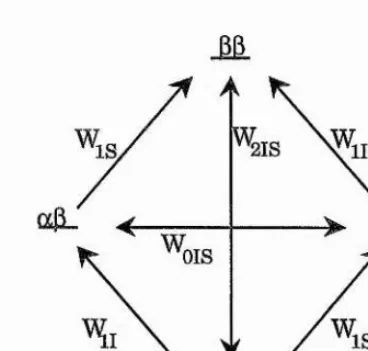
Consider a two spin system, I and S, that are not dipolai* coupled, but are close in space. Transition probabilities between states can be defined as shown (figure 1.11), with the populations of each energy level given by the Boltzmann distiibution.
'2IS
OIS
[image:49.614.210.394.169.345.2]a a
Figure 1.11 - Energy level diagram for two non-coupled spins, I and S, in close spatial proximity. Wi, Wg, and Wo represent the transition probabilities for single, double, and zero quantum processes, respectively. Spin states are labelled a or p.
The application of a radio-frequency pulse at the frequency of spin S saturates this spin causing the equalisation of the populations across the S spin transitions, and relaxation will proceed by various pathways including double and zero quantum spin transitions, W% and Wo respectively. W% transitions are promoted by magnetic fields fluctuating at ~2co, and Wqby
low frequency fluctuations. In slowly tumbling molecules relaxation by the Wqpathway will
Chapterl: Introduction 27
In a multi-spin system, the rate of intensity change for spin I is:
^ =
[
1.
2]
where 1% and are I spin intensities along the Z-axis at time zero and time t respectively. Ri is the relaxation rate of I, and M represents all other spins in the system. The initial rate, when Iz=Iz°, Mz=Mz®, and Sz=0, is given by:
[1.3]
dt
For homonuclear ^H-^H interactions, a is given by:
= -® ^ )] [14]
where h is Planck’s constant divided by 2tc, jio is the permeability of free space, rjs is the internuclear distance, and y is the gyromagnetic ratio of proton spins, I and S. J(co), the spectial density function is defined as:
i i J i
where Tc, the correlation time for isotropic moleculai* reorientation, is inversely proportional to rates of molecular motions, and is identical for each *H-^H vector in a rigid isotropically tumbling molecule.
Chapterl: Introduction 28
If ail constant terms in equation 1.4 aie gathered together as a single term, k, the equation simplifies to:
Gis=ki'is'^ [1.6]
Then, when an NOE enhancement is observed between two other spins (I and M) and the distance between I and S is known, the I-M distance can be estimated from a simple ratio calculation:
1^
[1.7] aIM
Due to the r'^ dependence small inaccuracies in measured NOE enhancements have negligible effects in calculated internuclear distances, and this reference distance method, also referred to as the isolated spin pair approximation (ISPA), may be used to model theoretical NOE intensities from static or dynamic structures provided that the following caveats are adhered to:
1. The accuracy of the calculated I-M distance relies upon that of the reference distance. 2. Inaccuracies are introduced by non-instantaneous saturation.
3. The method is only valid for the initial rate approximation.
4. Integration of spectia, particulaily with overlapping signals may be inaccurate.
5. Internuclear vectors connecting IS and SM must have the same effective correlation time
(Te). This will not be the case if the molecule exhibits anistropic tumbling or has flexibility
in the S-M distance.
6. In the event of internal motions, enhancements are heavily weighted by conformations with the closest contact, due to the r'^ dependence, with the internucleai* distance rather than the enhancement itself which is averaged.
7. Nuclei I, S, and M must have the same gyromagnetic ration, and must not be relaxed by a directly attached NMR ‘active’ nucleus.
Chapter I: Introduction 29
Providing these conditions aie satisfied, the I-M distance can be calculated to within ~10%. However, in flexible oligosaccharides inequalities of Tc become relevant, and internal mobility adds uncertainties to the proportionality of a and (Genest, 1989). For a flexible molecule in multi-site conformational exchange that is slow on the Tc time-scale, the effective internucleai' distance is simply a time-average of the separation at each individual conformation <r'^>'^^®.
1.4.3 Full Relaxation Matrix Calculations
In contrast to the ISPA method, full relaxation matrix calculations offer a more theoretically rigorous method for the interpretation of (R)NOESY data. In full relaxation matrix calculations the longitudinal relaxation of a complete spin-system is determined, thus the effects of spin-diffusion, the presence of heteronuclei, and chemical exchange may be incorporated in calculations for a fixed or dynamic model of the system using a suitable parametrised algorithm. The full relaxation rate matrix, R, is calculated (assuming cross correlation can be ignored) by solving a set of generalised Bloch equations, which govern the time dependence of the magnetisation of each spin I within the molecule, and is best calculated in the form of a matrix:
-Mj~ Ris '" Rin ~Mj-d Ms Rsi Rss ” Rsn Ms
dt : : ; :
Rn2 '" R,m_
[1.8]
with Mi=Mzi - Mzi°. Here Md® is the z magnetisation of spin I at equilibrium. Rn is given by:
[1.9]
the dipolar contiibutions to the spin-lattice relaxation rate pis are given by:
[
1.
10]
Chapter 1: Introduction 30
If all inter-proton vectors aie assumed to be rigid and to move isotropically, the spectral density function talces the form given in equation 1.5, and we can rewrite equation 1.8 as:
— M = ~ R M [1.11]
dt
where R is the relaxation rate matrix, and the solution is given by:
= [1.12]
These equations can be generalised for the analysis of 2D NOESY spectra in which case M(Tm) is a matrix proportional to the normalised NOE intensities recorded with a mixing time Tm and M(0) is the diagonal matrix containing the z magnetisations.
1.4.4 Spin-Spin Couplings Constants
The magnitude of spin-spin coupling constants is affected by the degree of atomic overlap, and is therefore related to the dihedral angle (0) between vicinal coupled spins (Kaiplus, 1959; Karplus, 1963). The generalised Karplus relationship, applicable to both homonucleai" and heteronucleai* spin-coupling constants, takes the form:
J=Acos^0 + BcosB + C