Identifying a vertical neutral position of the breast using
simple measures.
KNIGHT, Miranda K.
Available from Sheffield Hallam University Research Archive (SHURA) at:
http://shura.shu.ac.uk/20740/
This document is the author deposited version. You are advised to consult the
publisher's version if you wish to cite from it.
Published version
KNIGHT, Miranda K. (2016). Identifying a vertical neutral position of the breast using
simple measures. Masters, Sheffield Hallam University (United Kingdom)..
Copyright and re-use policy
See
http://shura.shu.ac.uk/information.html
Sheffield Hallam University Research Archive
Learning ana 1
1services
Collegiate Learning Centre
Collegiate Crescent Campus
Sheffield S10 2BP
102 113 362 0
ProQ uest N u m b e r: 10702839
All rights re s e rv e d IN FO R M A TIO N T O ALL USERS
T h e q u a lity of this r e p ro d u c tio n is d e p e n d e n t u p o n the q u a lity of the c o p y s u b m itte d . In the unlikely e v e n t that the a u th o r did not send a c o m p le t e m a n u s c rip t
and there are missing p a g e s , these will be n o te d . Also, if m a te r ia l had to be r e m o v e d , a n o t e will in d ic a te the d e le tio n .
uest
P ro Q u e s t 10702839
Published by P roQ uest L L C (2 0 1 7 ). C o p y r ig h t of the Dissertation is held by the A u th o r.
All rights re s e rv e d .
This w ork is p ro te c te d a g a in s t u n a u th o riz e d c o p y in g under Title 17, United States C o d e M ic ro fo rm Edition © ProQ uest LLC.
ProQ uest LLC.
7 8 9 East Eisenhow er P a rk w a y P.O . Box 1346
Identifying a Vertical Neutral Position of the Breast Using Simple
Measures
M iranda Knight
A thesis submitted in partial fulfilment of the requirem ents of Sheffield Hallam University
for the degree of M aster of Philosophy
Contents
Abstract....
IV
[image:5.612.108.522.74.661.2]Acknowledgements... VI
Table of Figures... VII
1
Introduction... 1
-1.1
Background
...
1
-1.2
Statement o f the purpose
... 4
-1.4
Scope o f the study
... 4
-1.5
Assumptions o f the stud y
... 4
-1.6
Limitations o f the study
...
5
-1.
7
Operational definitions
... 5
-1.8
Structure o f the thesis
...
5
-2
Literature R eview ... 7
-2.1
Anatomy o f the b re a st
... 7
-2.2
M astalgia
...
8
-2.3
Current treatments
...-
9
-2.4
Breast size and shape
...
1 0
-2.5
Kinematics o f the breast during movement
...
1 2
-2.6
Neutral p o sitio n
...
2 7
-2.7
Summary
...
3 2
-3
Detecting free-fall using the three-dimensional motion capture
system... 34
-3.3 Prediction
...
3 5
-3.4
M e th o d
...
3 7
-3.5
Results
...
4 4
-3.6 Discussion
4 7
-3.7 Chapter summary
...-
49
-4
Measuring freefall in a female bre a st... 50
-4.1
Introduction
...
5 0
-4.2
Aim and objectives
... -
51
-4.3
Methods
...
5 2
-4.4
Results
... 55
-4.5
Discussion
...
58
-4.6
Chapter summary
...-
60
-5
Assessing simple movements in identifying a neutral position. -
61
-5.1 Introduction
...
61
-5.2 Aim and objectives
...
61
-5.3 M ethods
...
6 2
-5.4
Results
... -
65
-5.5 Discussion
...
9 0
-5.6 Chapter summary
...
9 2
-6
Pilot w o rk ... 93
-6.1 In tro ductio n
...
93
-6.2 Results
...
9 4
-6.3 Discussion
...
9 8
6.4
Chapter summary
...
9 9
-7
Discussion... 1 0 0
-8
Conclusion... 1 0 2
-References
... 1 0 3
Appendix... 1 1 2
Abstract
During physical activity, many women suffer from breast discomfort due to excessive
breast motion. It has been hypothesised that movement-induced breast discom fort is
caused by straining the tissue of the breast. To understand the stress applied to the
breast tissue during exercise and in turn understand the motion of the breast in
engineering terms, the breasts need to be placed in a position where the tissue is neither
in tension nor compression. Haake and Scurr (2010) developed a method, the lift and
drop test, to locate this position and termed it the neutral position.
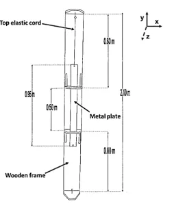
The three-dimensional motion capture system was assessed as to whether it was
capable in measuring accelerations o f -1 g, in a simple oscillating system. Eight cameras,
sampling at 200 Hz, captured the motion of the metal plate attached to a wooden
structure by either one or two elastic cords. Accelerations of -1 g were found when the
metal plate was unloaded, therefore the elastic cords were not under tension. The results
showed that the system was able to measure accelerations of -1 g, however, the motion
was too simple and therefore testing was needed to be completed on a w om en’s breast.
A participant (34D cup size) placed two markers on the body (right nipple and
suprasternal notch). Eight cameras placed in a semi-circle tracked the markers, during
the lift and drop exercises. The maximum negative acceleration found during the
exercise was -0.64 ± 0.04 g. The lift and drop exercise was deemed inappropriate in
locating the neutral position. Therefore, further work in identifying an appropriate method
in locating the neutral position was required.
Seven participants with breast sizes ranging from 34A to 36D placed three markers on
their body (right and left nipple and suprasternal notch). Ten cam eras tracked the
markers during running (10 k m .h r1); stepping off a low box (0.26 m) and a high box (0.51
m); vertical countermovement jump; and lifting and dropping the right and left breast.
The vertical countermovement jum p forced the breasts to oscillate nearly one and a half
times (1.3 ± 0.2), causing the breasts to move through a neutral position multiple times
in a single trial. During a single trial a higher and lower neutral position was recorded.
The accelerations between these positions were -0.13 g and -1.80 g and therefore the
neutral position was reconceptualised as a neutral zone. The exercise also produced low
discomfort scores of 0.6 ± 0.7.
Breast motion during running (10 k m .h r1) and walking (4 k m .h r1) of a participant with a
breast size of 34A was used to demonstrate the effects of breast motion with respect to
the neutral zone and perceived breast discomfort. The work showed that breast
discom fort was reduced when wearing a sports bra compared to no support. This could
be due to 1) the magnitude of vertical breast displacem ent being reduced; 2) breasts
lifted closer to the neutral zone; and 3) level of breast support increased.
This research indicates that the previously defined vertical neutral position, should
instead, be considered a neutral zone defined by upper and lower boundaries, which are
found most effectively by performing a counterm ovement jump. The vertical neutral zone
could allow for greater understanding of the stress applied to the breast tissue during
movement which in turn could inform the design of bras.
Acknowledgem ents
I would like to thank my supervisory team, Professor Steve Haake, Dr Jon W heat and Dr
Heather Driscoll, for their support, expert knowledge and guidance throughout my time
as a researcher. I want to acknowledge the handy work of Terry Senior for designing and
building the bungee structure and Amanda Brothwell and Carole Harris for administrative
support.
I want to thank all my friends, for their continual support, encouragement, enthusiasm
and proof reading. I have to acknowledge my amazing participants! W ithout them, this
research would never have been able to be undertaken. Finally, but no means least my
family, they have been my inspiration and drive to pursue this endeavour.
Table of Figures
Figure 2.1: Structure of the breast (Page and Steele 1999)... 8
-Figure 2.2: Miner's rule - diagrammatic representation of cumulative damage (Maddox
2003 )... 29
-Figure 2.3: Goodm an’s rule - left hand side of each line is classed as the safe zone and
the right hand side is the failure zone (Roymech 2013)... 30
-Figure 2.4: Flaake, Milligan and Scurr (2012) maximum acceleration vs. maximum strain.
... 3 1
-Figure 2.5: Stress-strain curve of the nonlinear elastic behaviour of a ligament (Bindra
2004 )... 3 2
-Figure 3.1: Sketch of the resting height, loaded and unloaded states of a mass attached
by a single elastic cord. The vertical direction is the y-axis of the global coordinate
system ... 36
-Figure 3.2: Sketch of upper and lower boundaries of free-fall defined by the resting height
of the metal plate when the two elastic cords are unloaded. The vertical direction is the
yaxis of the global coordinate system ... 37
-Figure 3.3: Sketch of the metal plate in the single elastic cord set-up. The vertical
direction is the y-axis of the global coordinate system. An additional elastic was attached
at the bottom of the metal plate... 38
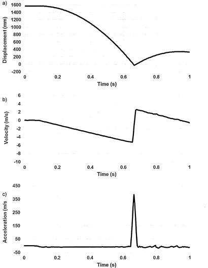
-Figure 3.4: a) Raw displacement of a retroreflective marker dropped from an unspecified
height differentiated to b) velocity, and then c) acceleration... 41
-Figure 3.5: Plot of the residual between a filtered and an unfiltered signal as a function
of the filter cutoff frequency (adapted from W inter 2005)... 43
-Figure 3.6: Vertical displacement of the metal plate with respect to the reference m arker
on the wooden frame during a typical single elastic cord set-up. (Blue triangle points =
metal plate positions where acceleration equals -1.00 g ± 0.04 g; the red line = resting
height of the metal plate when the top elastic cord was unloaded)... 45
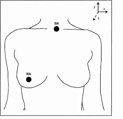
-Figure 4.1: Marker positions on the upper body (SN = suprasternal notch; RN = right
nipple) and orientation of the global coordinate system ... 54
Figure 4.2: Typical trial of a static lift and drop test with no breast support; a)
displacement versus time; b) acceleration versus time; and c) acceleration versus
displacement. Letters represent the following: O at initial lifted breast position, A initial
breast bounce, B - D subsequent oscillations 57
-Figure 5.1: Marker positions on the upper body (SN = suprasternal notch; RN = right
nipple; LN = left nipple) and orientation of the global coordinate system ... 63
-Figure 5.2: Motion of the left nipple of a 36D - sized participant during a typical gait cycle
in a bare breasted run. a) Breast and suprasternal notch vertical displacem ent (left axis)
and suprasternal notch vertical acceleration (right axis), b) Vertical breast displacem ent
with respect to the suprasternal notch and vertical breast acceleration. (B = maximum
height of the sternal notch during the flight phase; C - D = left leg support phase; E - A =
right leg support phase)... 67
-Figure 5.3: Mean ± SD of the resting height and vertical neutral position of the left breast
during five gait cycles in a bare breasted 10 k m .h r1 running trial for each participant...
6 8
-Figure 5.4: Motion of the left nipple of a 36D - sized participant during a typical counter
m ovement jump. a) Breast and suprasternal notch vertical displacement (left axis) and
suprasternal notch vertical acceleration (right axis), b) Vertical breast displacem ent with
respect to the suprasternal notch (left axis - solid line) and vertical breast acceleration
(right axis - dashed line). (A, G = stationary; B = pre-jump knee flexion; C to D = upwards
flight; D to E = downwards flight; F = knee flexion on landing; red lines = vertical neutral
position boundaries; 1 = tension in upper breast tissue; 2 = vertical neutral position; 3 =
compression in upper breast tissue)... 70
-Figure 5.5: Mean ± SD of a) the resting height and vertical neutral position in the initial
upwards direction; and b) initial (I) and final (F) neutral positions of the left breast during
five vertical countermovement jum p trials for each participant... 72
-Figure 5.6: Motion of the left nipple of a 36D - sized participant during a typical low box
activity, a) Breast and suprasternal notch vertical displacem ent (left axis) and
suprasternal notch vertical acceleration (right axis), b) Vertical breast displacem ent with
respect to the suprasternal notch (left axis - solid line) and vertical breast acceleration
(right axis - dashed line). (A, E = stationary; B to C = downwards flight; D = knee flexion
on landing)... 74
Figure 5.7: Mean ± SD of the resting height and vertical neutral position of the left breast during five low box trials for each participant... 75 -Figure 5.8: Motion of the left nipple of a 36D- sized participant during a typical high box activity, a) Breast and suprasternal notch vertical displacement (left axis) and suprasternal notch vertical acceleration (right axis), b) Vertical breast displacement with respect to the suprasternal notch (left axis - solid line) and vertical breast acceleration (right axis - dashed line). (A, E = stationary; B to C = downwards flight; F = knee flexion on landing; red lines = vertical neutral position boundaries; 1 = tension in upper breast tissue; 2 = vertical neutral position; 3 = compression in upper breast tissue) 78 -Figure 5.9: Mean ± SD of the resting height and vertical neutral position of the left breast during five high box trials for each participant... 79 -Figure 5.10: Motion of the left nipple of a 36D - sized participant during a typical lift and drop activity. Vertical breast displacement with respect to the suprasternal notch and vertical breast acceleration (O = initial lifted breast position; A = vertical neutral position; B = initial breast bounce; C - E = subsequent breast oscillation; red lines = vertical neutral position)... 81 -Figure 5.11: Mean ± SD of the resting height and vertical neutral position of the left breast during five lift and drop trials for each participant... 82 -Figure 5.12: Motion of the left nipple of participant 3 and 6 (cup size - 36D) during a typical bare breasted run at 10 km .hr1. a) Vertical breast displacement with respect to the suprasternal notch against time; and b) vertical breast acceleration against time. (Red and blue squares = foot contact)... 87 -Figure 5.13: Motion of the breasts of a 36D - sized participant during a typical gait cycle at 10 km .hr1. a) Vertical right breast displacement relative to the suprasternal notch and vertical right breast acceleration, b) vertical left breast displacement relative to the suprasternal notch and vertical left breast acceleration. (A = ipsilateral foot contact; B = first breast peak; C = second breast peak; D = contralateral foot contact)... 89 -Figure 6.1: Vertical left breast displacement with respect to the neutral position and vertical suprasternal notch displacement with respect to its mean position during a typical 10 km .hr1 run for participant 2 (34A) in a) no-bra; b) everyday bra; and c) sports bra conditions. (Dashed line = static resting height of the nipple without a bra) 95
Figure 6.2: Vertical left breast displacement with respect to the neutral position and
vertical suprasternal notch displacement with respect to its mean position during atypical
4 km.hr-1 walk for participant 2 (34A) in a) no-bra; b) everyday bra; and c) sports bra
conditions. (Dashed line = static resting height of the nipple without a bra)... 97
Table of Tables
Table 2.1: Summary of breast kinematic studies... 1 2 -Table 3.1: Resting heights of the metal plate with respect to the reference marker on the
wooden frame when the top and bottom elastic cord were unloaded... 40
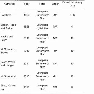
-Table 3.2: Filtering techniques used by previous authors 42
-Table 3.3: The regions of vertical displacements of the metal plate with respect to the
reference marker during free-fall across five trials 47
-Table 4.1: Maximum negative acceleration found in each trial from point O to A (max ±
Abs. RMS error)... 58
-Table 5.1: Mean ± SD of the vertical displacem ent as a percentage of the resultant
displacem ent occurring in the vertical global coordinate system for the suprasternal
notch and left nipple markers during the free-fall regions of the body over five gait cycles
in a bare breasted 10 k m .h r1 run for each participant... 69
-Table 5.2: Mean ± SD of the vertical displacem ent as a percentage of the resultant
displacement occurring in the vertical global coordinate system for the suprasternal
notch and left nipple markers during the free-fall region of the body over five vertical
counterm ovement jum p trials in each participant... 73
-Table 5.3: Mean ± SD of the vertical displacement as a percentage of the resultant
displacement occurring in the vertical global coordinate system for the suprasternal
notch and left nipple markers during the free-fall region of the body over five low box
trials in each participant... 76
-Table 5.4: Mean ± SD of the vertical displacement as a percentage of the resultant
displacement occurring in the vertical global coordinate system for the suprasternal
notch and left nipple markers during the free-fall region of the body over five high box
trials in each participant... 80
-Table 5.5: Mean ± SD of the vertical displacement as a percentage of the resultant
displacement occurring in the vertical global coordinate system for the suprasternal
notch and left nipple markers from the moment the breast was dropped under gravity
until the first breast bounce over five lift and drop trials in each participant... 83
Table 5.6: Summary of the five activities: flight times (mean ± SD), number of oscillations
(mean ± SD), whether the breast reached an acceleration of -1.00 g ± 0.04 g, consistently
found these accelerations in all the trials for each participant, above the resting height of
the breasts and the level of discom fort... 84
-Table 6.1: Perceived breast discomfort scores out of 10 (0 = comfort, 10 = d is c o m fo rt)
in bare breasted, everyday bra and sports bra conditions during a 10 k m .h r1 run for
participant 2 (34A)... 96
-Table 6.2: Perceived breast discomfort scores out of 10 (0 = comfort, 10 = discomfort) in
bare breasted, everyday bra and sports bra conditions during a 4 k m .h r1 walk for
participant 2 (34A)... 98
1
Introduction
1.1
Background
It has become apparent in recent years that breast pain, termed mastalgia, is one of the
most common breast complaints presented to doctors (Smith, Pruthi and Fitzpatrick
2004) affecting up to 60% of women (Ader and Shriver 1997). Many women, in particular
larger breasted women, found that during physical activity excessive breast motion
caused breast discomfort and embarrassment associated with physical appearance,
both of which cause barriers to women participating in physical activity (Robbins, Pender
and Kazanis 2003). This then has a knock on effect on reduced energy expenditure
associated with decreased physical activity, creating a vicious cycle that can contribute
to weight gain leading to increased breast mass (Valea and Katz 2007).
Inactivity and unhealthy eating habits are associated with weight gain, which are the
major underlying causes for modern diseases such as cardiovascular heart disease or
type 2 diabetes mellitus (Warburton, Nicol and Bredin 2006). With worldwide rates of
overweight and obesity levels nearly doubling since 1980 and forecast to increase even
further (World Health Organization 2014), there are only three options doctors can advise
patients to do: 1) change their diet; 2) uptake of physical activity; and/or 3) medical
intervention. The overall trend found in a systematic review of longitudinal studies of
long-term health benefits of physical activity is that there is a negative relationship
between physical activity and weight gain over time (Reiner et al. 2013).
Brown et al. (2013) found that 32% of 1285 females who took part in the London 2012
Marathon suffered from mastalgia. It was also noted that the frequency of m astalgia
increased with breast size. Abdel-Hadi (2000) found that wearing a fitted bra such as a
suggests that only 41% of women and 13% of adolescent females wear a sports bra
during exercise (Bowles, Steele and Munro 2008; McGhee, Steele and Munro 2010).
Today's sports bra has developed considerably since its evolvem ent from the corset.
Ancient Greek women wore a band of cloth over their breasts to prevent/reduce the
sagging and bouncing of them and in turn decrease breast discomfort. The reason for
this was to hold the breasts firmly in place and prevent them from bouncing (Fontanel
1997). The bra was developed from corsets and first introduced in the 1920s. Throughout
the decade, bras were designed to lift, enlarge, support, confide, flatten, reveal and
modestly cover women's breasts, making them the most important element in a w om en's
w ardrobe (Bressler, Newman and Proctor 1998).
In 1972, Title XI legislation was passed allowing women to take part in sport and physical
activity. In 1977, Lisa Lindahl, Poly Smith and Hinda Schreiber sewed two jockstraps
together and marketed it as the jockbra, which became the Jogbra (Bastone, 2014).
Since 1977, two forms of sports bras have been developed, encapsulated and
compression bras. The function of the compression bra is to flatten and redistribute the
mass evenly across the chest, thus minimizing motion. Encapsulated bras, on the other
hand, have cups to separate and support the breast mass in a more feminine shape. By
the 1990s the bra became the most com plex piece of lingerie ever created as it was
composed of 43 components and designed with a structure and function com parable to
those of a cantilever staircase or a suspension bridge (Bressler, Newman and Proctor
1998). However, with all this advanced technology, "they can put a man on the moon,
but they can't put a woman in a sports bra that is very com fortable" - (Miller 1998) -
suggesting that the anatomy and m ovement of the breast is highly complex.
In the last two decades, researchers have looked into correlating breast kinematics with
breast discomfort with a large proportion of literature suggesting excessive vertical
breast displacement causes discom fort (Mason, Page and Fallon 1999; White, Scurr and
-Smith 2009). McGhee and Steele (2010) showed that elevating the breasts on the chest
wall rather than decreasing vertical breast displacement, reduced movement-induced
breast pain. Additionally, Mason, Page and Fallon (1999) hypothesised that m ovement-
induced breast pain may be caused by tension on the supporting structures. Page and
Steele (1999) suggested that the mechanics of the breast need to be investigated to
achieve a better designed sports bra. Recent studies investigating stress of the breast
tissue during motion and discomfort evolved from related studies on the mechanics of
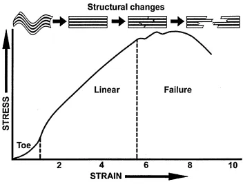
skin. It is known that the behaviour of skin is a non-linear viscoelastic material. The
stress-strain curve of skin can be divided into three phases (Dunn and Silver 1983; Silver,
Freeman and DeVore 2001):
• Strains up to 0.3, collagen offers little resistance to deformation and the behaviour
is dominated by elastic fibres.
• Strains between 0.3 to 0.6, collagen fibres begin to offer resistance to
deformations.
• Strains above 0.6, the individual fibres break.
Studies in breast research are conducted in a laboratory predominantly using motion
capture equipment. Therefore, stress and/or strain need to be calculated from
displacements. Displacements can be classified as a change of length, which are
associated with the local deformation of the body. Strain (e ) is defined as the deformation
of a solid due to stress (or) and calculated as follows (Eq. 1):
AL a
where, AL is the change in length of the material from its initial length, L0 and E is Young's modulus. Stress is linked to strain as stress is proportional to load and strain is
proportional to deformation and is expressed as (Eq. 2):
-a
E = - Eq. 2
£
Haake and Scurr (2011) defined L0 in Eq. 1 as the neutral position of the breast and was found using a static lift and drop activity. The neutral position is a position on the chest
wall where the breast tissue is neither in tension nor compression. The position can be
located during movement where the acceleration of the breast in the vertical direction is
about -1 g, known as free-fall. This position is required as the unsupported length of the
breast tissue is under both tension and compression due to gravity. Therefore, the
neutral position may be important in calculating the stress of the breast tissue and in turn
understanding the motion of the breast in engineering terms.
1.2
Statement of the purpose
The purpose of this thesis is to identify a vertical neutral position of the breasts through
the use of simple movements. In this analysis, the biomechanical variables included
vertical displacement and acceleration of the suprasternal notch and breast. Participants
also quantified a measure of perceived discom fort during the data collection.
1.4
Scope of the study
Eight volunteers participated in this study and had varied fitness levels but were able to
maintain a 2 minute run at 10 k m .h r1 on a treadmill. Participants’ breast size ranged from
34A to 36D and age ranged between 19 to 32 years. Data were collected in the
Biomechanics Laboratory in the Academy for Sport and Physical Activity, located in the
Collegiate Hall building at Sheffield Hallam University.
1.5
Assumptions of the study
The breast is assumed to resemble a cantilever beam represented by a spring-dam per
model. The nipple represents the vertical position of the centre of mass of the breast and
the cantilever beam theory assumes that the dynam ic motion of the breast above and
-below the static position is symmetrical. The vertical breast displacement was used as a
proxy for vertical strain of the breast tissue.
1.6
Limitations of the study
Due to the nature of the study, recruitment was difficult and therefore participants that
volunteered were from a limited population with variations in body/breast mass, breast
shape and gait pattern. However, as the vertical neutral position of the breast is specific
to an individual the analysis was based on a single subject design.
1.7
Operational definitions
The following is a list of definitions of terms that are used in this thesis:
Kinematics is the descriptive analysis of movement which encompasses displacement, velocity, acceleration and temporal relationships.
Neutral position is the point in the vertical direction (relative to the sternum) where the surface of the breast tissue is assumed to be neither in tension nor com pression and is
defined by breast acceleration o f -1 g during motion.
Resting height is the position of the breast on the chest wall (relative to the sternum) without support when standing stationary.
Strain is calculated from vertical breast displacements.
Perceived discom fort score is a subjective measure of breast discom fort collected pre- and post-trials, which was calculated as follows: post-trial discom fort score - pre-trial
discomfort score = Activity perceived discom fort score.
1.8
Structure of the thesis
The thesis contains seven chapters beginning with a literature review to identify the
underlying problem. A further three chapters of the experimental work were separated
-into first a chapter on error in calculating acceleration in simple motion using a motion
capture system and assessing whether the system is able to detect periods of free-fall.
The second chapter is to identify whether true fee-fall occurs in a female breast when
performing a static lift and drop test. The final experimental chapter assesses a range of
simple activities in their repeatability and suitability in locating the vertical neutral
position. This was followed by a pilot work section on the effects of the neutral position
2
Literature Review
2.1 Anatomy of the breast
The breast can differ between individuals with anatomical variation in the volume, width,
length, projections, shape and position on the chest wall (Avsar et al. 2010). The breasts
lie on the deep pectoral fascia, which in turn overlies the pectoralis m ajor muscle (Figure
2.1). The breast spreads vertically from the 2nd or 3rd rib up to the 6th rib and medially
from the sternal edge to almost the mediaxillary line (Drake, Vogl and Mitchell 2014).
The subm am m ary space - between the breast and the deep fascia - is loose connective
tissue. This allows the breast some degree of m ovement on the deep pectoral fascia.
A mature breast is composed of about 80% fat and 20% glandular tissue (Valea and
Katz 2007); however, breast composition can vary depending on the individual (Poplack
et al. 2004). The Cooper’s ligaments offer some internal support to the breast as they
extend from the skin to the underlying pectoralis fascia (Valea and Katz 2007). Stretching
of these ligaments is believed to be the cause of pain and sagging (Page and Steele
1999). The only additional support to the Coopers ligaments is the external overlying
skin; therefore, females wear extra external support such as a bra to prevent the
Adipose 1 Rib
Deep fa: Pectoral minor Pectoral major Interco
muscl Lobule
Ampulla Ampulla
Secondary Mammary
tubuies duct
m
y .'
Lactiferous [image:24.612.180.417.41.305.2]/ duct
Figure 2.1: Structure of the breast (Page and Steele 1999).
2.2
Mastalgia
Mastalgia is defined as a 'benign disorder arising from hormonal activity' according to
the Cardiff group classification of benign breast conditions (Hughes, Mansel and
W ebster 1987). Women can suffer from three types of mastalgia: cyclical mastalgia, non-
cyclical mastalgia and musculoskeletal pain. Cyclical mastalgia is linked to the menstrual
cycle. The pain is released by the onset of the menses - a change in hormone levels.
Non-cyclical mastalgia is unrelated to the menstrual cycle, but associated with
continuous or intermittent pain but with irregular exacerbations. The mean age of the
women at presentation is greater than that of cyclical breast pain, often in their 30's and
40's (BeLieu 1994). Pain which is located behind the breast in the muscle is known as
musculoskeletal pain. The most common of the three types of mastalgia is cyclical pain,
making up 67% of the women with mastalgia; 31% suffer from non-cyclical pain and
about 2% suffer from musculoskeletal pain (Gatley and Mansel 1990; Smith, Pruthi and Fitzpatrick 2004).
The results of previous research contradict the Cardiff Groups definition of mastalgia and suggests that mastalgia can occur due to exercise and therefore the term mastalgia encompasses any pain that occurs in the breast (Iddon and Dixon 2013). Gehlsen and Albohm (1980) and Brown et al. (2013) reported that up to 70% of female athletes complaining of movement-induced mastalgia during exercises that involved running and jumping. Mason, Page and Fallon (1999) hypothesised the aetiology of movement-induced breast pain to arise from tension on both the skin and fascia of the breast during motion. Similarly, Page and Steele (1999) hypothesised that breast pain would occur due to stretching of the support structures when repeatedly being loaded during physical activity, leading to sagging of the breast. Breast pain varies in severity from individual to individual therefore affecting the type of treatment that may be used.
2.3
Current treatments
Treatments for mastalgia often fail due to doctors and clinicians not fully understanding the condition and the breast anatomy and physiology (Abdel-Hadi 2000). Due to an unsatisfactory understanding of mastalgia, the medical community find it difficult to identify and treat (Blichert-Toft et al. 1979). Initially, 85% of patients with mastalgia are relieved of symptoms or could live with the pain when reassured that it is benign, with the remaining 15% of patients requiring medical treatment (Pye, Mansel and Hughes 1985).
There are a range of drugs which have been used in trying to cure mastalgia. Some of these drugs are used as treatments for breast cancer (e.g. tamoxifen), pituitary tumours (e.g. bromocriptine), and marketed as cancer remedies (e.g. evening primrose oil). Prescribing a drug requires taking into account the efficacy, cost and side effects,
-9-however treatm ents are usually prescribed with little or no scientific support (Smith,
Pruthi and Fitzpatrick 2004; Qureshi and Sultan 2005).
Even though danazol, bromocriptine, and tamoxifen provide good clinical responses to
treatment, they do have side effects associated with them. Danazol can cause menstrual
irregularity, depression, acne, hirsutism, deepening of the voice, change in libido and
m uscular pains. Bromocriptine side effects include increased fertility, nausea,
constipation and mental changes (Montgomery et al. 1979; Faiz and Fentiman 2000).
Tamoxifen has been associated with endometrial cancer, hot flushes and vaginal
discharge (Fentiman et al. 1986; Fisher et al. 1996). The relapse rate for danazol and
tamoxifen has been reported to be about 50% (Pye, Mansel and Hughes 1985), therefore
lowering their effectiveness as a treatm ent for mastalgia.
The majority of women do not want to take drugs for their symptoms due to the many
side effects associated with them (Qureshi and Sultan 2005). Abdel-Hadi (2000) found
that in a group of 100 females a mechanical support such as a sports bra can relieve all
symptoms of mastalgia in up to 85% of patients. Researchers have begun to investigate
the kinematics of the breast to try and aid designers in creating a more supportive bra.
2.4
Breast size and shape
With fluctuating asymmetry present in all parts of the body, it is possible for differences
to occur in size and shape between pairs of breasts. In morphology, static breast
asymmetry does exist, but the exact reason why is unclear (Losken et al. 2005). Russo
and Russo (2004) believe that breast development is significantly influenced by
hormones; which if unevenly distributed between left and right side of the body may alter
growth. Another reason may be due to asymmetrical torsos which causes the breasts to
sit on the chest wall at different positions. Variables that are frequently used to define
-marked asymmetries between left and right breasts are volume and vertical distance between the suprasternal notch and nipple.
McGhee et al. (2013) found that the breast volume of 15 women had a greater right breast volume than the left breast by a mean difference of 8 ml. Smith et al. (1986) showed that left breast volume was greater than right breast volume by a mean difference of 16.2 ml. Conversely, Loughry et al. (1987) found that 99.6% of 248 women had a difference in size between left and right breast volume, however there was no predominance as to which side is greater in volume. Hussain et al. (1999) found that breast volume is also dependent on where the women were in their menstrual cycle, with the largest breast volumes recorded during the last week before the onset of the menses. The unequivocal results in breast volume and the change in volume throughout the menstrual cycle suggest breast volume is dependent on the individual.
significant result found in this study compared to Brown et al. (1999) may be due to the
larger number of participants.
2.5
Kinematics of the breast during movement
Breast motion studies have been conducted since the late 1970s with an em phasis on
correlating breast kinematics with discomfort/pain. Research in this area has generated
some inconsistent findings due to small sample sizes, experimental set up, marker
positions and the way in which the biomechanical variables are calculated. Alm ost all the
research in breast kinematics focuses on or includes the vertical com ponent of breast
motion. The three main reasons are: 1) the vertical component makes up about 50% of
breast motion (Scurr, W hite and Hedger 2011); 2) it is most affected by gravity (McGhee
et al. 2013); and 3) it has been associated with movement-induced breast discom fort
(Gehlsen and Albohm 1980; Lorentzen and Lawson 1987; Mason, Page and Fallon
1999; Starr et al. 2005). The different approaches which have been taken are shown in
Table 2.1.
12-T a b le 2. 1 : S um ma ry of breast kin em a tic s tu d ie s 13 Q. -*—*
o
CD•N ^ N CQ
0 Q-CL 0 CL CL 0 CL Q.
z
0 O c 0 4— 0 M— 0 cc 0O a
S? J?
c: u
0 Jt: O £
E
I i
o £—I 0
§ ) ! C l*~
0 0 T3 0 0 ■g —- 0 CM 0 0 -g -4—* 0 00 o
<
E CO 00 E CO CO 03 cd E CO CO 03 E CO C3 L_ JC E CD CD 03 C 0 E Q. 'Z3 CT LU CO c o 2 oE S 0 E ° 5 ®
§ °
5
* o ci 0 o r a c _i > <E E co
c:
o
0 j-0 c g E c o 0 o o 0 o E 0 ^ E! 0
0 0
_ > > 0 . 0 m
0
0.
0
<- 0 C E —I 0
0 >•. 0 0 1
_
m0 0 hr N CD 0
0
03
<
0
DO 0
>, JO -§ 7=5
>4 OJ 4— ^
S> i l
LU O CD" Q
<
Z 0 00 •c 0
.Q o
CL
I_ JO
0 CL 0
D ■e
o 0 o
CL CL
CO ZU CO
00 00
O
CD’' Q
o<3 _
CD < O
CM h
-CO ^ 00
0
9
CM +1
0
c!
0
Q.
O LO o■sr 03ID
0
Q_
iz 0 o 0
<
>-o
o
g oo
0 03
o6
0 E i« r n CD <
t-N 0 0 0 £:
u _| 03
O . 03
[image:29.613.122.558.57.717.2]ZD
Q .
O o
> , 0
I I
CO CL
0 Q . Q . 0 C L C L 0 Q . Q . 0 O C 0 £0 a : E zs c 1— 0 CO E 13 C i_ 0 •4—* CO E o
E -g £ o
CO c
E E
0 3 r = C 0 0 0 'i_ -*—4 0 00 0 0 ; o GO CM O
<
E jx. CD CD 0 3 L J Z E a; CM K of L . J Z E o<: j z E o JZCO JOo
.. 0
tz <
c 0 E Q . 3 O ’ LU c o o
<
0 o 'c oc 0
I °o 5 £
0
TO
i_ 0 ETO
O CO8
0
“ > 2 .
0 0
O O
c C
0 0
c SZ 2
(0 ~ 0 V I U ---0 x : J^
o o c
-o g | .c E £
Q. o
CM 1q
E
TO rr.
o
- 0 F
CD . E £
— O t*=
0 •4—* 0
TO
m 0TO
>_ JO 0 •M1— o C L CO N -0 * ^o -Q S’ wQ - - e
3 o . o <0
Z CM
TO
•e co
o £ _
g - Q - J O
Q. o C
o ° o <2
M a ' z ^
o o 0 2
Z & £ 0
TO 0) N CO ’(/)
CD Q < O
Q o3 O 0 0 3
<
CM 0■s I
o o Z ■■£
TO Q .
0 3
ID lO CO
S3 TO ZJ 0 <
>-c 0 c 0
TO
E JZ2 ^
go 0 3
o CD 0 3
0 3 0 3 0 3
C" 03 C O 0 Q
£ 0 3 =
<9 ro ro
3
CL *-»
ID
o
> , 0
*-> o CO Q_
0 O c 0 £(1) q:
f t ED) — C •*-Cl) O < c 0) E Q. ‘3 cr UJ 0 >» -4—* to 5 m
CD 0) 1- N m 0
CD
CD
<
0
•s I
o' o 2 €
CD
CL
ro ±= CD
0 CL Q. E 3 c 1— 0 GO 0 o E 10 CO CD QC 0 cd t— 0 x E
0 CD
M— ( } 0 ^ oc
o £
I I
m £0 cd 1_ X3 CO •e o CL 0 00 0 CL CL M— C O O cd
1_ 0 E 0 c 0
"CD 0 0
_ J Q . cd
c3 § O ^
°a q
Q Q
£ F ° -0— CD CO CM
0
CD ■ =
0 JS 0 E S CD O O 0 P
1_ L_
0 0 ;g 'i — ■4—« CO CO o c >4 o 0 o 5 0 J5
> CD
QC ro
E 0 -4—* 0 >4 CO TO 0 0 . 0 CD i_ .Q 0 ■e o Q. 0 CO
1 D
O Q
CO £5 CM ™
CO
CD
0 fc JO cd S CO CM
0
CL
Q. iz
cd
E -g .2 -5 CO c
0 o CM o CM O CO s i
CL O
O E
cd .C ) >4 cd ■o >* 0 > LU
o3 Q Q Q
CD
CO 03
CM
_
£ O ■q. ™
S 3
O 0
0
CL CL
06
E
=3 JD E ■=
£ "E CO CO
0 0 ■g 10 0 o cd CL ■o 0 -4—» o 0 0 0 0 CO o 0 F
co E
1
*
! o § « «
o > CD
L—
(D E _ CD 3 E cd 0 o E .t=: p 0
ro .2> ro 2
O TJ O >
CL O CL O O —3 o -I—» O
CD CO^r
co
0" oS £ ®
S CO
Z3
CL
o
>* £ = I 55 q .
o o c 0 £ 0 oc
c
**-0 o < c 0 E Q. 'u cr 111 0 >» co 0 I— CD 0 O) < 0
O
I
o oZ '€
ro D_
° 0 :b 0
0 Q.
Q .
£ 03
.0 > C/D
*- 0 o3 Jt;
o 0 C/3
y C/3
0 .
_i o <
• M 0 '0 0 O) o> . in o q: i_ JZ E in 0 x E 0 0 0 T3
Cd 0 O D_ in
0
_ 2
■e -o
>< 01— — XI
§- "9 co 0 o ^
Q- 0
cr u/
^ -e o 0 -> CL LU 03
+' 00
3 ^
in
03
«-fc~ . 2 8 > OD
3 i 0 § C/3 > I CM
* N 0 CL CL 5 03 03 < 0 0 p 'i_«4—»
03 <N E co o cL 0 0 0 1_ 0 E 0 O ■o
CL 0
O '
CL
IO
scsj :<"5 N *S~T N *
0
CL CL
:?S
?S
ro os o
£ S 2
s - - s
Q . P £,o o a
c/3 c t: >_ v+— o 0 -t—* _Q •|_
o o o
‘i_ 0 T—
£ Q .
0 0
0
c . JZ -V—'
tL O 0 - 0 0
0 - 7 3 O . cs >>C/3
w ai -o ? S !
z ui i i
CO ^
^ CD CM + |
CO
_ °3 0
■M
1c £
!: t O) o v o
O b O 0 3 C/3 CM
& 0 < ? « o >, T- o
L JZ E j*. o of •a 0 i_ 0 i_ M— c — 0
0 0
H
^ 0
CO o
0
t i -Q « s = - §
0 - 0 . 0
§ - ■ £ 2 “ 0 O
O > CL Z LU 03
T- CO id ^
CM +1
in
o6 *- t= .-2 05 O 3 ^ 0 5
CL
•4—'
0
o
> * £
= I
CO S.
CD a c 0 s— CD H— CD a.:
£CD — E
C
**-CD O < c CD E Q_ 3 c r LU 0 _>% GO 2 m 0 CD < £ c CD Q . O L_ 03 CL
6 ) n ®
r- Q- T3 ■I E O CL 03 E
CO TD
0
CL CL
[9 o3
-ffi 0
C
D
2 - c CL O
=5 O
CO
C0 .Q
0 0
3 1_ 0 S Z-4-41
0 O O 0 4-41 i— 0 T—
sz
0 CL 0 -C -4-4 03*4_
44— c 00
O CO cd L. -C E . x CD 2 0
x E 0 0 5= o
£ "O
Q_ 0 O
L_
CL
10
nT -Q o >, 2
CL 03 -O
w 0 o
O > CL
Z
LUCO
o o -00 o3 0 . x 0 0 X 0 CL CL O o c 0 c i— 0
-4—>
CO
CD
0
■g o £
00 0
E . x CO +1 CO 00
Cd
o CNI o CO <Cd
o CL 0 c0
OJ 0 O
~ 0' o 0 O 0 3 fn
0 .p m O 8 R e
( - 0 0
0
_ -a $ 0 0 _q
0 £ 2 0 Q
_
L3- t -rj C 0 O cLU “ C O 0“
0 S
0 ^ 0 o 0
Q_ 15 0 CL
o +1 CO co o CNI
o<3
0
^ w °
0 r:
O ■-
0 |> 3
0 CL CL 0 C L_ <1) (/) 2 -c Q. O ^ O
CO
cCD c ■0
c
0
4—*
CO c
E
CM
i
1
1
\
1 1 ^ I
a: cr
§ „^ -4—* ^ — n
0
n0
-2 0 - ^ 0 CO U. CO LLT3 0
i_
0
L_ >4—
c
— 0
0 0
& I
^ 000 o
0 t -Q —
o >, 2
cl 0 _Q2 ® 0
O > CL
Z LU CO
O CL •M
ZD
o
0
r-O • -U-* CO
io
> , co
- ? ■*-’ o C/D Q .
0) Q . Q . 0) CL Q . 0 O C 0 0 M— 0 a: 0 c I_ 0 GO 0 i_ CL O C/D Jt; 0 o
° 3 " o j o * - r a o o o O£ ~
o T3 0 'c 0 ^ oj a . m
o c c * = to 2: c ro to .E 0 £
co
C 5= i- 0 0) — •£
w ° 3 o ) o 2 -c ~ -a . o 0
C/D O c i_ -=c ro co .E &— M— o tn -Q 'l_ sz •4—*
o o o
' u 0 T-£ CLCO
0 0 c . c-4—»
E o> —
c
*•-0
C/3
~ d)0 "TT
03 O
LO O
'ro $
o > ,
T - O
o
<
i o c
. .
0 7=1
> »
JCt
E — a;
0
^ 0
r - O C/3 E O CO
0 O
g 2
-E Q_
O 0
E 03 C 73 C 0 C/D
a: of
5 -*->o w
— 0 C/D LL
c 0 E Q . ‘o O ' LU TJ 0 i_ 0 i_ <+— c — CO
co 0
0
o
o- c O ro oo o
• o 0 l_ 0 l_ M— c
— CO CO 0
0
n
o ' c
^ CO
00 o 0 > -4—< 0 0 t_ CD •e o _
Q- 0
CL U O
-0
JQ
a3 - 'S
> o ■ CL
5 w LU CM
0 t -Q —
s >> § 0 - 0 - 0 § ■ ■ £ £ o ® ° O > D. Z LU C/D
0 0 CD co
CD I < 0 03 <
T - 00
io ^ CM +|
00 ^ in +i CM co c 0 Q.
O CM 00
0 0. o o C/D o5 0 •M JZ £ 0 03
"O T-0 o
Z CM
o3 CM
l b
=3 Q l -*—< O O * N C/3 H CQ : N N
>, CO - 1
+ -> o
CO
Q_<e T E
f £ °o c l
|5
0— —> r ~ t- C CO £ =
• c M" L— 0 sz s_ O 0 u CL CD
T3 C
0 0 i_ T3 0 0
0 E
ZS
l—
0 E -Q>o 0f— 0CL 3
CO
0 0 CLCL
o 0 E o 0 J>_-1—» CO O E _i "c
0 0 C 0 1 0 M— 0 o : CM j*:
TD
O
C 0 0 -Q 00 C 0 0
£ I £
.QP C £
n E
-C O 0 _
W CM
O 0
O £
c ^ 0 ^0 C —
0 TC3
CO 0
a i
c**-<5 o
1/5 m 0 ^ ■ ^ 0 °
LO !r >,
t
- -Q O
w 00 iy
_ 0 o
O u > co n u
L .
O
< j* :E
r^-tr
E CD I 00 cd c 0 E Q . 13 cr LU c o o > E 0 00 >4CO
co > 0 c CD <O CM O CO < DU
-E O
Cl co
° §
CM CO
0 "co 0 1_ CO ■e CO 0 L— -Q
o 0 O >
CL _Q CL 0
CL CL T3
CO > .
CO ■£ co i
_
o OCl o
0
>
Z CO Z LU
O
CL
0 0
i- N
CQ m
CD
CD I Q 0 CD <
03 o
LO M" CM
CD ■M■M""
CO 4—' c 0 Q . O CM 0 CL
£ 0
CM
>- ° ^
c f CD
5 z
n oej
CO
3 CL 4^ Z>
o
>, 0 - 1
•*-> o
C/3 Q_
£ (D dc
c l*
-«? o
o < c (D
E
Q . 'l3 c r LU 0) (/) CD i_ CQCD 0) >- N
CQ 0
CD CD < C CD Q . O tc CD CL ?n *
^ CD -C Q. 0) Q_ DC c
CD ^ £Z M=-1- 0 CD —
0 03
CD -C CD O* * -Q . 4—* O -n CDU «4—» ,« ° c c C/3 C CD CD
0
~ 0 CD
CD O
if) O
E
o DC o 0 0 CD1 o •M Q .M - 5 o
>-0 x= o o 0 ■<-0 o
CD £
O O
CD
o
1C s
CL (D -Q
o- -o 0
w - *
i: 0 o o > o.
Z LU C/3
+1 CNI CM CNI
CD
CD to 0
^ o 1 CNI
?N
K ts
^ 0 - C Q .
O ) Q . DC E
co C 5= * - CD
0 —
0 o3
2 -C Q - -2 ^ O C/3 c
x i i_ .25.2 £t — i — -d £
-c -c cd ro
- 5 o
>-0 x : o o
JZ 0 T— cd a 0
p 0 £
E CD i
0
~ 0
CD ^ CD O
if ) O
L. _£=
E
O DC g 'c o o 0<p 0
O CO "o. 0
o E
CNI CD£ O
0
t " CD 0 X )
■e o
CL
a . -a
o ?>*
o ® o O > Q . Z LU 03 0
+1 CNI CNI CNI
0
0 £,
_ 0 ^
~ O
13 Q. +-»
13
o
>• SB
= 1
55 £
o CJ c: 0 a3 u -0 (T
fe E
03 —d *
-5 o
o < c 0
E
Q. '=} c r LU 0 >* w 0 t _ CQ 0 O) < wo
|
o o 2 -50 Q.
5= 0 O 0 <
>-* N
o3 0
« £ g;
0 o ) .9
-;N •N •*
N * ?=S
0 ^ c 4= fc 0
0 — w 03 2 £ Q- ^ -o ^ o
CO c O) o i— * i_ 0 c c
0 0
~ d)0 -TT O) O io o
L_ JZ
E
.s*: 0 01 o 'c o l _ 0 _0 01 o Cl O 0■e -Q 0
+i
cm oJ CM
C/3 .d
IO
o
CM
o -- o
cc co
u- 5 o
*-C/3 £
O O 0 •*“
0
Q. 0* -Q § ■ ? .£ ^ 0 o
O > Q .
Z LU CO
E
o
cd
^ 0
Q.
03 q_
in c
0 ^ c &> ® ~
D i o 2 .C O o 0 . 0 -o O
° c c C/3 c 0 0
M- £ o «--ili
o o 0
T-0 03 < c 0
E
0 > 0E
1 1_ 0 c D oo
S
O. 0E
O O
■c o Q. Q. 3 C/3 O +1
CM ™ CM
CD
0 ID § O ; | " in 0
Q.
E
o
.
o>
U)
Q.
Q.
Q. CL
Boschma (1994) investigated the effects of full, medium and no supports on breast biomechanical variables in 15 participants with breast cup size of C to DD, running at 7.2 km .hr1. The motion of the nipple and sternum markers were recorded using three cameras, two of which were directed at the activity and one at the heel to record heel strike. The results showed that as support decreased, vertical breast displacement, perceived discomfort and stride rate increased while stride length and vertical trunk displacement decreased. From the results it was concluded that perceived discomfort was affected by cup size and bra style, rather than quantitative vertical breast displacement. This was supported by Lawson and Lorentzen (1990) who found that only one out of seven sports bras showed a significant correlation coefficient for comfort with quantitative breast displacements. The results suggest that comfort during exercise is more than just a feeling that the motion of the breast in the vertical direction is limited.
Alternatively, Mason, Page and Fallon (1999) used three participants with C to D cup size breasts. Markers on the suprasternal notch and nipple were tracked during gait at 7, 10 and 13 km .hr1 and an aerobic activity in four support conditions, using two 16 mm Photosonic high-speed cine cameras. The results suggested that perceived comfort followed the pattern of vertical breast movement, rather than maximum deceleration force. As the breast support decreased, breast displacement increased resulting in increased perceived discomfort.
White, Scurr and Smith (2009) found from the mean discomfort scores obtained from eight participants with size D cup bras that the compression bra was the most comfortable as it produced the least amount of resultant breast displacement out of all the support conditions (encapsulated bra, fashion bra and bare-breasted). Markers were placed on the right and left clavicles and ASIS to convert the global coordinate system to a local coordinate system with the origin at the right clavicle. Therefore, the right nipple
-resultant displacements were made independent and eliminated the six-degrees-of-freedom movement of the body.
Conversely, McGhee, Steele and Power (2007) found that increase in breast comfort was attributed to the significant reduction in mean peak vertical breast velocity in deep-water running compared to treadmill running. Sixteen participants (C to J cup size) ran on a treadmill above ground and 2.4 m under water at a self-selected pace. Markers were placed on the sternum, level with the articulation of the 3rd rib, on the participants' nipple and superior aspect of the breast immediately above the nipple. The markers were tracked with either a camcorder digital video camera or a poolcam video camera.
McGhee and Steele (2010) used 20 participants with cup size of C to F. The participants had markers placed at the suprasternal notch and on the nipple. The markers were tracked using two OptoTRAK 3020 sensors during an average speed run of 8.3 km .hr1 on a treadmill, while wearing either an encapsulated bra; compression bra; or experimental bra (encapsulated sports bra, incorporating elevation and compression). They found that the experimental bra produced lower subjective ratings of perceived breast movement, breast discomfort and bra discomfort than the other support conditions. It was noted that there was no significant difference in the vertical breast displacement in each condition. The results suggest that elevating the breasts higher on the torso reduced the tension and loading on the anatomical structures of the breast. One possible reason for this is that these structures are further away from their end of range compared to the other support conditions.
McGhee, Steele and Munro (2010) used 15 participants with cup sizes of D to G. Markers were placed on the suprasternal notch, left nipple and left heel. The markers were tracked using two OptoTRAK 3020 sensors during an 8 - 9 km .hr1 run on a treadmill, in two support conditions (an everyday bra and bare-breasted). They found that the mean vertical component of the maximum net bra-breast force of the left breast during the
-downward phase was on average, significantly less when the participants wore the
sports bra compared to the fashion bra and was associated with less movement-induced
breast discom fort with no change in rate of perceived exertion.
A common theme in this literature is the level of discomfort a women experiences during
the exercises. Mason, Page and Fallon (1999) with Heil’s (1993) designed a visual
analogue scale (VAS) in the form of a Likert scale. The scale ranges from ‘0’ (comfort)
to ‘10’ (pain), with ‘5’ representing uncomfortable. They presented the scale to the
participants immediately after the treadmill exercise. This scale and method have been
adopted by many researchers (e.g. White, Scurr and Smith 2009; Scurr, W hite and
Hedger 2010). The main advantage of V A S ’s is that they are easy to implement. A
potential disadvantage of this approach of rating breast discomfort during an activity is
that there was no measure of a baseline level before each activity. The rest period
provided between each activity may have been insufficient for perceived breast
discomfort to return to baseline levels before undertaking the next activity.
McGhee and Steele (2010) modified the scale to show ‘ 10’