http://wrap.warwick.ac.uk/
Original citation:
Qureshi, H. and Rajpoot, Nasir M. (Nasir Mahmood) (2010) Comparative analysis of
spatial and transform domain methods for meningioma subtype classification. In:
Medical Image Understanding and Analysis (MIUA 2010), Coventry, UK, 6-7 July 2010.
Published in: Proceedings of Medical Image Understanding and Analysis 2010 pp.
209-213.
Permanent WRAP url:
http://wrap.warwick.ac.uk/47475
Copyright and reuse:
The Warwick Research Archive Portal (WRAP) makes this work by researchers of the
University of Warwick available open access under the following conditions. Copyright ©
and all moral rights to the version of the paper presented here belong to the individual
author(s) and/or other copyright owners. To the extent reasonable and practicable the
material made available in WRAP has been checked for eligibility before being made
available.
Copies of full items can be used for personal research or study, educational, or
not-for-profit purposes without prior permission or charge. Provided that the authors, title and
full bibliographic details are credited, a hyperlink and/or URL is given for the original
metadata page and the content is not changed in any way.
A note on versions:
The version presented in WRAP is the published version or, version of record, and may
be cited as it appears here.
Comparative Analysis of Spatial and
Transform Domain Methods for Meningioma
Subtype Classification
Hammad Qureshi School of Elect. Eng. & Comp. Science, NUST, H-12, Islamabad, Pakistan. Nasir Rajpoot Dep. of Comp. Sc., Univ. of Warwick,
Coventry, CV4 7AL, United Kingdom.
Abstract
Pattern recognition in histopathological image analysis requires new techniques and methods. Various techniques have been presented and some state of the art techniques have been applied to complex textural data in histological images. In this paper, we compare the novel Adaptive Discriminant Wavelet Packet Transform (ADWPT) with a few prominent techniques in texture analysis namely Local Binary Patterns (LBP), Grey Level Co-occurrence Matrices (GLCMs) and Gabor Transforms. We show that ADWPT is a better technique for Meningioma subtype classification and produces classification accuracies of as high as 90%.
1
Introduction
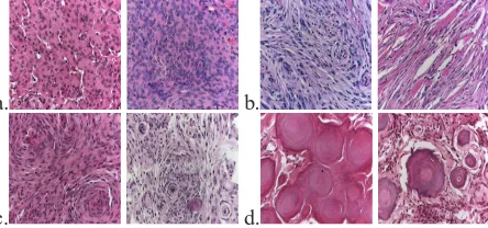
Meningioma subtype classification is a real-world problem from the domain of Histological Image Analysis. Meningiomas are tumours of the Meninges (covering of the brain and the nervous system). Histological images are real world data and are considerably different from synthetic textural data. Histological images have a uniquely complex texture which repre-sents a new set of issues. The texture in histological images such as Meningiomas is more or less non-homogenous i.e. different areas in an image may have different textural proper-ties which in turn may represent different patterns. Hence, textural analysis and subsequent recognition is not straightforward. Moreover, intra-class variation amongst the samples be-longing to the same class is high and to make matters worse inter-class differences amongst the samples is low. This could be seen in the Meningioma subtype images depicted in the Figure1.
Diagnosis of Meningiomas is still carried out by human experts. Its hampered by the fact that the reviewing of the histological slides is time consuming, prone to error and the inter-rater variability amongst the experts is considerable [2] which makes the therapy regimens biased. Definition of diagnostic criterion for all tumour entities within the World Health Organization (WHO) Classification of Tumours [4] has been problematic. Hence, there is a need for an automated computer based technique to introduce more objectivity in to the analysis. Most Meningiomas are benign [8] which means that neuropathologists are spend-ing most of their time analysspend-ing and diagnosspend-ing benign tumours. Consequently, their is an urgent need to develop automated techniques to aid the neuropathologist.
c
a. b.
[image:3.595.90.312.31.135.2]c. d.
Figure 1: Various Meningioma Images belonging to each subtype a. Meningiothelial, b. Fibroblastic, c. Transitional, d. Psammomatoes
Some of the results on Meningioma subtype classification have been presented in [10] [9] [12] [5] [11] [1]. Many techniques have been used in literature for texture classification. Randen and Husoy [13] presented a paper on comparing various texture analysis techniques for Brodatz texture classification. In this paper we compare the novel Adaptive Discrimi-nant Wavelet Packet Transform (ADWPT) with Gray Level Co-occurrence Matrix (GLCM), Gabor Transform (GT) and Local Binary Patterns (LBPs) for Meningioma subtype classifi-cation. This paper presents comparative results between these techniques.
2
Methods
2.1
Gabor Transform
Gabor analysis of the textures was carried out as proposed by Ma and Manjunath [6]. Four scales and six orientations were used to provide texture representations at various scales and orientations. Energy feature is used to construct the feature set. The mean and variance as suggested by Ma and Manjunath was also computed and classification results generated.
2.2
Local Binary Patterns
LBP [7] with a radius of 1 and 8 neighbourhood pixels was used in the analysis. Other radii and number of pixels were also used with no apparent improvement in results.
2.3
Adaptive Discriminant Wavelet Packet Transform
ADWPT was carried out up to the fourth level. The subband selection for the most discrim-inant decomposition was obtained using the Fisher Discrimdiscrim-inant. A detailed discussion of ADWPT is presented in [10] and [11].
2.4
Gray Level Co-occurrence Matrix (GLCM)
GLCM analysis was carried for four directions i.e. 0o, 45o, 90oand 135owith distances set
2.5
Classification using Support Vector Machines (SVMs)
A gaussian kernel is used and a search for the best parameter is carried out. Matlab version of SVMs [14] developed by Chang and Lin [3] are used for classification.
3
Results and Discussion
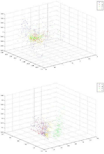
Figure2shows the projections on the first three principal components performed after PCA analysis of the features acquired for the two best feature sets i.e. GLCM and ADWPT re-spectively. The other figures have not been included due to lack of space. The 3D plots show that ADWPT performs much better than LBP, GLCM and Gabor Transform. In case of ADWPT, psammomatous is separated well with transitional also found on the edge forming a relatively separate cluster. GLCM produces comparative results to Gabor but is not able to differentiate psammomatous well. LBP performs the worst with no clusters seen.
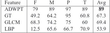
[image:4.595.116.300.325.380.2]The classification results given in Table1 again prove that ADWPT provides the best differentiation amongst the meningioma subtypes followed by Gabor and GLCM with LBP providing the worse results. There were a total of 960 meningioma images with 240 images per subtype. 20% of the data is used for testing i.e. 1 patient per subtype while the rest used for training. Daubechies 8-tap filter was the wavelet filter used.
Table 1: 5-fold cross validated classification accuracy results using Support Vector Machines for LBP, GLCM, Gabor Transform and ADWPT (Fishers Discriminant) (F=Fibroblastic, M=Meningiotheliamatous, P=Psammomatous, T=Transitional)
Feature F M P T Avg
ADWPT 79 89 97 89 89
GT 49.2 64.2 95 60.8 67.3 GLCM 68.3 74.2 75 60 69.4 LBP 12.5 65.6 66.7 70.9 53.9
The results in table1clearly show that ADWPT performs much better than GLCM, GT and LBP for meningioma subtype classification. The selection of subbands using the AD-WPT provides a mechanism for selecting the optimal wavelet packet representation. This enables the extraction of good features for classification. GT and GLCM acquire classifi-cation accuracies of around 67% and 69% respectively which is lower than ADWPT. LBP provides the worst classification accuracies of 53.9%.
4
Conclusion
[1] O.S. Al-Kadi. Texture measures combination for improved meningioma classification of histopathological images. Pattern Recognition, 2010.
[2] P.C. Burger. What is an oligodendroglioma? Brain Pathol (2002), 12:257–259, 2002.
[3] Chih-Chung Chang and Chih-Jen Lin. LIBSVM: a library for support vector machines, 2001. Software available at urlhttp://www.csie.ntu.edu.tw/ cjlin/libsvm.
[4] P. Kleihues and W. K. Cavenee. World Health Organization Classification of Tumours.
Pathology and Genetics. Tumours of the Nervous System. IARC Press, 2000.
[5] B. Lessmann, V. Hans, A. Degenhard, and T. W. Nattkemper. Feature space exploration of pathology images using content-based database visualization. In Proceedings SPIE
Medical Imaging, 2006.
[6] W.Y. Ma and B.S. Manjunath. Texture features and learning similarity. In Computer
Vision and Pattern Recognition, 1996. Proceedings CVPR ’96, 1996 IEEE Computer Society Conference on, pages 425–430, Jun 1996. doi: 10.1109/CVPR.1996.517107.
[7] T. Ojala, M. Pietikainen, and T. Maenpaa. Multiresolution gray-scale and rotation invariant texture classification with local binary patterns. Pattern Analysis and Machine
Intelligence, IEEE Transactions on, 24(7):971–987, Jul 2002. ISSN 0162-8828. doi:
10.1109/TPAMI.2002.1017623.
[8] A. Perry, D.H. Gutmann, and G. Reifenberger. Molecular pathogenesis of menin-giomas. Journal of neuro-oncology, 70(2):183–202, 2004.
[9] H. Qureshi, N. Rajpoot, K. Masood, and V. Hans. Classification of meningiomas us-ing discriminant wavelet packets and learnus-ing vector quantization. In Proceedus-ings of
Medical Image Understanding and Analysis, 2006.
[10] H. Qureshi, O. Sertel, N. Rajpoot, R. Wilson, and M. Gurcan. Adaptive discriminant wavelet packet transform and local binary patterns for meningioma subtype classifica-tion. In Proceedings 11th Medical Image Computing and Computer-Assisted
Interven-tion (MICCAI’2008), 2008.
[11] Hammad Qureshi, Nasir Rajpoot, Tim Nattkemper, and Volkmar Hans. A robust adap-tive wavelet-based method for classification of meningioma histology images. In
Pro-ceedings MICCAI’2009 Workshop on Optical Tissue Image Analysis in Microscopy, Histology, and Endoscopy (OPTIMHisE), 2009.
[12] Hammad Qureshi, Roland Wilson, and Nasir Rajpoot. Optimal wavelet basis for wavelet packets based meningioma subtype classification. In Proceedings 12th Medical
Image Understanding and Analysis (MIUA’2008), 2008.
[13] T. Randen and J. H. Husoy. Filtering for texture classification: a comparative study.
IEEE Transactions on Pattern Analysis and Machine Intelligence, 21(4):291–310,
1999.