1535-9778/10/$12.00 doi:10.1128/EC.00291-09
Copyright © 2010, American Society for Microbiology. All Rights Reserved.
Comparative Transcript Profiling of
Candida albicans
and
Candida dubliniensis
Identifies
SFL2
, a
C. albicans
Gene
Required for Virulence in a Reconstituted
Epithelial Infection Model
䌤
†
Martin J. Spiering,
1‡ Gary P. Moran,
1Murielle Chauvel,
2,3Donna M. MacCallum,
4Judy Higgins,
1Karsten Hokamp,
5Tim Yeomans,
1§ Christophe d’Enfert,
2,3David C. Coleman,
1and Derek J. Sullivan
1*
Microbiology Research Unit, Division of Oral Biosciences, Dublin Dental School and Hospital, University of Dublin, Trinity College, Dublin 2, Ireland1; Institut Pasteur, Unite´ Biologie et Pathoge´nicite´ Fongiques, Paris, France2; INRA USC2019, Paris, France3;
Aberdeen Fungal Group, School of Medical Sciences, Institute of Medical Sciences, University of Aberdeen, Aberdeen AB25 2ZD, United Kingdom4; and Smurfit Institute of Genetics, University of Dublin, Trinity College, Dublin 2, Ireland5
Received 8 October 2009/Accepted 11 December 2009
Candida albicansandCandida dubliniensisare closely related species displaying differences in virulence and genome content, therefore providing potential opportunities to identify novelC. albicansvirulence genes.C. albicans gene arrays were used for comparative analysis of global gene expression in the two species in reconstituted human oral epithelium (RHE).C. albicans(SC5314) showed upregulation of hypha-specific and virulence genes within 30 min postinoculation, coinciding with rapid induction of filamentation and increased RHE damage.C. dubliniensis(CD36) showed no detectable upregulation of hypha-specific genes, grew as yeast, and caused limited RHE damage. Several genes absent or highly divergent inC. dubliniensiswere upregulated inC. albicans. One such gene,SFL2(orf19.3969), encoding a putative heat shock factor, was deleted inC. albicans. ⌬⌬sfl2 cells failed to filament under a range of hypha-inducing conditions and exhibited greatly reduced RHE damage, reversed by reintroduction ofSFL2into the⌬⌬sfl2strain. Moreover,SFL2 overexpres-sion inC. albicanstriggered hyphal morphogenesis. AlthoughSFL2deletion had no apparent effect on host survival in the murine model of systemic infection,⌬⌬sfl2strain-infected kidney tissues contained only yeast cells. These results suggest a role forSFL2in morphogenesis and an indirect role inC. albicanspathogenesis in epithelial tissues.
Candida dubliniensisis the closest relative of the important opportunistic human pathogenCandida albicans(8, 21, 44). In spite of their close relationship, the two species show signifi-cant differences in virulence and epidemiology (42, 43). Al-thoughC. dubliniensishas historically been associated with oral infections in HIV-infected patients (33), it is generally less pathogenic thanC. albicans, as judged by differences in car-riage rates and prevalence in the human host (7, 27, 28, 45) and by differences in virulence in vitro and in murine infection models (8, 12, 23, 41). However, the reasons for these virulence differences are poorly understood and are the focus of inves-tigations to determine their genetic basis.
Among traits important for virulence and variable between
C. albicansand C. dubliniensis are adherence to human epi-thelial tissues and production of hydrolytic enzymes, as well as
resistance to antifungal agents, oxidative stress, and phagocy-tosis by cells of the host immune system (8, 12, 20, 38, 41, 47). A trait critically important forCandidavirulence especially in endothelial and epithelial infection models is the ability to undergo the yeast-to-hypha transition (hyphal morphogenesis) (16, 26, 31). Importantly, hyphal morphogenesis in response to many stimuli is consistently slower inC. dubliniensisthan inC. albicans(8, 41). Some specific triggers that induce hyphal mor-phogenesis inC. albicans, such as increased CO2/HCO3⫺and growth in mammalian tissues (14, 37, 52), do not induce hy-phae inC. dubliniensis(14, 23, 41). The transcriptional regu-lator Nrg1 has been identified as a key reguregu-lator suppressing filamentation inC. dubliniensis, and it has been suggested that this suppression of hyphal growth may partially explain whyC. dubliniensisis less virulent in the human host thanC. albicans
(23).
The close phylogenetic relationship betweenC. albicansand
C. dubliniensis may offer opportunities to identify virulence genes inC. albicansin infection models where the two species exhibit differences in virulence. A conceptually similar ap-proach, using twoC. albicansstrains differing in their ability to invade tissue in an organ model of mammalian infection cou-pled with microarray-based analysis of gene expression, iden-tifiedDFG16as being required for pH sensing during tissue invasion (46). The approach adopted in the present study rep-resents a progression from a study by Moran et al. (22), which
* Corresponding author. Mailing address: Microbiology Research Unit, Division of Oral Biosciences, Dublin Dental School and Hospi-tal, University of Dublin, Trinity College, Dublin 2, Ireland. Phone: 353 1 612 7275. Fax: 353 1 612 7295. E-mail: Derek.Sullivan@dental .tcd.ie.
‡ Present address: Center for Advanced Research in Biotechnology, UMBI, Rockville, MD 20850.
§ Present address: Allergy Standards, Ltd., Trinity Enterprise Cam-pus, Dublin 2, Ireland.
† Supplemental material for this article may be found at http://ec .asm.org/.
䌤Published ahead of print on 18 December 2009.
analyzed the genome contents ofC. albicansandC. dublinien-sisby comparative genomic hybridization toC. albicansgene arrays. Although the vast majority of genes are highly con-served between the two species, approximately 200C. albicans
genes (representing⬃4.0% of its genome) are absent or highly diverged inC. dubliniensis, including known virulence genes and genes of unknown function (11, 22). Availability of the genome sequences of C. albicans (4, 13) andC. dubliniensis
(11) in database-accessible formats (2, 11) has enabled mining of the differences in the genetic repertoire of the two species. For comparative study of C. albicans and C. dubliniensis
gene expression, a suitable human infection model has to fulfill several criteria: chiefly ease of use, reproducibility, and suffi-cient recovery of fungal material for analysis. Reconstituted human oral epithelium (RHE) represents such a model, having been used extensively to studyCandidavirulence (reviewed in reference 36), including microarray-based transcriptional pro-filing (52). The RHE infection model permits quantitative measurements of virulence, and we have recently demon-strated thatC. dubliniensisis far less virulent thanC. albicans
in the RHE model (23, 41). Here we report the results of gene expression profiling inC. albicansandC. dubliniensis coincu-bated with RHE tissue, representing the first comparative analysis of global transcription in these two species in an in-fection model. We further describe the identification of oneC. albicans gene, which we have named SFL2 (orf19.3969;
IPF8627.2), which is critical for filamentationin vitroand vir-ulence in the RHE model.
MATERIALS AND METHODS
Strains and culture conditions.TheC. albicansandC. dubliniensisstrains used in this study are listed in Table 1. Growth media were from Oxoid (Basingstoke, Hampshire, United Kingdom), and amino acids were from Sigma-Aldrich Ire-land, Ltd. (Tallaght, Dublin, Ireland). All strains were cultured on yeast extract-peptone-dextrose (YPD) agar or in YPD broth at 37°C and 200 rpm, unless indicated otherwise. All liquid media for fungal cell cultures for microarray studies or in transformation experiments were filter sterilized. Hexose solutions (10⫻concentration) used in agar media were filter sterilized and added to molten agar media shortly before pouring.
Chemicals and enzymes.All chemicals used were analytical grade or molec-ular biology grade and supplied by Sigma-Aldrich, Ambion (Warrington, United Kingdom), or Roche Diagnostics (Mannheim, Germany). Ultrapure Milli-Q water (Millipore Ireland B.V., Cork, Ireland) was used in all experiments. PCRs for cloning of DNA constructs were performed with Expand high-fidelity enzyme (Roche), and diagnostic PCRs were carried out with GoTaq enzyme (Promega, Madison, WI).
RHE inoculation and coincubation withCandidacells and RHE tissue damage measurements.Inoculation and coincubation of RHE withCandidacells were performed as previously described (41). In brief,Candidacells were grown in semisynchronized cultures (i.e., in 25°C and 37°C serial YPD cultures) (36) and harvested by centrifugation, the cell density was adjusted in phosphate-buffered saline (PBS), and RHE tissues were inoculated as described previously (41).
[image:2.585.41.551.81.409.2]Candidacultures on polycarbonate filter (PCF) membranes (used as RHE sup-port matrix) were initiated in the same way as the RHE cultures.CandidaRHE andCandidaPCF cultures and RHE-only controls were incubated in MCDB 153 maintenance medium (containing 0.1% glucose; Skinethic Laboratories, Nice,
TABLE 1. Candidastrains used in this study
Strain Description Parent strain Source or reference
C. albicans
SC5314 Wild type 9
CaMS1 ⌬orf19.4445::SAT1/orf19.4445 SC5314 This study
CaMS1-1 ⌬orf19.4445::FRT/orf19.4445 CaMS1 This study
CaMS16 ⌬orf19.4445::FRT/⌬orf19.4445::SAT1 CaMS1-1 This study
CaMS24 ⌬orf19.7304::SAT1/⌬orf19.7304::SAT1 SC5314 This study
CaMS24-2 ⌬orf19.7304::FRT/⌬orf19.7304::FRT CaMS24 This study
CaMS46 ⌬SFL2::SAT1/SFL2 SC5314 This study
CaMS48 ⌬SFL2::SAT1/SFL2 SC5314 This study
CaMS46-2 ⌬SFL2::FRT/SFL2 CaMS46 This study
CaMS48-2 ⌬SFL2::FRT/SFL2 CaMS48 This study
CaMS49 ⌬SFL2::FRT/⌬SFL2::SAT1 CaMS46-2 This study
CaMS49-1 ⌬SFL2::FRT/⌬SFL2::FRT CaMS49 This study
CaMS50 ⌬SFL2::FRT/⌬SFL2::SAT1 CaMS46-2 This study
CaMS50-1 ⌬SFL2::FRT/⌬SFL2::FRT CaMS50 This study
CaMS58 ⌬SFL2::FRT/SFL2::SAT1 CaMS49-1 This study
CaMS60 ⌬SFL2::FRT/SFL2::SAT1 CaMS50-1 This study
CEC955 ura3⌬::imm434/ura3⌬::imm434 his1::hisG/HIS1 arg4::hisG/ARG4 ADH1/adh1::(ADH1p-cartTA SAT1 TETp-caGFP)
BWP17 A. Firon and C. d’Enfert, unpublished
CEC1352 ura3⌬::imm434/ura3⌬::imm434 his1::hisG/HIS1 arg4::hisG/ARG4 ADH1/adh1::(ADH1p-cartTA SAT1 TETp-caGFP)RPS1/RPS1::(URA3 TETp-SFL2)
CEC955 This study
CEC1147 ura3⌬::imm434/ura3⌬::imm434 his1::hisG/HIS1 arg4::hisG/ARG4 ADH1/adh1::(ADH1p-cartTA SAT1 TETp-caGFP)RPS1/RPS1::(URA3 TETp-GFP)
CEC955 A. Firon and C. d’Enfert, unpublished
C. dubliniensis
CD36 Wild type 44
Wu¨284 Wild type 24
France) in a CO2incubator at 37°C, with 5% (vol/vol) CO2and 100% humidity,
and sampled for RNA or lactate dehydrogenase (LDH) measurements (see below) at regular time points (30, 90, 360, or 720 min). Activity of human LDH in RHE culture medium was measured with the CytoTox 96 nonradioactive cytotoxicity assay (Promega) (41). The LDH assay product, formazan, was mea-sured by spectrophotometry, and concentrations were determined with its ex-tinction coefficient (15,600 M⫺1cm⫺1).CandidaRHE cultures and RHE
con-trols (0.5 cm2
) were fixed, sectioned, stained, and examined by light microscopy as previously described (41). Cell morphology inCandidaPCF cultures was examined by light microscopy of cell suspensions obtained by rinsing the PCF membranes.
RNA extraction.For RNA extractions, 2 ml of a solution containing 2 parts (vol/vol) RNAlatersolution (Ambion) and 1 part filter-sterilized 10% (wt/vol) saponin (Sigma-Aldrich) in PBS was added to a 4-cm2
CandidaRHE orCandida
PCF culture. To recover the fungal cells, membranes were rinsed 3 or 4 times with the RNAlater-saponin solution, and the cell suspensions from 2 or 3 Can-didaRHE orCandidaPCF cultures were transferred to a 50-ml centrifuge tube (Sarstedt, Wexford, Ireland) along with the cut filter membranes and immedi-ately frozen and stored at⫺20°C. For total RNA extraction, cell suspensions containing the filter membranes were thawed at room temperature, vortexed for
5 to 10 s, and centrifuged (3,200⫻gfor 5 min). The membranes were removed, the tubes were centrifuged as before, and the supernatant was removed by careful aspiration. RNA was extracted with the RNeasy minikit (Qiagen, West Sussex, United Kingdom) with cell disruption performed in a Mikro-Dismem-brator S system (Sartorius Stedim Biotech, Go¨ttingen, Germany) for 2 min at 2,000 rpm.Candidacultures (⬃8 ml) used as the 0-min control (inoculum) in PBS (described above) were harvested by centrifugation as before, taken up in 2 ml RNAlatersolution, and stored at⫺20°C; RNA was extracted fromCandida
cell pellets as described above. Total RNA was resuspended in nuclease-free water and DNase I treated with the DNAfreeTurbo kit (Ambion), measured by 260/280 spectrophotometer readings, and integrity was checked on 1.2% Tris-borate-EDTA (TBE) gels. Absence of DNA in RNA samples after DNase treatment was routinely checked by PCR with primers ACT1F and qP-ACT1R (Table 2).
[image:3.585.47.539.72.488.2]Amplification and Cy labeling of RNA and hybridization toCandidagene arrays.The Amino Allyl MessageAmp II aRNA amplification kit (Ambion) was used for RNA amplification and labeling, following the manufacturer’s protocol. One microgram of total RNA was used for reverse transcription, and amino-allyl-labeled RNA (aaRNA) was linearly amplified by T7-basedin vitro transcrip-tion (49). aaRNA was Cy labeled withN-hydroxysuccinimide esters of Cy3 and
TABLE 2. Primers used in this study
Name Sequence (5⬘33⬘)
CdADH1 F1...TCCCCGCGGTTGAGATGAGACCGTa
CdADH1 R1 ...GCTCTAGACATAATTGTTTTTGTATTTGb
M13F/5⬘orf19.3969...AGTATAGAAATTTTTTCCATATCTTTCCAATTAGTACAACCATTCCTACAATTAATCTACCT ATTCAGTTTTGATTTCCGGTAAAACGACGGCCAGT
M13F/5⬘orf19.7304...TACTCTTGCTTCCCCTCCCTACTCTTGCTTCCCCTCCCTACTCTTACTCCCCCTCCCTACTCCA TTCCACCAACCACTTAGTAAAACGACGGCCAGT
M13F/orf19.3969complR ...GATACTGATAATATGAATAAATGATGTTGTATAATATATAGAGTTTTATTGTATTAGAATT TTTCAATATAAAATAAAAAGTAAAACGACGGCCAGT
M13F/orf19.4445R1...CCATTCACCACTAGAACAAGTATCAGTAAAACGACGGCCAGT
M13R/3⬘orf19.3969 ...AGCTAGTTGAAGAAATTAAAAAGTTATATCTCTCCCTCTATAATCTTTGTTCATATTTCTTA GTTATCTCTCTATACGTTGGAAACAGCTATGACCATG
M13R/orf19.4445F2...CAGAGAATCAGCAACGCGCTATTGGAAACAGCTATGACCATG
M13R/3⬘orf19.7304 ...TGAAATCAATGATAAATTGTTAAAAAAATATGCATAACTTCACAACTAATCACCACCACTA CTACTTAATAATACCCTTCGGAAACAGCTATGACCATG
Nourse-split1 ...GATTGATCTGTCGGCAGTGGTTTC Nourse-split2 ...CAAATTCGATGAGACTGTGCGCGA
orf19.3969complF ...AGTATAGAAATTTTTTCCATATCTTTCCAATTAGTACAACCATTCCTACAATTAATCTACCT ATTCAGTTTTGATTTCCG
orf19.3969complR/M13F ...ACTGGCCGTCGTTTTACTTTTTATTTTATATTGAAAAATTCTAATACAATAAAACTCTATAT ATTATACAACATCATTTATTCATATTATCAGTATC
orf19.3969GTWF ...GGGACAAGTTTGTACAAAAAAGCAGGCTTGATGAGTAAGAAAAATCCTGGTGATCCTCGT orf19.3969GTWR ...GGGACCACTTTGTACAAGAAAGCTGGGTTTTATTCATATTATCAGTATCATCATCACT orf19.4445F1...GAATGTTGGAAGTAGTCGAAATCGTG
orf19.4445F1nest...CCGTGTTAAATGTGTGGATCCTATTGCC
orf19.4445F2/M13R...CATGGTCATAGCTGTTTCCAATAGCGCGTTGCTGATTCTCTG orf19.4445R1/M13F...ACTGGCCGTCGTTTTACTGATACTTGTTCTAGTGGTGAATGG orf19.4445R2 ...TAACCCACACAAAACACAGCCAAC
orf19.4445R2nest ...GGTGTATCCCAACTCGTGCAATTCCTG qP-ACT1F ...AGCTCCAGAAGCTTTGTTCAGACC qP-ACT1R...TGCATACGTTCAGCAATACCTGGG qP-Cd36_54430F ...GCAACCACTACAACAACCGCTACA qP-Cd36_54430R ...CTGGTGGTGCCGGTATTTGTTGAA qP-orf19.7304F ...GGTCCAATTGTCATTGGTATTGGCA qP-orf19.7304R ...AATAGCCACGCATGCACCATTGGA qP-RPS7ACalbF ...GTTGCTCAAGCTTTCGTTGATTTGG qP-RPS7ACdubF...GTTGCTCAAGCTTTTGTTGATTTGG qP-RPS7AR...GCTTGTAAACTTGGTGGTGGAACG qP-SFL2F...CCACACCAACAACCAGAAATGGCT qP-SFL2R ...TGTTGGACAGTAGACCCAGGTTGT SAT1check1...AATCCAGACAGTCGAGTTAGACAGA SAT1check2...GAGCACAGGATGACGCCTAACAT
SFL2 F2 ...GACCTGTCGACGCAACAATGAGTAAGAAAAATCCc
SFL2 R2...TGAAGATCTTCATTTATTCATATTATCAGTATCAd
Cy5 dyes (GE Healthcare, Bucks, United Kingdom) and used in hybridizations toC. albicansgene arrays.Candida albicans70-mer oligoarrays (NRC, Canada) representing 6,320 open reading frames (ORFs) spotted in duplicate on glass slides were used in gene expression profiling ofC.albicansorC. dubliniensis
RHE cultures 30 min postinoculation (p.i.) versus inoculum cultures (0 min). These experiments were carried out in 3 biological replicates (fresh RHE and
Candidacultures set up on separate occasions) with dye swaps performed on biological replicates.C. albicans-spotted cDNA gene arrays (Eurogentec, Sera-ing, Belgium) representing 6,039 ORFs were used in expression profiling of
CandidaRHE cultures 90 min p.i., relative toCandidaPCF cultures 90 min p.i., performed in two biological replicates with dye swaps carried out within each replicate. Approximately 5l of Cy-labeled aaRNA (1g of each treatment) was incubated at 70°C for 5 min, chilled on ice for 1 min, added to 55l digoxigenin (DIG) EasyHyb solution (Roche Diagnostics), and immediately loaded onto a microarray slide in a hybridization chamber (Corning, NY). Slides were covered with a plastic HybriSlip (Schleicher & Schuell, Keene, NH), incubated stationary in a hybridization oven at 42°C for 16 to 18 h, and washed in 50-ml washing solutions at room temperature. The following washes were performed: 10 min with 1⫻SSC (0.15 M NaCl plus 0.015 M sodium citrate) plus 0.2% SDS, 10 min with 0.1⫻SSC plus 0.2% SDS, and 5 min with 0.1⫻SSC. Slides were briefly dipped in fresh 0.1⫻SSC and sterile Milli-Q water, dried by centrifugation at 500⫻g, and scanned immediately.
Microarray data analysis.Microarray slides were scanned with a GenePix 4000B scanner (Axon Instruments, Sunnyvale, CA) at a resolution of 10m, using the auto PMT setting. Fluorescent intensity data were extracted using GenePix Pro 6.1 software (Axon Instruments). GenePix result (gpr) files were uploaded into ArrayPipe (http://www.pathogenomics.ca/arraypipe/) (10) for fur-ther analysis. Log2-transformed ratios of Cy5 to Cy3 intensities were calculated
for each detected feature, effects of background subtraction and normalization methods were assessed with MA plots, and variation among technical replicates were assessed by interslide ratio plots. Analysis settings giving normal distribu-tion of intensity ratios and acceptable variadistribu-tion among replicates were back-ground subtraction with the normexp algorithm (35) and loess normalization on each subgrid. To identify consistently expressed genes, only those genes whose expression was detected in at least 2 biological replicates were included in further analysis. Statistical significance of differences in log2ratios from 0 (no change)
within groups was determined with empirical Bayes (eBayes) moderated one-samplettests (39) and between groups by two-sample Student’sttests.
Sequence retrieval and analyses.Genomic, exon-only, and predicted protein sequences were retrieved from theCandida Genome database (http://www .candidagenome.org/) orCandida dubliniensisGeneDB (http://www.genedb.org /genedb/cdubliniensis/) forC. albicans(Assembly 21) andC. dubliniensis, respec-tively, and used in primer design for real-time PCR and PCR-based cloning and for BLAST searches. Real-time PCR primers were designed in SciTools at IDT (http://www.idtdna.com/SciTools/SciTools.aspx?c⫽US) using the default set-tings; wherever possible, the length of PCR products was restricted to 80 to 150 bp. Primers were purchased from Sigma-Aldrich.
Extraction of fungal genomic DNA.Candidagenomic DNA for PCR-based cloning, diagnostic PCR, or quantitative PCR (qPCR) was extracted as described previously (40) with modifications. To a cell pellet from a 2-ml overnight culture in a 1.5-ml microcentrifuge tube was added 12 acid-washed glass beads (0.7 to 1 mm; Sigma-Aldrich) and 0.3 ml lysis buffer (40 mM Tris-acetate, 20 mM sodium acetate, 1 mM EDTA, 1% SDS [pH 7.8]); the mixture was vortexed for 1 min and incubated for 30 to 45 min at 65°C, and DNA was extracted as described previously (40).
RT and qPCR for quantitative analysis of gene expression.Primers used in real-time quantitative PCR (qPCR) are listed with prefix “qP” in Table 2. Reverse transcription (RT) was performed with 250 ng of total RNA, 0.05M gene-specific primer, and Superscript II reverse transcriptase (Invitrogen, Carls-bad, CA) in 10-l volumes following the manufacturer’s protocol. Reverse tran-scription real-time qPCR (RT-qPCR) for quantification of gene expression was carried out with Power or Fast Sybr green kits (Applied Biosystems, Foster City, CA), with 25l containing 0.4M each primer and the ABI 7700 sequence detector or the ABI 7500 Fast real-time PCR system (Applied Biosystems). Each reaction was carried out in duplicate. Cycling conditions were 1 hold at 95°C for 10 min and 40 cycles of 95°C for 30 s, 50°C for 30 s, and 70°C for 1 min. Amplification efficiency of each gene primer set was determined with 7 serial DNA concentration steps (within 0.1 to 100 ng of genomic DNA). All primer sets had amplification efficiencies that were not significantly different from that for the endogenous control gene,ACT1(P⬎0.05; extra-sum-of-squaresFtest of regression slopes in GraphPad Prism 4.0c; GraphPad Software, Inc.; http://www .graphpad.com/prism/Prism.htm).
As found in previous studies (6),ACT1expression varied among different
growth conditions, but its variability was lower than that ofPMA1andTFB4, also evaluated as normalizing genes (M.J.S., unpublished observation). To account for the variation inACT1expression in the normalization of gene expression, the variation inACT1expression among nine treatments (inoculum cultures, as well as RHE and PCF cultures at four time points) was quantified with equally loaded RNA. Threshold cycle (CT) values forACT1obtained from two independent
experiments carried out in three technical replicates were used to calculate an adjustment factor (AF) for each treatment (TR) by the equation AFACT1(TR)⫽
2CT ACT1(TR)⫺CT ACT1(median), whereC
T ACT1(TR)is theCTforACT1in the
treat-ment andCT ACT1(median)is the medianCTofACT1of the 9 treatments. AF
values were calculated for eachC. albicans andC. dubliniensis andACT1 -normalized and AF-adjusted expression (⫽normalized expression [NE]) of a test gene (G) in each treatment determined from theCTs forGand forACT1
by the equation NEG(TR)⫽2⫺
[CT G(TR)⫺CT ACT1(TR)]⫻AF
ACT1(TR)⫺ 1
.
Candida gene knockout constructs. Gene knockout (ko) constructs for
orf19.7304andSFL2(orf19.3969) were generated by a PCR-based method (50). TheSAT1flipper cassette (34) was PCR amplified from M13 forward and reverse priming sites in plasmid pSFS2A, with primers having 80 bases at their 5⬘ ends that were identical to the 5⬘upstream or 3⬘downstream regions, respec-tively, of the targeted gene. PCR amplification was with primers M13F/5⬘ orf19.3969 and M13R/3⬘orf19.3969 to replaceSFL2and M13F/5⬘orf19.7304 and M13R/3⬘orf19.7304 to replaceorf19.7304with theSAT1flipper. All PCRs were performed in 50-l reaction mixtures, using a cycling regimen of 1 hold at 95°C for 2 min and then 7 cycles of 95°C for 30 s, 58°C for 30 s, and 70°C for 4 min followed by 28 cycles of 95°C for 30 s and 68°C for 5 min.orf19.4445was deleted by a split-marker approach. Its 5⬘- and 3⬘-flanking regions were PCR amplified from SC5314 genomic DNA with primers orf19.4445F1 and orf19.4445R1/M13F and orf19.4445F2/M13R and orf19.4445R2, respectively. Partial, overlapping
SAT1 cassettes were generated by PCR with pSFS2A and primers M13F/ orf19.4445R1 and Nourse-split2 and primers Nourse-split1 and M13R/ orf19.4445F2. The 5⬘and 3⬘orf19.4445flanks were fused to the partialSAT1
cassettes in a thermocycler reaction (1 cycle of 95°C for 2 min and then 10 cycles of 95°C for 30 s and 70°C for 5 min), followed by PCR with primers orf19.4445F1nest and Nourse-split2 (orf19.44455⬘-flanking region with a 3.8-kb
SAT1fragment) or primers orf19.4445R2nest and Nourse-split2 (orf19.44453⬘ -flanking region with the 0.9-kbSAT1fragment). The amplified deletion con-structs were purified with the GeneElute PCR clean-up kit (Sigma) and concen-trated by ethanol precipitation. DNA pellets (2 to 5g) were taken up in 5l sterile ultrapure water for transformation.
Candidatransformation and confirmation of transformant genotypes. Can-didacells were transformed by electroporation as previously described (23) and selected on YPD plates with 200g/ml nourseothricin (Jena Bioscience GmbH, Jena, Germany). Correct integration ofSAT1into the targeted gene locus was checked with primers SAT1check1 or SAT1check2 and primers annealing out-side the gene region targeted for knockout.
C. albicansis diploid; therefore, the two alleles of each gene were removed by sequential deletion, utilizing the reusableSAT1flipper marker (34). The number of alleles in heterozygous and homozygous ko and complemented strains was checked by qPCR with genomic DNA (1 ng per 20-l reaction) and the appli-cable “qP” primers (Table 2) annealing within the gene regions targeted for deletion, using the same reagents and instrumentation as described for RT-qPCR. Amplification ofACT1was used as a normalizing control for loading, and wild-type (WT) SC5314 was used as the calibrator for determination of the number of alleles in each transformant.
Complementation ofSFL2-deleted transformants.To reintroduceSFL2into
⌬⌬sfl2strains, theSFL2ORF was amplified from SC5314 genomic DNA by PCR (35 cycles of 95°C for 30 s and 68°C for 3 min) with primers orf19.3969complF containing 80 bases with 100% identity to the region immediately upstream of the deletedSFL2region and orf19.3969complR/M13F, containing 80 bases 100% identical to theSFL23⬘untranscribed region (UTR) gene region—deleted in⌬⌬sfl2strains—and M13F sequence in reverse complement orientation. TheSAT1cassette was PCR am-plified as before with primers M13F/orf19.3969complR and M13R/3⬘ orf19.3969, containing 80 bases 100% identical to the intergenic region immediately down-stream of the deletedSFL23⬘UTR region. Both fragments were targeted to the
SFL2locusviathe 80-nucleotide-longSFL2-flanking sequences; the 98-bp se-quence overlapping theSFL2fragment with theSAT1cassette was absent in the
⌬⌬sfl2strains and permittedin vivorecombination between the two fragments. The two DNA fragments were purified as described above, mixed in equimolar amounts to give 2 to 5g DNA in 5l, and used to transformCandidacells. Transformants were selected by nourseothricin resistance, and reintroduction of
SFL2was checked by PCR and qPCR as described above.
SFL2overexpression inC. albicansand expression inC. dubliniensis.AnSFL2
tech-nology (Invitrogen). TheSFL2ORF devoid of its stop codon was amplified fromC. albicansgenomic DNA using primers orf19.3969GTWF and orf19.3969GTWR and a cycling regimen of 1 hold at 95°C for 3 min and then 7 cycles of 94°C for 15 s, 52°C for 15 s, and 72°C for 2.5 min followed by 26 cycles of 94°C for 15 s, 60°C for 15 s, and 72°C for 2.5 min. The PCR product was cloned in the pDONR207 vector using the Gateway BP clonase (Invitrogen) according to the supplier’s instructions. Using Gateway LR clonase (Invitrogen), theSFL2ORF was then transferred into the CIp-Op2 expression vector, a derivative of CIp10 (25) that harbors a doxycycline-inducible promoter (30) and a Gateway cloning cassette (A. Firon and C. d’Enfert, unpublished results). A control plasmid was con-structed using the ORF for green fluorescent protein (GFP). The resultingSFL2
and GFP overexpression plasmids were linearized with StuI to promote targeting at theC. albicans RPS1locus and transformed intoC. albicansstrain CEC955, yielding strains CEC1352 and CEC1147, respectively.
To express CaSFL2inC. dubliniensisstrains CD36 and Wu¨284, the following procedure was used. Plasmid CdpNIM1 was produced by replacing theC. albi-cans5⬘ADH1DNA sequence in pNIM1 (30) with the orthologousC. dubliniensis ADH1sequence, obtained by PCR using the primers CdADH1 F1 and CdADH1 R1 (which contain SacII and XbaI restriction enzyme sites, respectively). The 3⬘
ADH1sequence was left unchanged in pNIM1, because it is highly similar inC. dubliniensis. This plasmid was then digested with SalI and BglII to replace the GFP-coding sequence with theSFL2(orf19.3969) ORF (containing SalI and BglII cutting sites on either end), PCR amplified fromC. albicansSC5314 using primers SFL2 F2 and SFL2 R2. The resulting CdpNIM1SFL2 construct and the GFP control, CdpNIM1, were linearized with SacII and KpnI for electropo-ration into CD36 or Wu¨284. Transformants were screened by PCR for the presence ofSFL2(CD36/pNIM1SFL2 and Wu¨284/pNIM1SFL2) or GFP (CD36/ pNIM1GFP and Wu¨284/pNIM1GFP), and maintained on YPD containing 100
g/ml nourseothricin.
Phenotypic analysis of gene deletion strainsin vitroandin vivo.For germ tube induction tests,Candidacells were grown in YPD overnight at 30°C or 37°C and inoculated into 10% (vol/vol) fetal calf serum (FCS) in water (41). Germ tubes were identified by light microscopy at a 400⫻magnification as filaments non-constricted at the septa. To generate microaerophilic conditions, cells were grown in YPD overnight at 30°C with shaking (200 rpm) and collected by centrifugation. Approximately 100 cells in 5l PBS were added to 50l of yeast extract–2% sucrose (YPSuc) liquid agar (2%; cooled to 50°C), spotted on 10 ml YPSuc agar, overlaid with 15 ml YPSuc agar, and grown for 24 to 48 h at 25°C or 37°C. Spider and Pal’s agar media were prepared as described previously (1, 18), and cells were incubated at 30°C or 37°C for 5 days. The effects of a pH shift on mor-phology were tested by using cells precultured overnight in liquid Lee’s medium (15) at pH 4.5 and 30°C, which were diluted 1:500 into fresh Lee’s medium at pH 6.5 and 37°C.
C. albicansmorphological responses to CO2were tested by adding cells from
an overnight YPD shaking culture (200 rpm) grown at 37°C to give a density of 2⫻106cells/ml in 25 ml yeast nitrogen base (YNB) medium (Sigma-Aldrich)
with 0.1% glucose and an amino acid mixture based on the composition of artificial saliva (51). Cells were then incubated in static cultures at ambient (0.038%) or 5% CO2at 37°C and examined by light microscopy. To test the
sensitivity of gene knockouts and the WT to specific stresses, they were grown in YPD broth at 30°C to mid-exponential phase, and 10-fold serial dilutions of these cells spotted onto YPD plates and YPD plates supplemented with either 1 M NaCl or 5 mM H2O2. Growth was monitored after 24 and 48 h at 30°C (42°C for
the heat shock assay).
Virulence of deletion mutants in the mouse systemic model of infection was measured as described previously (23). For each fungal strain, six female BALB/c mice (6 to 8 weeks old) (Harlan, United Kingdom) were inoculated intravenously with 1.2⫻104
to 1.4⫻104
cells/g of body weight and survival was monitored over 28 days. Mice were humanely terminated when they had lost 20% body weight, showed signs of distress, or were no longer able to freely access food and water. Kidneys, spleen, and brain were removed from culled mice, and fungal burdens were determined by homogenizing tissues in sterile saline and plating on YPD agar to determine viable cell counts. In a second experiment, for each strain, five female BALB/c mice were inoculated intravenously with 2.8⫻104
to 3.2⫻104
cells/g of body weight. Mice were culled on day 3 postinfection, and kidneys, liver, lung, spleen, and brain were sampled for fungal organ burdens. In both infection models, half of each kidney was also fixed in formalin, to allow paraffin sections to be produced. Kidney sections (5m) were stained by Grocott’s methenamine silver stain and poststained with light-green SF yellowish stain, or periodic acid Schiff stained with hematoxylin poststaining.
Microarray data accession numbers.Raw and processed microarray data have been submitted to GEO (http://www.ncbi.nlm.nih.gov/geo/) under accession no. GSE13318 and GSE13345.
RESULTS
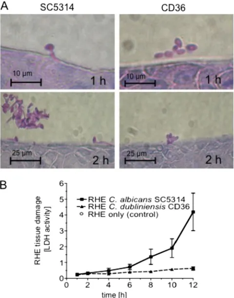
[image:5.585.301.541.69.374.2]Candida albicans andCandida dubliniensisshow major dif-ferences in growth morphology very early during RHE infec-tion.As shown previously,C. albicansis more virulent in the RHE infection model thanC. dubliniensisbecauseC. dublini-ensisis unable to form hyphae in this model (23, 41). These earlier studies focused on RHE colonizationⱖ12 h postinocu-lation (p.i.) when differences in hyphal formation were most apparent. To identify genes involved in the very early stages of this morphological switch inC. albicans, we conducted a time course study of RHE infection to determine the time points at which the differences in growth morphology between the two species first became evident. To this end, RHE tissues were inoculated withC. albicans SC5314 orC. dubliniensisCD36, incubated in 5% CO2 at 37°C, and sampled at regular time points 1 to 12 h postinoculation for tissue histology and to measure RHE tissue damage (41).C. albicanscells had begun to form germ tubes by 1 h p.i., whereasC. dubliniensis cells grew in the yeast phase (Fig. 1A), in which they remained for the whole duration of the experiment (12 h). These results
indicated that major differences in growth morphology be-tweenC. albicans and C. dubliniensisoccurred within 1 h of RHE colonization. RHE tissue damage (measured as LDH activity release into the RHE medium) byC. albicansstarted to increase within 4 to 12 h p.i., while damage of RHE tissues inoculated with C. dubliniensis was identical to that in the uninfected control (Fig. 1B).
C. albicans, but notC. dubliniensis, shows early upregulation of virulence genes on RHE.The above results suggested that significant differences in gene expression underlying the mor-phological differences between the two species may already be present within⬍1 h of RHE colonization. Therefore, we chose 30 min p.i., at which time the vast majority of cells grew as yeast (data not shown) for comparative analysis of global gene pression with whole-genome microarrays. Total RNA was ex-tracted from cells of both species on RHE 30 min p.i. and from the 0-min reference controls (Candidacells from the same cell pools used to inoculate the RHE; see Materials and Methods). Cy-labeled amplified RNAs (aaRNAs) fromC. albicansorC. dubliniensisRHE cultures 30 min p.i. were cohybridized with aaRNAs from the respective 0-min control cultures toC. albi-cans oligonucleotide microarrays (NRC, Canada). In three biological replicates, 2,654 and 1,301 consistently expressed genes were identified inC. albicansandC. dubliniensis, respec-tively.
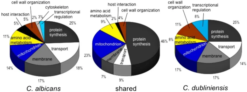
To identify genes highly expressed in both species in the RHE, the following selection criteria were applied: ⱖ2-fold upregulation in RHE 30 min p.i. and statistically significant (P ⬍ 0.05; eBayes t test) difference from 1 (no change in expression). This yielded 268 genes (10.1% of all expressed genes) inC. albicansand 82 (6.3%) genes in C. dubliniensis, with 47 genes upregulated in both species (see Tables S1 and S2 in the supplemental material). The majority of upregulated genes in the two species fell into the categories of protein synthesis, cellular transport, membrane, amino acid metabo-lism, or mitochondrial genes (Fig. 2). The array probes con-tainedC. albicans sequences; so given some sequence diver-gence betweenC. albicansandC. dubliniensis genes (11, 22), detection of fewer expressed genes inC. dubliniensiswas ex-pected. To distinguish between lack of detection due to
se-quence divergence and that due to absence of expression inC. dubliniensis, we estimated the minimum sequence identity be-tween probe and target sequences for detection of expression inC. dubliniensis by using the array probe sequences for all genes (shown in Tables S1 and S2 in the supplemental mate-rial) in BLASTN searches of C. dubliniensis GeneDB (http: //www.genedb.org/genedb/cdubliniensis/blast.jsp). The major-ity of genes (76%) showing consistent expression in C. dubliniensishad probe-target similarities ofⱖ90% alongⱖ68 nucleotides (see Table S2 in the supplemental material), indi-cating that this level of probe-target similarity permitted reli-able detection of expression ofC. dubliniensisgenes with theC. albicansoligoarray.
Ribosomal protein genes were the largest group showing upregulated expression in RHE 30 min p.i.: 83 (30% of all upregulated genes) and 37 (44%) ribosomal protein genes wereⱖ2-fold upregulated inC. albicans and C. dubliniensis, respectively (see Tables S1 and S2 in the supplemental mate-rial). RT-qPCR confirmed the expression for two ribosome-biogenesis genes in both species:RPS7A, whose dynamics in increase of expression were very similar to those of the hy-phally regulatedC. albicansgeneECE1(Fig. 3A) and those of
NOP1(Fig. 3B) in both species. Gene expression profiles were consistent with the respective morphologies of the two species in the RHE. Even at this early stage, C. albicans exhibited upregulation of several hyphally regulated and virulence-asso-ciated genes, includingECE1,HWP1,HYR1,ALS3,IHD1, and
RBT1(see Table S1 in the supplemental material), encoding hypha-specific proteins or cell surface adhesins, commonly up-regulated in human tissue models (32, 46, 52). With the excep-tion ofIHD1, probes for all of these genes had⬍90% similarity toC. dubliniensissequences, and no upregulated expression of any hyphally regulated genes was detectable inC. dubliniensis, with the exception of one gene, NIP7, encoding a hyphally induced ribosomal protein (see Table S2 in the supplemental material). TheC. albicansgene set was also significantly en-riched for genes encoding protein mannosyltransferases: i.e.,
PMT1,PMT2,PMT4, andPMT6were⬎2-fold upregulated in
[image:6.585.80.503.68.227.2]C. albicans, but not inC. dubliniensis(sequence similarity to the PMT gene probes was ⬎90%, except for being 89% for
PMT4[see Table S1 in the supplemental material]). The num-bers of genes showing significant (P ⬍ 0.05) and ⱖ 2-fold-downregulated expression in the RHE were 176 (6.6% of all expressed genes) forC. albicansand 49 (1.9%) forC. dublini-ensis, with 16 genes showing significant downregulation in both species. The majority (⬎50%) of downregulated genes had unknown functions, and several encoded enzymes for in-tra- and extracellular metabolite transport (ABC transport proteins as well as peptide and hexose transporters [see Tables S3 and S4 in the supplemental material]).
Besides interactions with epithelial cells in the RHE tissue, other factors in the RHE environment, including CO2 levels and microaerophilic conditions, could affect morphology and gene expression in theCandidacells. Therefore, to control for these conditions (29), we also grewC. albicansandC. dublini-ensison the polycarbonate filters (PCF) used as support matrix for the RHE. C. albicans—but not C. dubliniensis—formed hyphal elements when incubated on the polycarbonate filters under the same conditions and in the same growth medium as the RHE cultures (not shown). To profile gene expression, cells of eachCandidaspecies were incubated on RHE or PCF for 90 min. aaRNAs from RHE and PCF cultures were cohy-bridized to Eurogentec’sC. albicanscDNA array, and in two biological replicates, consistent expression of 4,613 genes for
C. albicansand 1,368 genes forC. dubliniensiswas detected. In total, 222 (4.8%) genes in C. albicans (see Table S5 in the supplemental material) and 122 (8.9%) genes inC. dubliniensis
(see Table S6 in the supplemental material) showed signifi-cantly (P ⬍ 0.05) and ⱖ1.5-fold upregulated expression on RHE 90 min p.i. relative to PCF 90 min p.i.
There were 78 genes that displayed upregulation on RHE relative to PCF in both species (35% and 64% of all upregu-lated in genes inC. albicansandC. dubliniensis, respectively). Among these were several heat shock protein genes (i.e.,
HSP12, HSP60, HSP70, HSP78, andHSP104 [see Tables S5 and S6 in the supplemental material]), suggesting similarities in stress response in the two species. Also upregulated in both species were genes encoding enzymes for utilization of C2 compounds (i.e., fatty acid-oxidation, glyoxylate cycle, and gluconeogenesis), such asPOX1-3,ICL1, andPCK1, which was confirmed by RT-qPCR analysis (data not shown).
[image:7.585.135.451.67.355.2]C. albicansshows early upregulation of several genes absent or divergent inC. dubliniensis.Following the initial character-ization of the C. albicans and C. dubliniensis gene sets, we focused on uncharacterized genes that may be absent from or divergent in C. dubliniensis (22), as increased expression of these genes inC. albicansin the RHE may provide clues to their possible involvement inCandidavirulence in this model. In this analysis, genes were preselected based uponⱖ1.5-fold upregulation (P⬍0.05) inC. albicansand on expression sig-nificantly different from expression inC. dubliniensis(P⬍0.05; Student’sttest). In the RHE experiment 30 min p.i. versus the 0-min experiment, 146 genes fulfilled these criteria (data not shown), 20 of which had unknown functions and 3 of which
were predicted to be absent or very divergent inC. dubliniensis
(Table 3). One of these genes,HYR1, encoding a hyphal cell wall protein, showed ⱖ2-fold upregulation in C. albicans in RHE. The other two, orf19.3969 (IPF8627) and orf19.7304
(IPF19812), had only predicted or unknown functions, respec-tively. Because of sequence relationships withSFL1described below, we assignedorf19.3969the name “SFL2.”
In the RHE-PCF 90-min p.i. comparison, we identified 24 genes with unknown function that were predicted to be absent from or divergent inC. dubliniensis(10.8% of all upregulated genes inC. albicans) and that hadⱖ1.5-fold upregulation on the RHE relative to PCF inC. albicans(Table 4). Among these genes wasHYR1, also upregulated in RHE 30 min p.i. relative to 0 min (see above), while SFL2was not among the genes showing differential expression in RHE relative to PCF 90 min p.i. (confirmed by RT-qPCR analysis) (Fig. 3), suggesting sim-ilar regulation of this gene under the two conditions.
RT-qPCR confirmed very early upregulation ofSFL2after inoculation onto RHE. C. albicans SFL2 was ⬎3-fold in-creased (P⬍0.05;ttest) 30 min p.i. compared to expression at 0 min and declined 90 to 720 min p.i.:SFL2expression was also upregulated in cells grown on PCF 30 min p.i., but tended to be
lower than expression in RHE 30 min p.i. (Fig. 3). The putative
SFL2 orthologue in C. dubliniensis, Cd36_54430, identified based on both sequence homology and synteny (in theCandida
Genome Database) (2), showed no appreciable increase in ex-pression under any condition, and its exex-pression levels appeared to be more than 1 order of magnitude lower than expression ofC. albicans SFL2. Expression oforf19.7304inC. albicansincreased rapidly within 90 min on both RHE and PCF, with a gradual decline approximately 360 to 720 min p.i. (Fig. 3).
SFL2is required forC. albicansfilamentation in response to multiple environmental signals.Upregulated expression ofSFL2
[image:8.585.39.544.90.391.2](orf19.3969) andorf19.7304in the RHE suggested their possible involvement inC. albicansvirulence. BLAST alignments of the predicted protein sequences forSFL2with its closestC. dublini-ensisorthologue,Cd36_54430, andorf19.7304with its closest or-thologue,Cd36_34500, indicated only 50% identity (58% similar-ity) and 68% identity (78% similarsimilar-ity) between them, respectively. Therefore, we focused onSFL2 and orf19.7304 for functional analysis by targeted gene knockout. Also included in these tests was orf19.4445, as its expression increased in C. albicansupon transfer to the RHE (not shown) and was 3-fold greater than expression on PCF 90 min p.i. (Table 4).
TABLE 3. C. albicansgenes of unknown function or with divergent orthologues inC. dubliniensisshowing significantaandⱖ1.5-fold
upregulation inC. albicansin RHE 30 min p.i.
Gene common nameb
C. albicans
systematic name
C. dubliniensis
closest putative homologue systematic name
% Sequence identity to probe (length of alignment关bp兴)c
Product description
Normalized expression ind
:
C. albicansSC5314 C. dublinienisCD36
Fold change
eBayest
testP
valuee
Fold change
eBayest
testP
valuee
Student’st
testP
valuef
IPF16210 orf19.1782 Cd36_24080 0 Unknown function 4.79 0.0034 ND NA NA
HWP1* orf19.1321 Cd36_43360 46 (64) Hyphal wall protein 4.74 0.0135 ND NA NA
IPF22064 orf19.5136 Cd36_72830 93 (70) Unknown function 3.45 0.0076 ND NA NA
IPF24039 orf19.3266 Cd36_25930 88 (70) Unknown function 3.07 0.0039 ND NA NA
IPF22684 orf19.4590 Cd36_41920 97 (63) Unknown function 2.46 0.0008 ND NA NA
HYR1* orf19.4975 Cd36_51670 0 GPIg-anchored putative
cell wall protein
2.39 0.0041 ND NA NA
IPF22235 orf19.5009 Cd36_12710 89 (69) Unknown function 2.35 0.0050 ND NA NA
IPF24177 orf19.3142 Cd36_46140 91 (70) Unknown function 2.19 0.0084 ND NA NA
IPF17436 orf19.4600.1 Cd36_41810 96 (56) Unknown function 2.18 0.0155 ⫺1.30 0.0551 0.0349 IPF26752 orf19.556 Cd36_30580 96 (69) Unknown function 1.95 0.0011 1.10 0.3595 0.0230
IPF18467 orf19.3394 Cd36_62010 90 (70) Unknown function 1.94 0.0113 ND NA NA
IPF8627* orf19.3969 Cd36_54430 0 Predicted HSF-type DNA-binding protein
1.93 0.0026 ND NA NA
IPF24338 orf19.2995 Cd36_02840 87 (70) Unknown function 1.91 0.0196 ND NA NA
IPF26046 orf19.1287 Cd36_53740 93 (43) Unknown function 1.90 0.0183 ND NA NA
IPF25066 orf19.2259 Cd36_21320 88 (70) Unknown function 1.89 0.0395 ND NA NA
IPF15822 orf19.2457 Cd36_05580 90 (62) Unknown function 1.86 0.0166 ND NA NA
IPF18322 orf19.3141 Cd36_46150 95 (65) Unknown function 1.86 0.0229 ND NA NA
IPF20385 orf19.6748 Cd36_87330 90 (70) Unknown function 1.60 0.0275 ND NA NA
IPF19812* orf19.7304 Cd36_34500 79 (67) Unknown function 1.60 0.0112 ND NA NA
IPF14519 orf19.4703 Cd36_34320 0 Unknown function 1.60 0.0252 ND NA NA
IPF23019 orf19.4241 0 Unknown function 1.59 0.0177 ND NA NA
IPF3432 orf19.6177 Cd36_87690 84 (70) Unknown function 1.57 0.0103 ND NA NA
IPF24748 orf19.2558 Cd36_26640 0 Unknown function 1.54 0.0482 ND NA NA
IPF25503 orf19.1829 Cd36_73360 0 Unknown function 1.53 0.0496 ND NA NA
a
Statistically significant upregulation of expression (eBayesttest;P⬍0.05) inC. albicans.
bⴱ, absent from or divergent in
C. dubliniensis(22).
c
Percent sequence identity of 70-mer probes onC. albicansoligoarray (NRC, Canada) toC. dubliniensisorthologue sequences. Identity was determined by BLASTN. The length of the longest alignment with the probe is given in parentheses where applicable.
d
Genes are ordered from highest to lowest fold changes in expression inC. albicans. ND, not detected; NA, not applicable.
e
Fold change⫽1.
f
Significant difference in fold change betweenC. albicansandC. dubliniensis.
g
SFL2,orf19.7304, andorf19.4445were deleted separately in SC5314 by replacement of each gene with the SAT1 flipper cassette (34). Several heterozygous (⌬) and homozygous (⌬⌬) knockout (ko) strains were generated (Table 1), and their phenotypes were tested in a range of filamentation-inducing conditions. Deletion oforf19.7304 andorf19.4445had no de-tectable effects on cell morphology in 10% fetal calf serum (FCS) and embedded growth in agar (not shown) and were not investigated any further in this study. However, cells of the ⌬sfl2 and ⌬⌬sfl2 ko strains displayed filamentation defects under several conditions. When cells of the⌬⌬sfl2ko strains were precultured at 30°C in YPD and inoculated into 10% FCS and incubated at 37°C, they formed germ tubes at a rate similar to the wild type (WT) (data not shown). However, in the absence of a temperature shift (37°C preculture), germ tube formation in the⌬⌬sfl2strains was reduced by approximately 50% and 7% compared to that in the WT after incubation in 10% serum for 30 min and 45 min, respectively.
SFL2ko mutants were also impaired in hyphal formation in response to a pH shift. Following a shift from pH 4.5 to pH 6.5 in Lee’s liquid medium, after 3 h of incubation, approximately
80% of WT cells had produced germ tubes or true hyphae, whereas⌬sfl2cells exhibited only 10 to 20% germ tube formation, and⌬⌬sfl2cells failed entirely to form hyphae in response to this pH shift (data not shown). Reintroduction of a WTSFL2allele into the⌬⌬sfl2strains restored filamentation to levels similar to those of the heterozygous⌬sfl2strains (data not shown).
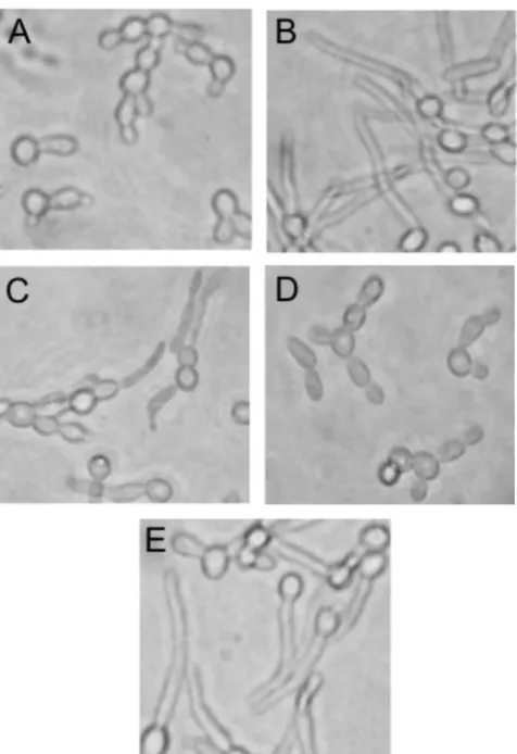
We then tested morphological responses to increased CO2, which induces hyphae inC. albicans(14, 37). To detect CO2 -specific morphogenic responses, we used a YNB medium with 0.1% glucose and supplemented with amino acids (YNB-Gluc-AA [see Materials and Methods]) in which the WT formed germ tubes only in 5% CO2and not in ambient CO2 (Fig. 4). In YNB-Gluc-AA and 5% CO2, the ⌬sfl2 strain
formed fewer and less-elongated germ tubes than the WT, the ⌬⌬sfl2 strain did not form any germ tubes, and the ⌬⌬sfl2
strain with reintroducedSFL2 showed germ tube formation similar to that of the⌬sfl2strain.
[image:9.585.46.540.90.407.2]We next tested the morphological responses of the mutant strains to microaerophilic conditions that induce filamentation inC. albicans (5). Cells were embedded in YPSuc agar and incubated at 25°C or 37°C; identical results were obtained at
TABLE 4. C. albicansgenes of unknown function or with divergent orthologues inC. dubliniensiswith significantaandⱖ1.5-fold upregulation
inC. albicanson RHE relative to expression on PCF 90 min p.i.
Gene common nameb
C. albicans
systematic name
C. dubliniensis
closest putative homologue
systematic name
% Sequence identity to probe (length of alignment关bp兴)c
Product description
Normalized expression ind
:
C. albicans
SC5314 C. dublinienisCD36
Fold change
eBayest
testP
valuee
Fold change
eBayest
testP
valuee
Student’st
testP
valuef
IPF4032 orf19.6168 Cd36_80830 75 (300) Predicted spindle pole body component
3.21 0.0000 ND NA NA
IPF8957 orf19.4894 Cd36_09640 92 (326) Unknown function 3.21 0.0000 1.45 0.0088 0.0298
IPF3092 orf19.4445 Cd36_06760 85 (325) Unknown function 3.03 0.0000 ND NA NA
HYR1* orf19.4975 Cd36_51670 57 (357) GPIg-anchored putative cell
wall protein
2.95 0.0000 ND NA NA
IPF4696 orf19.5282 Cd36_11100 86 (381) Unknown function 2.72 0.0001 ND NA NA
IPF12799 orf19.2515 Cd36_81030 89 (280) Unknown function 2.35 0.0007 ND NA NA
IPF1548 orf19.951 Cd36_50410 72 (251) Unknown function 1.90 0.0042 ND NA NA
POL93* orf19.6078 0 Predicted gypsy-like reverse transcriptase
1.86 0.0091 ND NA NA
IPF7182 orf19.3439 Cd36_61560 86 (268) Unknown function 1.78 0.0082 ND NA NA
IPF15784 orf19.715 Cd36_32010 83 (342) Unknown function 1.70 0.0025 ND NA NA
IPF13890* orf19.4918 Cd36_44430 0 Unknown function 1.69 0.0015 ND NA NA
IPF7456 orf19.2047 Cd36_15710 88 (322) Unknown function 1.67 0.0046 ND NA NA
IPF12629 orf19.5552 Cd36_63350 92 (366) Unknown function 1.67 0.0057 ND NA NA IPF13017 orf19.1785 Cd36_24050 71 (259) Unknown function 1.65 0.0021 ND NA NA IPF18468 orf19.4467 Cd36_03660 93 (327) Unknown function 1.61 0.0173 ND NA NA
IPF3468* orf19.4055 0 Unknown function 1.60 0.0027 ND NA NA
IPF4704 orf19.5278 Cd36_11140 88 (281) Unknown function 1.60 0.0038 ND NA NA
IPF10005 orf19.6196 Cd36_06620 84 (306) Unknown function 1.59 0.024 ND NA NA
IPF11796 orf19.2791 Cd36_07020 90 (329) Unknown function 1.57 0.0047 ND NA NA IPF22331 orf19.5026 Cd36_12870 88 (328) Unknown function 1.57 0.0034 ND NA NA IPF13231* orf19.3877 Cd36_31750 74 (302) Predicted LPF family protein 1.56 0.0152 ND NA NA IPF11858 orf19.1277 Cd36_45480 87 (273) Unknown function 1.54 0.0041 ND NA NA IPF12435* orf19.5619 Cd36_63710 75 (290) Predicted LPF family protein 1.52 0.0100 ND NA NA IPF19617 orf19.1350 Cd36_22460 75 (279) Unknown function 1.51 0.0105 ND NA NA
aStatistically significant upregulation of expression (eBayesttest;P⬍0.05) inC. albicans. bⴱ, absent from or divergent inC. dubliniensis(22).
cPercent sequence identity of cDNA probes onC. albicansgene arrays (Eurogentec) toC. dubliniensisorthologue sequences. Identity was determined by BLASTN.
The length of the longest alignment with the probe is given in parentheses where applicable.
dGenes are ordered from highest to lowest fold changes in expression inC. albicans. ND, not detected; NA, not applicable. eFold change⫽1.
both temperatures, and only results for embedded growth at 37°C are shown. The WT strain formed long elongated fila-ments (Fig. 5A and B). In contrast, only 20% of⌬sfl2colonies produced some filaments, and all⌬⌬sfl2colonies were com-pletely smooth and formed only short chains of yeast cells or pseudohyphae. Reintroduction of the SFL2 wild-type allele into the ⌬⌬sfl2 strain restored hyphal formation to a level similar to that of the⌬sfl2strain.
When incubated on Pal’s agar at 30°C for⬍72 h,C. albicans
grows as smooth colonies; however, when grown at 37°C for ⬎72 h, it forms smooth colonies with a hyphal fringe (D.J.S., unpublished data). Following growth for 96 h at 37°C on Pal’s agar, wild-type SC5314 formed colonies with a hyphal fringe, as expected, whereas⌬⌬sfl2 colonies lacked a hyphal fringe (Fig. 5C). On Spider agar, the wild-type strain formed wrinkled colonies with a hyphal fringe (data not shown), while the⌬sfl2
strain exhibited wrinkled colonies without a fringe and the ⌬⌬sfl2strain grew as fringeless, completely smooth colonies. The⌬⌬sfl2strain complemented with a wild-type copy ofSFL2
formed wrinkled colonies on Spider agar that were similar to those of the⌬sfl2strain (data not shown).
Deletion ofSFL2had no detectable effect on growth under aerobic conditions in standard media: the doubling time of the ⌬⌬sfl2strain during mid-log phase (60 to 330 min) in YPD at 37°C was 60.6⫾2.1 min (median⫾standard error [SE];n⫽2) and essentially identical to that of the parental WT strain (60.8⫾ 2.7 min). Growth of⌬⌬sfl2cells in response to heat, salt, or oxidative stress was similar to that of the WT (data not shown). Overexpression of SFL2 from a doxycycline-inducible pro-moter in C. albicans resulted in the formation of filaments in liquid YPD at 30°C, while the control (expressing GFP from the same promoter) grew only as yeast (Fig. 6). Moreover, when the
SFL2-overexpressing strain CEC1352 was grown on solid YPD or YPD plus 1% FCS at 37°C, colonies appeared wrinkled, whereas the colonies of the GFP-overexpressing strain remained smooth. Expression of CaSFL2inC. dubliniensisCD36 or Wu¨284 under the control of the doxycycline-inducible promoter and grown in YPD and doxycycline resulted in elongated cells and
pseudohy-FIG. 4. Growth ofC. albicans SFL2knockout and complemented strains under high-CO2conditions. Shown are cells of wild-type strain
SC5314 in ambient (0.038%) CO2(A) and 5% CO2(B) and cells of the
[image:10.585.45.283.67.414.2]⌬sfl2 strain (CaMS46-2) (C), ⌬⌬sfl2 strain (CaMS49-1) (D), and ⌬⌬sfl2strain with reintegratedSFL2(CaMS58) (E) in modified YNB medium after 3 h of growth in 5% CO2at 37°C.
[image:10.585.137.448.530.677.2]pha-like elements, whereas the GFP-expressing strains grew solely as yeast cells (data not shown).
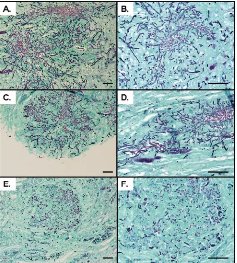
SFL2is required for RHE tissue colonization and damage by C. albicans, but not for virulence in the mouse model of systemic infection.To see ifSFL2 deletion affected coloniza-tion and tissue damage to the RHE, theSFL2ko strains were tested in this model. Compared to the WT, which exhibited extensive filamentation in the RHE, the⌬sfl2 strain showed only few hyphal elements, and cells of the⌬⌬sfl2strains were completely impaired in hyphal morphogenesis in the RHE; ⌬⌬sfl2 strains with reintegrated SFL2 showed filamentation levels similar to those of the⌬sfl2strain (Fig. 7). RHE damage mirrored the morphological phenotypes: i.e., sequential dele-tion of eachSFL2 allele resulted in a gradual reduction of tissue damage: LDH release caused by⌬sfl2 strains was ap-proximately 40% of that of the WT, and LDH release by ⌬⌬sfl2strains was about 50% of LDH release by⌬sfl2strains and significantly lower than that of the WT (P⬍0.01; analysis of variance [ANOVA] and Tukey’s test), with RHE damage similar to that in the uninfected control. RHE damage by ⌬⌬sfl2strains with reintegratedSFL2was similar to that of the ⌬sfl2strains.
[image:11.585.137.451.67.236.2]Finally, we tested⌬⌬sfl2strains inin vivomouse models of systemic infection, using the conventional 28-day model and a 3-day infection model. In these experiments, virulence of the ⌬⌬sfl2strain CaMS49-1 was very similar to that in the WT: survival times of mice inoculated with⌬⌬sfl2or WT (SC5314) cells were 16.7⫾3.9 and 15.3⫾2.9 days (mean⫾SE;n⫽6), respectively (P⬎0.05; Kaplan-Meier and log rank statistics), with no significant differences (P⬎0.05; ANOVA) detected forCandidaburdens in the kidney, spleen, or brain (data not shown). To examine whether changes in organ burdens were evident at an earlier time point during infection, mice were infected and then sampled at 3 days postinfection. Again, there was no significant difference inCandida burdens for the kid-ney, lung, liver, spleen, and brain (data not shown). Interest-ingly, histology of kidney sections obtained from the 3- and 28-day infection models revealed that the⌬⌬sfl2strain was defective in hypha formation, growing almost exclusively as yeast cells
FIG. 6. Overexpression ofSFL2inC. albicans. Strains CEC1352 (Table 2; indicated byTETp-SFL2), overexpressingSFL2from a doxycycline-inducible promoter, and CEC1147 (Table 2; indicated byTETp-GFP), expressing GFP from a doxycycline-inducible promoter, were grown for 18 h in liquid YPD at 30°C or for 5 days on solid YPD or YPD plus 1% FCS at 37°C in the presence or absence of doxycycline (50g/ml).
[image:11.585.302.541.290.622.2]with only a few short filaments (Fig. 8). Consistent with thein vitro phenotypes, ⌬sfl2 strain-infected renal lesions showed an intermediate phenotype, with some lesions containing long hyphae and others containing short filaments or yeasts (Fig. 8).
SFL2 encodes a putative DNA-binding heat shock factor protein. BLASTP searches of the NCBI nonredundant (nr) protein database with the inferred Sfl2p sequence (714 amino acids) gave a significant match with Hsr1p (E⫽1e⫺51, 64% identity; alignment of 129 amino acids at the Sfl2p protein N terminus), described as a heat-shock-related transcription factor (HSF) in Candida tropicalis (CAC12663), and WU-BLAST2 searches of Saccharomyces cerevisiae YeastDB (at http://seq .yeastgenome.org) gave significant (E⬍1e⫺18) matches to sev-eral HSF-type proteins, including Sfl1p (19 to 62% identity) and Mga1p (25 to 28% identity). A highly conserved HSF-type DNA-binding domain (pfam00447; E ⫽ 6e⫺16) and an HSF-type DNA-binding domain signature (PS00434; at amino acids 57 to 81) were detected in the N-terminal region of Sfl2p, also present at the same location (amino acids 58 to 82) in its putativeC. dubliniensisorthologue, Cd36_54430p. Sfl2p shared about 24% identity and the HSF-type signature with its closest relative inC. albicans, Sfl1p (orf19.454), which is a negative regulator of hyphal morphogenesis (3, 17).
DISCUSSION
The opportunistic pathogensC. albicansandC. dubliniensis
have a close phylogenetic relationship and share several
mor-phophysiological traits. Despite this, they display large differ-ences in virulence, withC. albicansbeing much more patho-genic in the human host and in human infection models than
C. dubliniensis. Here, to identify novel virulence-associated genes inC. albicansand begin to characterize commonalities and differences in gene expression between the two species, we compared global gene expression inC. albicans and C. dub-liniensisin the RHE model of the oral mucosa. Both species exhibited coordinated upregulation of primary metabolism genes in RHE 30 min postinoculation, indicating conserved responses in general metabolism in the two species. However, whereas C. albicans showed rapid filamentation and caused increased RHE tissue damage, as well as upregulated expres-sion of several known hyphally regulated genes,C. dubliniensis
grew only as yeast cells, caused very limited RHE damage, and lacked detectable upregulation of hyphal genes. C. albicans
also displayed upregulation of several genes of unknown func-tion that were absent or significantly divergent inC. dublini-ensis. One such gene, SFL2 (orf19.3969), showed significant upregulation inC. albicanson the RHE 30 min p.i. and high (⬃50%) sequence divergence with its likely orthologue inC. dubliniensis (Cd36_54430). When SFL2 was deleted in C. albicans, cells were unable to form hyphae under a variety of conditions, including growth under microaerophilic or high-CO2 conditions, in response to pH shifts, and in mouse kid-neys. Overexpression ofSFL2resulted in increased filamenta-tion in C. albicans and the production of pseudohypha-like cells inC. dubliniensis. Deletion ofSFL2inC. albicanshad no effect on mouse survival in the systemic infection model; how-ever,SFL2 deletion led to decreased RHE colonization and damage, demonstrating a possible role forSFL2in virulence in this oral mucosal infection model.
The greater virulence of C. albicansand the limited viru-lence of C. dubliniensiswere consistent with earlier observa-tions (12, 41) and with lower carriage and prevalence of C. dubliniensisin the oral cavities of healthy individuals (42, 43).
C. albicansand C. dubliniensisdisplayed broadly similar pat-terns of coordinately upregulated expression of ribosomal and other primary metabolism (e.g., protein, amino acid biosynthe-sis, and mitochondrial) genes (Fig. 2; see Tables S1 and S2 in the supplemental material), indicating high levels of metabolic activity in both species in the RHE. So given their differences in virulence in the RHE, it was surprising that growth initia-tion—as revealed by the temporal dynamics and magnitude of expression of the primary metabolism genes—appeared to be equally rapid inC. albicansandC. dubliniensis. Moreover, both species exhibited very similar upregulation in the RHE of C2 utilization genes, such as genes for fatty acid -oxidation, glyoxylate cycle, and gluconeogenesis, as well as heat shock protein genes (see Tables S5 and S6 in the supplemental ma-terial). This pattern of gene expression in mammalian tissues has previously been described only forC. albicans(46, 52), and upregulation of C2utilization and stress response genes also in
[image:12.585.44.285.69.337.2]C. dubliniensis suggests common pathways for physiological adaptation to the RHE environment in the two species. Thus, we hypothesize that only a small set of physiological cues in the RHE, triggering filamentation inC. albicanswhile failing to do so inC. dubliniensis, may be responsible for the difference in virulence. Filamentation of C. albicans cells growing on the polycarbonate filters used as RHE support matrix indicated