Published Online September 2011 (http://www.SciRP.org/journal/ojrad)
Positron Emission Tomography-Computed Tomography
(PET-CT) in Head and Neck Pathology
1
Luis A. Tamara, 2Ines Velez
1
Veterans Administration Medical Center, Houston, Texas, USA 2
College of Dental Medicine, Nova Southeastern University, Florida, USA E-mail: [email protected]; [email protected]
Received August 20, 2011; revised September 18; accepted September 27, 2011
Abstract
Cancer of the head and neck is often devastating and the morbidity associated with its treatment is substantial. Positron Emission Tomography-Computed Tomography (PET-CT) combines the power of biological / mo-lecular imaging with the anatomic detail of CT in order to provide a very sensitive and specific imaging tool for the evaluation of head and neck pathology. PET can aid the clinician in establishing diagnosis, staging, (It has been shown to be more accurate than CT), assessing, prognosis and determining response to therapy. Lymphoma, melanoma, multiple myeloma, leukemia, salivary gland tumors, odontogenic carcinomas, soft tissue sarcomas, thyroid, parathyroid, lacrimal gland and bone / cartilage tumors are some of the entities where PET-CT may be useful. (Tumors of salivary glands and of odontogenic tissue are particularly difficult to diagnose due to the relative infrequency when compared with other tumors and the extremely vast his-tologic variation). It is important to note that carcinoma metastasis, is the most common malignancy found within the mandibular bone. PET-CT and skeletal scintigraphy are both very sensitive and specific in these types of patients.
Keywords:PET-CT, Positron Emission Tomography, Head and Neck Cancer.
1. Introduction
The National Cancer Institute estimates than more than 11 million Americans with history of cancer are alive. 1.500.000 new cancers are expected to be diag-nosed each year, as well as more than 1.500 cancer deaths per day [1]. More than one person per minute dies of cancer in the US. Malignancy accounts for 1 of every 4 deaths in America. These statistics did not include bas-al cell and squamous cell carcinoma of the skin [1].
The complex anatomy and histology of the head and neck makes diagnosis and treatment of the area very challenging. Cancer of the head and neck is often devas-tating and the morbidity associated with its treatment substantial. In an effort to achieve the best possible re-sults, specialists caring for head and neck patients should be well versed in all the available diagnostic and thera-peutic modalities. Positron Emission Tomography- Com- puted tomography (PET-CT) is a highly sensitive and specific tool that can aid the clinician in establishing di-agnosis, staging, assessing prognosis and determining re- sponse to therapy.
By far, the most common ca ncer in head and neck, excluding basal cell carcinoma, is squamous cell carcinoma (SCC). It is the 9th most common type of cancer. Within the oral cavity, SCC has 50% five-year death rate [1]
2. Patho-Biology of Cancer .
formation of malignant growths. The mutations accumu-late, and give rise to malignant tumors which are com-posed of heterogeneous population of cells, with differ-ent genetic aberrations. The strongest tumoral cell is the source of a large portion of the tumor and will make it radio-resistant and / or chemo-resistant.
Malignant cells produce innumerable chemical prod-ucts that, forming an abnormal molecular signaling net-work, are able to cause important changes in the tissue. The lethality of a tumor is related to these molecules. The chemical communication between cells is responsi-ble for the characteristics of each type of cancer and for the complex and difficult process of metastasis. There is multilateral molecular signaling between tumoral and non-tumoral cells and the extra-cellular matrix. Among many other cancer products, there are growth factors, transcription factors, signaling molecules, neuro-endo- crine substances, mitogenetic proteins, proteolytic en-zymes, and angiogenetic products [2].
This ability of multi-chemical production is the hallmark of malignancy and therefore diagnosis and treatment may be based on the metabolic function of the neoplasm.
The potential to metastasize is one of the most impor-tant characteristics of malignancy. Basically, the cell involved in the metastatic process, experiences a long journey characterized by numerous obstacles. Malignant cells are separated from the primary tumor; they adhere to a blood or lymphatic vessel wall, invade through the wall between the endothelial cells and travel along the blood stream defending themselves against the immune system’s components. At the end of the journey, they transport viably to a distant organ and start reproducing in the new environment and forming another tumor. An-giogenesis is induced, so there is enough nutritional sup-ply to sustain expanding growth. The whole process re-quires numerous molecular cell products in order to be able to degrade physiologic barriers, stimulate replication, avoid immune surveillance, activate angiogenesis, etc.
Collections of distant manifestations that result from the substances produced by the tumor or in reaction to the tumor are called para-neoplastic syndromes. These manifestations may present in any organ or system such as endocrine, neuromuscular, musculoskeletal, cardio-vascular, cutaneous, hematological, gastrointestinal, re-productive, renal and respiratory and may be the first manifestation of the tumor. 75% of cancer patients ex-perience a para-neoplastic disorder. (Figure 1)
[image:2.595.314.533.84.264.2]Medical surveillance is of primary importance for can- cer patients not only because of the metastatic potential
Figure 1. Para-neoplastic pemphigus in a patient with non- Hodgkin lymphoma.
but also because it is known that a patient with history of cancer is at higher risk (14%) than the general population, for developing multiple primary cancers. These are new tumors that are biologically distinct from the original primary malignancy. The risk of developing subsequent cancers depends on genetic susceptibility, immunologic condition, exposures such as chemicals and microorgan-isms, and carcinogenic effects of cancer treatments.
Medical surveillance is of paramount importance to cancer patients, due to the possibility of metas-tases and the higher risk for developing subsequent primary malignancies. PET / CT is one of the best method for surveillance as the whole body can be visualized in a single test.
The main biochemical characteristic of a malignancy is the aerobic glycolysis (the Warburg effect). Hypoxic tumor cells are highly metabolic and glucose avid, as it becomes the primary source of ATP production [3]. This is one of the basic properties of the tissue that makes PET / CT (F18-FDG) useful in cancer diagnosis and staging.
to differentiate between normal cells and abnormal cells such as malignant or inflammatory. (Figure 2)
3. Squamous Cell Carcinoma.
Squamous cell carcinoma SCC is a malignant prolifera-tion of squamous epithelial cells. This malignancy is g- enerated within the basal cell layers of the epithelium. Anatomically, squamous epithelium covers the external surface of the body and lines internal cavities. This tissue is also lining some of the structures that communicate internal organs with the exterior [4]. SCC comprises about 95% of oral cancers; it is the most common oro-pharyngeal / naso-pharyngeal malignancy (Figure3), and it is the second most common cancer of the skin. (Figure4)
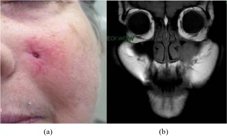
Patients with tumors of paranasal sinuses usually pre-sent at advanced stage and therefore, prognosis is extre- mely poor. (Figure5) PET-CT is the most sensitive way to detect these tumors and its metastases.
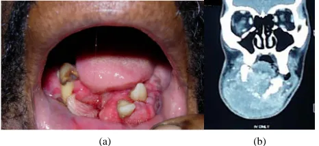
[image:3.595.308.539.93.281.2]There are, centrally in mandible and maxilla, remnants of odontogenic epithelium called rests of Malassez. These odontogenic rests, as well as the squamous epithelium that lines numerous types of cysts, can suffer squamous metaplasia and later give origin to squamous cell carci-noma. (Figure6)
[image:3.595.308.538.315.453.2]Figure 2. Glucose continues the oxidation pathway within the cells. FDG undergoes no further oxidation.
Figure 3. Nasopharyngeal carcinoma.
Figure 4. Squamous cell carcinoma of the skin.
(a) (b)
Figure 5. (a) Squamous cell carcinoma of left maxillary si- nus- invading through the skin; (b) Same patient. MRI Left maxillary sinus mass.
(a) (b)
Figure 6 (a) Intra-osseous squamous cell carcinoma ori- ginated in Malazess rests; (b) Intra-osseous squamous cell carcinoma originated in an odontogenic cyst
SCC may appear in the conjunctiva and the nasolac-rimal duct, where it is diagnosed early because obstruc-tion symptoms and early metastasis to the nasal cavity.
[image:3.595.58.286.408.535.2] [image:3.595.308.539.503.606.2] [image:3.595.58.286.572.702.2]host are responsible for the development of this type of tumor. There are multiple genetic defects, such as those found in syndromes, which give the host the susceptibil-ity for malignancies. Numerous immune system condi-tions, from immunodeficiency to autoimmune disease, also play a role in the development of squamous cell carcinoma. Chemicals such as tobacco and alcohol, mi-croorganisms particularly HPV 16-18-32-33-35 and ra-diation have been identified as being associated with oral cancer. The type of carcinogen, the frequency of expo-sure, the dose and the length of time it interacted with the tissue, relate to the development of this condition. This is usually a disease of the elderly [4].
[image:4.595.308.541.85.192.2]Oral squamous cell carcinoma can have extreme vari-able clinical manifestations depending on several factors such as location, differentiation and time of evolution. The clinical appearance may vary from a flat superficial lesion (Figure 7) to a huge ulcerated destructive and invasive mass. (Figure8) SCC may appear white or red, be flat or nodular, be crater-form or verrucous, (Figure9a-b) exo-phytic or endoexo-phytic, be large or small, ulcerated or not, fast or slow-growing, be indurated or not, painful or painless. Weight loss is a common finding and according to the location, nasal obstruction, epistaxis and dysphagia are seen. Nasopharyngeal carcinoma is a type of undiffer-entiated squamous cell carcinoma with extremely poor prognosis. Imaging manifestations of SCC are variable, most of the time a diffuse destructive process. (Figure 10)
Figure 7. Early squamous cell carcinoma. Innocuous-looking lesion.
(a) (b)
Figure 8 (a) Huge ulcerated destructive and invasive s- quamous cell carcinoma; (b) Same case. Destructive and invasive mass.
(a) (b)
[image:4.595.309.539.243.354.2]Figure 9 (a) Squamous cell carcinoma. Fast growing ve- rrucous mass; (b) Verrucous squamous cell carcinoma as-sociated to an invasive tumor.
Figure 10. 3D image. Diffuse resorption of bone.
[image:4.595.308.536.403.601.2] [image:4.595.61.283.431.693.2]immunosuppres-sive therapy, radiation and spread of virus, especially HPV are some of the risks factors for multiple primary squamous cell carcinomas of the head and neck [5].
Proliferative verrucous leukoplakia is a field canceri-zation condition, of unknown etiology, which has the unique quality of relentless transformation through dif-ferent stages of hyperkeratosis, verrucous hyperplasia, verrucous carcinoma and eventually invasive carcinoma. These lesions tend to develop all over the oral mucosa. (Figure12)
The malignant tumors’ ability of multi-chemical pro-duction and signaling between the tissues is responsible for most of the cancer symptoms, such as weight loss, anemia, failure of the immune system, metastasis and death. Some patients may also die from complications of the surgery, chemo or radiotherapy. It is estimated that about one patient per hour die from oral squamous cell carcinoma in the United States.
Furthermore, every known type of cancer has the possi-bility of being found on head and neck [2]. Aside from squamous cell carcinoma, lymphoma, melanoma of skin and mucosa, multiple myeloma, leukemia, salivary gland tumors, odontogenic carcinomas, soft tissue sarcomas, thy-roid, parathythy-roid, lacrimal gland and bone / cartilage tumors are some of the entities where PET-CT may be useful [7-9]. It is important to note that carcinoma metastasis, from thy-roid, breast, lung, prostate, uterus or kidney, is the most
(a) (b)
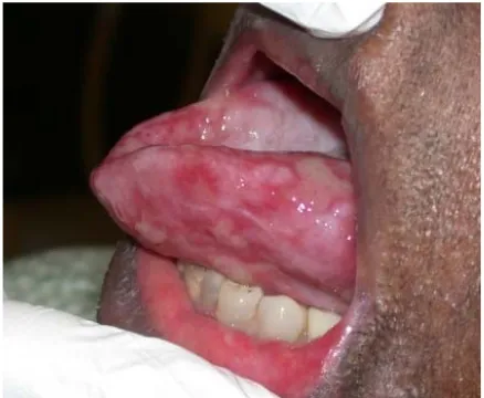
Figure 11. (a) Squamous cell carcinoma of the right la- teral border of the tongue; (b) Left lateral tongue. This lesion appeared simultaneously with the lesion on the right side.
(a) (b)
Figure 12. (a) White slightly elevated lesion. Histologic di-agnosis: hyperkeratosis; (b) Same patient. Two years later. Histologic diagnosis: verrucous carcinoma.
common malignancy found within the mandibular bone. PET-CT is also very sensitive and specific in these types of patients. (Figure 13 and Figure 14) .
It has been found that 75.000 newly diagnosed cases and 21,000 deaths from lymphoma occur every year. Extra -nodal Non Hodgkin lymphomas are present in head and neck more frequently than in any other site. The mucosa, at the junction of the hard and soft palate, is the most common place, followed by the Waldeyer’s ring. These lymphomas are almost always B-cell lymphomas and are part of the so called MALTOMAS (mucosal as-sociated lymphoid tissue tumors). MALTOMAS may also arise, among other areas, in stomach, lung, parotid and lacrimal glands.
(a) (b)
[image:5.595.308.539.266.367.2]Figure 13. (a) Hodgkin lymphoma. Several enlarged lymph nodes cervical chain; (b) MALT lymphoma extension fro- m palate.
Figure 14. Panoramic radiography showing bone destruc-tion by lymphoma.
Lymphoma, melanoma of skin and mucosa, multi-ple myeloma, salivary gland tumors, odontogenic carcinomas, soft tissue sarcomas, thyroid, para-thyroid, lacrimal gland and bone / cartilage tu-mors are some of the entities where PET-CT may be useful. It is important to note that carcinoma metastasis, is the most common malignancy found within the mandibular bone. PET-CT is both very sensitive and specific in these types of patients.
[image:5.595.309.537.415.528.2] [image:5.595.57.288.423.540.2] [image:5.595.55.288.586.682.2]e -ti it ti -f si d li m uscle,
sm oth muscle, nerve fibers, bone and cartilage, vessels, bl
prognosis. They ar
xistent immune diseases, with head and neck manifesta ons, such as Sjogren syndrome and Hashimoto thyroid-is. On the other hand, Hodgkin lymphoma which is ini-ally a nodal disease, frequently exhibits the first mani estations in cervical lymphatic chains.
Sarcoma is a general term applied to malignancies ari-ng from mesenchymal tissue. Due to the increasiari-ng un-erstanding of human cells and tissue, some tumors be-eved to be “sarcomas” are not originated in
mesenchy-al tissue. Fibrous and adipose tissue, skeletmesenchy-al m o
ood, hemopoietic and lymphatic tissues, are present in the head and neck and may give origin to sarcomas.
Melanoma, one of the most aggressive cancers, has the ability to metastasize into any organ. Near 70.000 cutane ous melanoma cases are diagnosed per year. Mucosal me- lanomas metastasize more frequently and faster than skin melanomas and have an extremely poor
e not rare in the upper oral mucosa and in the eye.
The head and neck area contains specialized tissues that are not present in other parts of the body such as thyroid, parathyroid, salivary glands and odon-togenic tissue. Tumors of salivary glands and of odontogenic tissue are particularly difficult to di-agnose due to the relative infrequency when com-pared with other tumors and the extremely vast histologic variation. PET-CT could be of great value in these cases.
The progress in diagnosis and early staging of cancer, as well as the improvement on treatment, reflects an in-creased surviva d s o w o H t
q female patients1. Skin and m
l rate. As the survivor population increases ue to the evolving nature of the scientific field, medical urveillance is of paramount importance and PET-CT is ne of the best tools for this purpose.
Among the most common primary sites of patients ith multiple cancers are head and neck areas, especially ro-pharynx with squamous cell carcinoma, and Non-
o- dgkin lymphoma. Oro-pharyngeal cancer exhibits he largest Estimated Absolute Risk (EAR) for
subse-uent cancers in male and
ucosal melanoma and leukemia are primary malignant entities associated with moderate risk of multiple cancers. Secondary / multiple malignancies are effectively found with PET-CT SCAN.
PET-CT is of paramount importance in clinical staging, and has been shown to be more accurate than CT. It makes a significant difference identi-fying malignant normal size nodes.
Initial staging is indispensable for the choice of ther-apy and the assessment of the prognosis, and it is based on the size of the primary tumor and the metastasis. There are several staging systems. The most known the TNM system which evaluates T: the size of the primary tumor, N: absence or presence of regional lymph node metastasis and M: distant metastasis. PET CT is of pa- ram
s c
f
s field. Schwartz
et
ount importance in clinical staging, and has been hown to be more accurate than CT. It makes a signifi-ant difference identifying malignsignifi-ant normal size nodes.
Accurate staging is indispensable to define the type o urgery and the area of the radiotherapy
al. reported sensitivity of 96% and specificity of 98.5% for detection of nodal metastasis during the initial staging of SCC with FDG-PET / CT.
Cancer of unknown primary constitutes one of the most difficult cases for the clinician. PET / CT should be performed as the initial test in cases of unknown primary and the biopsies should be di-rected according to PET / CT results.
One of the main advantages of FDG-PET / CT during the initial staging of head and neck cancer is its capacity to identify distant disease, metastatic or synchronous, in any part of the body, all in the same test [10]. FDG-PET / CT is also of paramount importance for treatment plan-ning. FDG uptake is a prognostic indicator. Tumors with h
c u
p r
p
urgery remains the best therapy for squamous cell ca
neck (2% of head and neck cancer diagnosis). [11] The igh FDG uptake have poorer prognosis and higher re-urrence rate as compared with tumors with low FDG ptake. One advantage is that PET-CT data can be im-orted into the radiation treatment planning computer fo recise radiation dose and volume.
S
rcinoma. Sometimes combination with radiotherapy and chemotherapy is indicated. Removal of the entire lesion with its draining lymphatics and lymph node chains, including the tissues contained in the same area of the neck, (neck dissections) are needed in many cases of head and neck carcinoma. Sometimes the dissection includes the tail of the parotid, the sternocleidomastoid muscle, the spinal accessory nerve and the mandibular branch of the facial nerve. These surgeries leave variable, often severe, levels of functional compromise and de-formities for life. It has been suggested by Kovacs et al [10] that patients with positive lymph nodes in FDG-PET / CT, should undergo neck dissection and the sentinel lymph node biopsy technique should be performed in patients with negative FDG-PET / CT.
alig-apy more accu-most common sites for the primary neoplasm are the tonsillar area and the base of tongue. The treatment, which causes significant morbidity, consists of wide- field radiation that covers pharynx, larynx and bilateral neck. If the primary is localized, risks of complications decrease considerably. FDG-PET / CT is useful in iden-tification of the primary site. Wong et al published that in 27% of cases of unknown primary carcinoma, after nega-tive CT, MRI and endoscopy, the primary tumor was lo-calized with FDG-PET / CT. This fact has a significant impact regarding the morbidity and mortality. Menda and Graham [12] (2005) stated that PET / CT should be per-formed as the initial test in cases of unknown primary and the biopsies should be directed according to PET / CT results. Additional metastases are also discovered by PET-CT which may change the treatment and / or the ra-diation field [13]. On the other hand, the most common intraosseous malignancy in the mandibular bones is me-tastasis of carcinoma specially from lung, uterus, kidney, thyroid and prostate. These malignancies may be identi-fied by PET-CT.
Patients with unresectable cancers are initially treated with chemo-radiation. Cumulative data show that PET / CT has resulted in improved evaluation of response to therapy both in the curative and neoadjuvant settings with relatively high sensitivity, specificity, positive and negative predictive values. Interpretative pitfalls can be found in the presence of inflammatory response, tumor stunning and adjunct therapies. When considering a change in management strategies, histologic confirmation of PET / CT findings may be appropriate in selected cases [13].
4
.In summary, why PET / CT in the
evalua-tion of head and neck cancer?
Within the last decade, PET-CT has exploded into the c linical arena as a powerful imaging tool for the evalua-tion of many malignancies. Head and neck malignancies are no exception and PET-CT consistently performs bet-ter than the standard anatomic imaging techniques CT and MRI.
The reasons for this superior performance are multiple and can be outlined as follows:
1) PET-CT can localize primary head and neck m nancies that have not been identified by clinical / endo- scopic exam or other imaging techniques. (Figure15) 2) PET-CT can assess response to ther
rately than CT or MRI. It is not as affected by ana-tomical distortion from prior surgery or radiation therapy. It relies on biological tumoral activity rather than size changes. (Figure16)
gure 15. SCCA UNC Chapel Hill Department of nuclear edicine. SCCA of tongue. PET-CT can identify metastatic Fi
m
[image:7.595.341.507.85.152.2]lymph node involvement in normal size nodes.
Figure 16. SCCA UNC Chapel Hill Department of nuclear medicine. PET allows more accurate staging. If this patient had been staged with CT alone, he would have been under staging.
Figure 17. SCCA UNC Chapel Hill Department of nuclear medicine.
In this regard PET-CT consistently outperforms dedicated CT imaging. (Figure17)
4) PET-CT combines the power of biological / molecular imaging with the anatomic detail of CT in order to provide a very sensitive and specific imaging tool for the evaluation of head and neck malignancies.
3) PET-CT can be used in radiation therapy planning for precise target delineation of the biological tumor vo-lume.
PET-CT combines the power of biological / mo-lecular imaging with the anatomic detail of CT in order to provide a very sensitive and specific im-aging tool for the evaluation of head and neck ma-lignancies. The use of PET-CT in head and neck cancer may make a significant difference in the treatment and prognosis of many patients.
5. References
[image:7.595.346.501.212.293.2] [image:7.595.347.502.349.428.2][2]
gulatable tissues express a unique insulin-sensitive glu-cose transport protein”. Nature 333: 183-185, 1988. Atlanta: American Cancer Society, 2009.
D.E. James, R. Brown, J. Navarro, P.R. Pilch. “Insulin re-
doi:10.1038/333183a0
3] D.E. James, M. Strube , M. Mueckler. “Molecular clon-ing and characterization of an insulin regulatable glucos transporter”. Nature 338: 83-87, 1989.
[
e
doi:10.1038/338083a0
4] M.H. Ross, E.J. Reith. “Epithelium” in: Histology. New York, Ed Harper & Row: 1985
[
] C.B. Begg. “Methodological and Statistical Considera-tudy of Multiple Primary Cancers”. In: adows AT, Robimson E, Multiple Primary cancers. Philadelphia, PA: Lippincott Williams and Wi
gnant epithelial
Mc-ositron Emission tomograph
.7.626 [5
tions in the S Neugut AI, Me
l-kins; 1999:13-26
[6] R. Marx, D. Stern. “Premalignant and mali
tumors of mucosa and skin”. In: Oral and Maxillofacial Pathology. Quintaesscence, First edition, 2003; 283 -373.
[7] F. R. Miller, D. Hussey, M. Beeram, T. Eng, H.S.
Guff, A.O.Randall. “P y in
the management of unknown primary head and neck car-cinoma”. Archives of Otolaryngology Head and neck Surgery.131:626-629. 2005.
doi:10.1001/archotol.131
aging of head and neck [8] H. Schoder, H.W.D. Yeung, M. Gonen, D. Krauss, S.M.
Larson: “Positron Emission im
cancer, including thyroid carcinoma.” Seminars in Nu-clear Medicine. 34:180-197. 2004.
doi:10.1053/j.semnuclmed.2004.03.004
[9] A.F. Kovacs, N. Dobert, J.Gaa.: “Positron emission to-mography in combination with sentinel lymph node bi-opsy reduces the rate of elec
treatment of oral and oropharyn
tive neck dissections in the geal cancer”. Journal of Clinical Oncology 22:3973-3980, 2004.
doi:10.1200/JCO.2004.01.124
[10] D.L. Schwartz, E. Ford, J. Rajendran, B. Yueh, M. D. Coltrera, F. Virgin, et al “FDG-PE
preradiotherapy staging of head and nec
T / CT imaging for k squamous cell carcinoma”. International Journal of Radiation Oncology Biology and Physics 61:129-136, 2005.
doi:10.1016/j.ijrobp.2004.03.040
[11] W.J. Wong, M. Saunders: “The impact of FDG PET on the management of occult primary head a
Clinical Oncology 15:461-466. 2
nd neck tumors”. 003.
doi:10.1016/j.clon.2003.07.006
[12] Y. Menda, M. Graham. “Update on FDG / PET and Posi-tron emission tomography / computed tomography imag-ing of squamous head and neck cancers”. Seminars in Nuclear Medicine 35:214-219. 2005.
doi:10.1053/j.semnuclmed.2005.05.001
[13] L. Kostakoglu. “FDG-PET Evaluation of response to treatment”. PET Clinics. 3: 37-75. 2008.
doi:10.1016/j.cpet.2008.09.001
[14] G.W. Goerres, D.T. Schimdt, F. Bandhauer. “Positron emission tomography in the early follow up of advanced head and neck cancer”. Archives of Otolaryngology Head and Neck Surgery 130: 105-109. 2004.