J. Exp. Biol. (1967), 47. 165-178 165 With 5 text-figures
Printed in Great Britain
CONTROL OF THE CIRCADIAN RHYTHM OF ACTIVITY
IN THE COCKROACH
II. THE ROLE OF THE SUB-OESOPHAGEAL GANGLION AND VENTRAL NERVE CORD
BY JOHN BRADY*
Zoological Laboratory, Downing Street, Cambridge
{Received 17 March 1967)
INTRODUCTION
Only a very few physiological clocks have been located accurately in any organisms; two of them in invertebrates are well documented. One of these is the large neuron called the ' parabolic burster' in the parieto-visceral ganglion of Aplysia (Strumwasser, 1965, etc.) and is electrical; its function is unknown. The other is the neurosecretory clock found in the sub-oesophageal ganglion of Periplaneta (Harker, 1956, etc.) and is hormonal; it functions in the control of the circadian rhythm of locomotor activity. The existence of this sub-oesophageal ganglion clock and various other aspects of Harker's work have been challenged by Roberts (1966).
An investigation was undertaken to re-examine some of Harker's more important results in the light of Roberts's criticisms. The part of this work concerning the implication of the neurosecretory cells of the brain and corpora cardiaca in the control of cockroach activity rhythms has been reported earlier (Brady, 1967ft). The present paper concerns experiments on the sub-oesophageal ganglion clock itself and a search for other clocks in the ventral nerve cord. A synthesis of the conflicting results of Harker, Roberts and the author is attempted.
MATERIAL AND METHODS (a) Culture conditions and activity recording
The Periplaneta americana used in this study came from the Zoological Society of London and were reared on crushed whole oats and apple in a large culture tank at approximately 280 C. For the early implantation experiments this tank was kept under natural daylight, but for the majority of the time was under an artificial light cycle of L D i 2 : i 2 (i.e. light 12 hr.:darkness 12 hr.) with the LD transition occurring at 12.00 G.M.T. Where relevant the environmental phase-relationships are indicated in the text. Adult males were used exclusively in the experiments.
All observations were carried out in the light-controlled and temperature-controlled incubators of Brown & Unwin (1961) and the activity of intact animals recorded in either photocell box actographs (Brown & Unwin, 1961) or running-wheel actographs
166 JOHN BRADY
(Roberts, i960) (see Brady, 19676). Headless or severely operated animals were fre-quently too inactive to provide assessable records in either of these actographs so various modifications were used. The first of these was the one used by Harker for all her later observations (Harker, personal communication) and simply consisted of reducing the floor area of a photocell box with a rough cylinder of perforated zinc; an alternative was the use of a narrow Perspex box whose dimensions were very similar. By these modifications the normal floor area of about 7x13 cm. was cut down to about 3x13 cm.—the width of the infra-red light beam being about 2-5 cm.
(a) No. A. 13- S-P-B, in DD, days 0 to 5
(6) No. C.3: S-P-B, in DD, days - 3 to 0 to +3
(c) No. A.36: S-P-B, in DD, days 1 to 5
(d) No. A.70: H-W, in DD, days 1 to 4
(c) No. A.73: T-B, in DD. days 1 to 6
(f) No. A.74: T-B, in DD, days 1 to 4
•I'-.TH '.'."J:ufl •.Jh'^^'^V'''J'.,'V'^'/V:'"'.''T£"i"ri1 *,.•," 17'.'^V/'.;'J'7'"T^";'.'"'.'."'jjyi-'.'.'j''' '.'• • i ' . i ' ' ^ i ' ' I ' i T i ^ y i S c :
Fig. 1. Activity records of headless Periplaneta implanted with sub-oesophageal ganglia from rhythmic donors, (a) Record of the donor to (b) (C.3). Donor cockroaches maintained in LD 12:12 with the LD transition occurring at thick vertical line, recipient cockroaches maintained in LD with the transition occurring 5-10 hr. later. All records taken in DD after implantation on day o. S-P-B, small photocell box; H-W, headless-wheel; T-B, twitch-box. Each hori-zontal line represents 24 hr. activity, successive days are displayed in order beneath each other. (6) Beheaded at O and implanted at V ( = day o).
Circadian rhythm in cockroach. II 167
animal twitched, a spool was fitted to the spindle and arranged to wind up a cotton thread; the total number of revolutions could thus be checked against the number of marks on the record. This wheel provided satisfactory records of many operated animals (e.g. Fig. 4 c) and of some headless ones, but in others tended to be rather faint (e.g. Fig. 1 d). For convenience in the text these three modified actographs are called, respectively: 'small photocell box', 'twitch-box' and 'headless-wheel'.(b) Operational techniques
Sub-oesophageal ganglia for implantation were dissected out of non-anaesthetized donors pinned down on their backs. The ganglia were kept carefully free of contamina-tion from the salivary duct, and were handled only by their connectives to the pro-thoracic ganglion. They were implanted through V-shaped slots cut in the abdominal tergites of the recipient cockroaches, which had previously been beheaded (after tightly ligaturing the neck to prevent contamination of the haemocoel by gut contents). This procedure is identical with that employed by Harker (personal communication), but modifications were also tried as described in the Results. Implants of other ganglia were carried out similarly. Records were taken of the activity of implanted animals in constant darkness (DD) for at least 3 days (more usually 4-6 days) after the operation. The implantations were always carried out in the light phase of both host and donor. Hosts were previously kept under natural daylight, and the donors under artificial LD 12:12 with the LD transition occurring at 1200 G.M.T. The rhythms of the two sets of animals were thus between about 5 and 10 hr. out of phase with each other (except in two cases where they were both in phase).
Ventral nerve-cord connectives were cut with fine tungsten hooks: the circum-oesophageal pair through a hole to the frons, and those to the pro-thoracic ganglion through the ventral neck membranes. It was always possible to see the ends of the nerves draw apart when they were completely cut through, so only sample post-mortem examinations were made after these operations. Operational wounds were not sealed with wax since clotting of the blood appeared to do the job better.
Neurosecretory cells were destroyed in situ by means of a micro-cautery using a radio-frequency oscillator (designed by J. A. Popple). This could be set to burn out areas less than 100 fi in diameter. The neurosecretory cells in the sub-oesophageal ganglion are relatively large and are usually clearly visible as whitish dots in the living ganglion (Brady, 1967a). They were cauterized through slits cut in the articulation membranes of the labium, the animal being anaesthetized with CO2 and held down
on its back in plasticine with a rubber ligature across its neck (Brady, 19676). After recording from such cauterized animals in DD they were sacrificed and their sub-oesophageal ganglia were examined histologically. Tissue was fixed in aqueous Bouin and sections were cut at IO/J; the paraldehyde-fuchsin (PF) staining schedule of Cameron & Steele (1959) was followed with minor modifications. Neurosecretory cells are here defined as being only those cells which contain much PF-positive material; for a full description of these cells in the cockroach sub-oesophageal ganglion see
168 JOHN BRADY
RESULTS
Implantation of sub-otsophageal ganglia
Forty-eight headless cockroaches were implanted with sub-oesophageal ganglia and their activity recorded in DD. Only ten of the donors of these implants were tested previously, all were rhythmic (in LD). The other 23 donors (15 single and 4 double implants) were not tested individually but were selected from four different batches of cockroaches from the main stock tank. Samples of three of these four batches, when tested by running-wheel in LD, gave the following proportions rhythmic (denominator = no. tested): 10/10, 12/14, 34/34; a sample from the fourth, tested in a photocell box, gave 4/5. The probability that the donors were rhythmic is therefore
Table 1. Summary of sub-oesophageal ganglion (SOG) implantation experiments
on headless Periplaneta americana
Form of implant One SOG
Two synchronous SOG
SOG plus corpora cardiaca SOG with nerves
left long Totals Acto-graph type* S-P-B H-W T-B I S-P-B \H-W fS-P-B \H-W fS-P-B \T-B No. of animals implanted 7 4 3 z 2 i 1 8 1 29
Post-implantation records: nos. falling into different classesf Apparently rhythmic Doubtfully rhythmic Diffusely rhythmic Apparently arrhythmic 1 o o o I o o o o I o o o o o o 3t 5 z 3 2 1 o 1 4 1 19
• S-P-B, small photocell box; H-W, headless-wheel; T-B, twitch-box. t For description of classes of record see text.
X Two of these with donors in phase with previous rhythm of host.
high. A rather lower rate of apparent rhythmicity was generally observed when animals were tested in photocell boxes, but the reaction of cockroaches to the two types of actograph is very different (Brady, 19676) and it is felt that the wheel must provide the truer picture since it gives the higher rate, and can scarcely create rhythmicity where none exists inherently.
In order to be sure that the numbers of apparently arrhythmic implanted animals were not biased, 19 records were discarded from the 48 trials for a variety of reasons, namely: record so faint or so intense that a rhythm might have been obscured; host survived for less than 3 days; a possibility that a recorder might not be functioning perfectly. The results of the remaining 29 implants with assessable records are sum-marized in Table 1.
Orcadian rhythm in cockroach. II 169
It must be remarked that animal C. 3 (Fig. 1 b) was the very first implant performed by the author, and the clarity of the apparent rhythm is unique in this material. There is, however, no other known reason to mistrust this record. The four cases of' doubtful' rhythms showed a major burst of activity on the day after the operation at about the same time as the donor's peak, but this was not repeated on subsequent days. The four ' diffusely' rhythmic animals had records indicating that the majority of their activity had been confined to rather extensive periods of the day, but that these periods were roughly coincident for 2 or 3 days (e.g. Fig. 1 c). The remaining 19 implanted animals gave no indication of any induced rhythm.
As will be seen from Table 1, implantations were performed in a variety of forms and three different types of actograph used to record the activity. Four attempts were made to increase the titre of sub-oesophageal ganglion hormone by implanting pairs of ganglia from donors selected from synchronized batches of cockroaches. Two further attempts were made to do this by implanting the donor's corpora cardiaca together with the sub-oesophageal ganglion (not nervously connected); this was done with the work of Harker (1960 c) in mind. The number of successes, however, remained very low.
It seemed possible that the failures might be caused by the implanted ganglia being mishandled in some way which impaired their ability to release the hormone. Maddrell (1966) has shown that in Rhodnius neurosecretion may be released through the walls of some abdominal nerves. It is possible that a similar mechanism may exist in Periplaneta for the release of neurosecretory material from the sub-oesophageal ganglion. Nine attempts were made to take account of this by dissecting out the ganglia with all their nerves, including the connectives, kept as long as possible. This was done by pulling these free from the donor with forceps, rather than by cutting them short with scissors. In five out of the nine special care was taken to keep intact the two pairs of very small nerves which leave the posterior part of the ganglion (Brady, 1967 a); a section of salivary gland had to be included to ensure that this had been done. No improvement emerged, however; four of the nine showed very faint signs of a rhythm but the hosts of two of these were in phase with the donor anyway.
Cautery of neurosecretory cells in the sub-oesophageal ganglion
Headless cockroaches are generally very inactive and it seemed possible that records of implantation experiments might be improved if the host animals were less severely damaged. Harker (1960 c, 1964) reported that probably only four neurosecretory cells in the sub-oesophageal ganglion are involved in the production of the secretion controlling rhythmic activity. She has identified these cells as being the two lateral pairs (Brady, 1967 a). An attempt was therefore made to render potential host cock-roaches arrhythmic by using a micro-cautery to destroy only these relevant cells, leaving the rest of the head intact. The surprising results of this experiment are summarized in Table 2, from which it will be seen that at least six animals remained clearly rhythmic in DD after the successful removal of both pairs of lateral neuro-secretory cells (Fig. 2).
170 J O H N BRADY
rhythmic. The remaining 7 were possibly rhythmic but were not clearly so. The autopsies carried out on 9 of the 11 clearly rhythmic animals indicated that in most cases a few neurosecretory cells remained apparently intact. It should be emphasized, however, that although it is possible to say when a neurosecretory cell has been
Table 2. Post-mortem examinations on the sub-oesophageal ganglia of Periplaneta americana remaining rhythmic after the cauterization of the neurosecretory cells in this
ganglion S t a t e of N^ ^ ^ after cautery^
Animal no. A.41 AA.35 AA.36 AA.27t
AA.3i§
AA 32§ A-42§ A43 §
AA 39
Days between operation and
sacrifice
8
24
4 4 8 8 7 9 7
Type of actograph*
P-B R-W R-W R - W R-W R-W P-B P-B R-W
Anterior Lateral Large Small Large Small
Ventro-anterior Medial
Two further animals (B. 43 and AA. 66) were clearly rhythmic but died before they could be autopsied, their ganglia had been extensively cauterized. Seven more animals were cauterized but were not autopsied because their activity records were not unequivocally rhythmic.
The bilaterally arranged pairs of neurosecretory (N.S.) cells have been described elsewhere (Brady, 1967a).
• P-B, Photocell box; R-W, running-wheel.
t , Both cells of a pair destroyed; + + , both cells remain apparently intact; — 1 - , one member of pair destroyed.
§ Activity record illustrated in Fig. 2.
% Activity recorded in LD.
(a) No. AA 31: R-W, in LD days —3 to 0, in DD days 0 to 7
— v
-(b) No AA.32: R-W, in DD, days 0 to 6
(c) No A 42: P-B, in DD, days 1 to 6
(d) No. A.43. P-B, in DD. days 1 to 8
Circadian rhythm in cockroach. II 171
destroyed, because it is absent from its normal site, it is not possible to say when a cell is functionally intact. A cell which appears intact on histological grounds may in fact not be functioning physiologically, and thus more cells may have been effectively destroyed than the data in Table 2 suggest.
The medial cells, being situated near the centre of the ganglion, proved particularly difficult to cauterize without destroying much of the ganglion. However, it appears unlikely that they are involved in the control of the rhythm since they are sometimes absent from the ganglia of intact specimens (Brady, 1967a) and were in any case removed from at least three animals which remained rhythmic after cautery. If the medial cells are excluded from consideration it may be said that at least four animals were found to be rhythmic in DD after the removal of all the apparently potential sources of rhythmic neurosecretion from their sub-oesophageal ganglia. Finally, in one case certainly (AA.39), and possibly two others (B.43, AA.66), a very clear rhythm was retained in the absence of any PF-positive cells. The fact that two animals were rendered arrhythmic is probably of little significance in view of the effect of damage to the ventral nerve cord reported below.
(a) No. A . 8 5 : S-P-B, in DD, days 0 to 4
(b) No. A . 8 7 : S-P-B, in DD, days 0 to 5
Fig. 3. Activity records of headless Periplaneta implanted with abdominal ganglia plus attached neurohaemal organs from rhythmic donors. Details as in Fig. 1.
Implantation of abdominal ganglia
It seems possible that the sub-oesophageal ganglion may not be the only hormonal clock in the cockroach. This is suggested (a) by the fact that all the ganglia of the ventral nerve cord have pairs of neurosecretory cells which appear histologically identical with those in the sub-oesophageal ganglion implicated by Harker, and (b) by the results of cauterizing the sub-oesophageal ganglion cells. The abdominal ganglia appeared ideal candidates for implantation in the search for further clocks. They have recently been shown to exist in association with neurohaemal organs (Brady & Maddrell, 1967) and so the problem of interfering with the neurosecretory release mechanism might be avoided. Moreover, Harker only tested tissues from the head (1956).
172 JOHN BRADY
Cutting the nerve-cord connectives
The only procedure so far discovered which makes cockroaches apparently arrhyth-mic is beheading (Harker, 1956; Roberts, 1966). This suggests that essential features of the clock mechanism may be present in the head. Both Harker and Roberts consider that the key factor in this cephalic control of the rhythm is the secretion of a hormone. If this is so, cutting the ventral nerve cord in the neck might be expected not to affect it. Such an operation should leave the thoracic ganglia physiologically in the same state as if the animal were beheaded, and should leave the neurosecretory cells in the head in much the same state as if implanted. The only important difference from headless animals might be that the nervous connections between the brain and sub-oesophageal ganglion were retained, and that some unknown inhibitory system might be function-ing between the two.
(a) No. B.30. T-B, in DD, days 1 to 6 1 dV>~flIvv7- iji'-tfiw' '"''
(b) No. AA.40: T-B, in DD, days 1 to 4
• • • • • '
-i -i a I I I U I III»IIIII«II -i -i -in 11 ft.1.-1
(c) No. A.52: H-W, in DD, days 0 to 4
(d) No AA.42: T-B, in DD, days 0 to 4 L'Jlf':
Fig. 4. Activity records of Periplaneta in DD after severance of their ventral nerve-cord connectives; (a, b) with the post-sub-oeaophageal ganglion connectives cut, (c, d) with the circum-oesophageal commissures cut. Other details as in Fig. i.
The nerve cord was cut between the sub-oesophageal and pro-thoracic ganglia in eleven cockroaches whose post-operational activity was measured in DD. The survival times of these animals were of the same order as beheaded animals, i.e. from about 3 days to a week or more. Seven of them yielded records of more than 2 days duration, excluding both the day of operation and the day before death or moribundity. Five of these seven had from 4 to 9 days such record, and three of these five were still alive when removed from the actograph. The intensity of activity was of a similar low level to that observed after beheading. One animal appeared to show a free-running rhythm for the first 3 post-operative days but was subsequently arrhythmic for a further 3 days until sacrificed for autopsy (its connectives were found to have been properly cut). None of the other six animals showed any sign of a rhythm (Fig. \a, b). That this was not merely the result of trauma from the operation is apparent from the fact that animals remain rhythmic after cautery of the suboesophageal ganglion, which involves a more extensive operation at the same site.
Orcadian rhythm in cockroach. II 173
bisection), ending in death from 3 to 6 days later. This very intense activity, although possibly obscuring an underlying periodicity, did not express itself in any overtly rhythmic pattern (Fig. 4 c, d). Again, that this result was not only a simple response to trauma is indicated by brain cautery experiments (Brady, 19676), which involve a much more extensive operation at the same site, frequently without loss of rhythm. It seems likely that this hyperactivity results from the removal of some inhibitory system between the brain and sub-oesophageal ganglion.Since one of these 11 animals (7 + 4) appeared to show a rhythm for a few days in the absence of a nervous link between the head and the rest of the body, it cannot be concluded that the expression of the rhythm was abolished by this operation. How-ever, if rhythmicity is controlled hormonally from the head, and if headless animals do respond readily to implants of sub-oesophageal ganglia (Harker, 1956, etc.) one would not expect such a high proportion of animals with severed nerve cords to appear arrhythmic. It is clear that this operation seriously interferes with the expression of the rhythm, even though the hormonal system is left virtually intact. It seems reasonable to assume that the ventral nerve cord is an important link in the normal chain of command which controls the rhythm. The fact that one animal appeared to retain its rhythm does not, of course, give any indication as to where the source of this control may have been situated.
DISCUSSION
Implantation of sub-oesophageal ganglia
Only 29 of the 48 headless implanted animals have been accepted for assessment of their post-operative activity. Two out of the 29 gave records which suggested an induced rhythm, but only 1 of these had an intense record (Fig. 1 b). A rhythm as clear as that observed in this latter animal would probably have been detected, if present, in about another 10 of the 19 discarded records, so that the proportion of 'successes' is perhaps nearer 2 out of 40 than 2 out of 29.
The interpretation of the further 8 indefinitely rhythmic records is much less clear. The duration of peak activity is normally about 2 hr. If a cockroach produces random bursts of activity of about this length on average once every 24 hr. for the first day or two after being beheaded, the probability of such a burst coinciding with the donor's activity peak is fairly high—a chance of at least 1 in 12 in fact. Moreover, if there is more than one burst of activity per day, or the activity lasts longer than 2 hr. (both of which frequently occur, see Fig. 1), the probability of coincidence is obviously increased. The four ' doubtfully' rhythmic hosts may well only have been showing the result of this sort of behaviour. It is also conceivable that the two ' successes' above were similarly the result of chance, but the probability of such coincidences occurring at random on two successive days are considerably lower, though not necessarily lower than 2 out of 40. The four ' diffusely' rhythmic animals appeared rhythmic because they had activity patterns which coincided for 2 or 3 days. However, this activity extended over periods of many hours each day so that it is almost impossible to deduce whether they indicate a rhythm inherent in the host, a rhythm induced by the implant, or again chance coincidence.
174 JOHN BRADY
40). Roberts reports no successes in 19 similar trials, but a close examination of his six published records suggests that some rhythms may have been obscured by the method of presenting the results. If his \ hr. data are aggregated into 3 hr. groups, in one case (Roberts, 1966, fig. 3, bottom right) it appears that there was a major burst of activity between 9 p.m. and midnight on day 1, followed by a similar burst between 6 and 9 p.m. on day 2. The presentation of data beyond about day 3 is largely irrelevant since Harker (1956) states that the induced rhythm usually disappears during the second and third day. Roberts's records are shown up to day 8, which distracts the eye from the significant part at the beginning. But his implants were in any case in-phase with their recipients so the possibly obscured successes may not be very significant. Harker has done many hundreds of these experiments and she states that the majority gave records as clear as that shown in Fig. 1 b of the present paper (Harker, personal communication). It seems likely that the rarity of Roberts's and my own successes must stem from unknown errors in our technique, even though several of my own implants were closely supervised by Dr Harker herself. One is nevertheless left with the doubt that the few apparent successes were perhaps only the results of chance, or arose from the headless animal responding to unexcluded environmental cues from the daily rhythm of the laboratory. This latter possibility could not be checked by the usual method of observing whether the rhythm went into a free-run, because the number of post-operational days was too few.
Other clocks
A tendency has arisen to speak of the sub-oesophageal ganglion neurosecretory cells rather as if they were the only hormonal clock in the cockroach concerned with the control of the activity rhythm (e.g. Harker, 1964, pp. 63-4). This presumably arose from the fact that headless cockroaches appear to be arrhythmic until implanted with sub-oesophageal ganglia. However, it has now been shown that the intact cock-roach can remain rhythmic in the absence of any PF-positive cells in the sub-oeso-phageal ganglion. The explanation of Harker's observations on these cells must lie in the fact that she identified them by cauterizing implanted ganglia (Harker, personal communication). Clearly in the intact animal another mechanism takes over when these cells are removed, so that they cannot be the only source of control of the activity rhythm. This raises the interesting question of whether other rhythmic centres exist outside the head. If they do, then the effect of implanting sub-oesophageal ganglia may merely reflect the state of these centres in the host and bear little relation to any inherent rhythmicity in the implant. Unfortunately, the experiments on implanting abdominal ganglia and their neurohaemal organs proved negative, so that as yet no evidence has emerged to implicate other structures.
Orcadian rhythm in cockroach. II 175
takes at least two activity cycles for completion, whereas a lesser shift should be com-pleted within a single cycle.
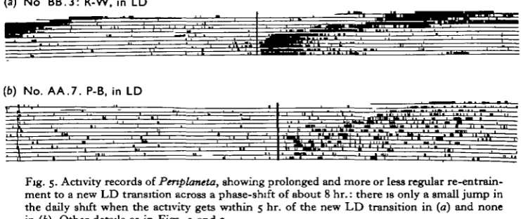
It is difficult to fit this hypothesis to the records of re-entrainment observed by the author. No specific experiments were carried out, but re-entrainment to new environ-mental cycles involving earlier LD transition times was observed in several Periplaneta, with the activity recorded in both photocell boxes and running-wheels. Six animals gave very clear records of gradual re-entrainment, and a further six, although having rather noisy or indistinct initial records, showed the last 2 or 3 days of phase-shifting fairly clearly. In seven cases the total shift was more than 5 hr.; in one of these (Fig. 5 a), and possibly two others, there was some slight sign of an acceleration in phase-shifting when the activity peak came close to about 5 hr. from the new LD transition. In no case out of the total of twelve animals, however, did the animal reach the final steady-state at anywhere near this point. Indeed, rather the reverse tended to occur with the daily shift growing steadily less the closer the new environmental signal
(a) No BB.3: R-W, in LD
(b) No. AA.7. P-B, in LD
•l M l l L ' l ' l - l l f i
[image:11.451.38.409.258.414.2]re.
Fig. 5. Activity records of Periplaneta, showing prolonged and more or less regular re-entrain-ment to a new LD transition across a phase-shift of about 8 hr.: there is only a small jump in the daily shift when the activity gets within 5 hr. of the new LD transition in (a) and none in (hi). Other details as in Figs. 1 and 2.
was approached (Fig. 5 a). Roberts has illustrated similar records from another cock-roach, Leucophaea (1962, 1965), and the effect is also observed in mammals (deCoursey, i960), in birds (Enright, 1965) and in fish (Davis, i960), so that it appears to be a quite general characteristic of circadian locomotor rhythms.
176 JOHN BRADY
observed gradual shifting. Before it can cover such results the model clearly requires some modification.
It seems possible that the jumps in phase-shifting, which Harker describes, were the result of the sort of phenomenon that Fig. 5 b was selected to show. In this record the underlying phase-shifting appears to continue at the previous rate, even though a burst of activity nearer the LD transition occurs a few days before the steady-state of re-entrainment is reached. Such apparent jumps also occur when the new LD transi-tion occurs at a later time than before. In these cases the presence of light during the animal's normal peak activity time presumably inhibits activity until the lights go off. A similar result can occur in other animals, for example Aschoff (1965) has illustrated a case in the chaffinch.
A tentative synthesis
On purely a priori grounds the brain would appear to be the most likely organ in which a master-clock mechanism should reside. The brain's function in insects is mainly sensory and integrative, and almost entirely non-motor (Huber, 1965); it lies strategically between the photo-receptors in the head (Roberts, 1965) and the loco-motor effectors in the thorax. At the moment, the simplest synthesis of the work of the author, Roberts and Harker seems to suggest a higher clock mechanism in the brain controlling lower, segmental clock mechanisms in the ventral nerve cord. The higher centre would be electrical, possibly of the type described by Strumwasser (1965), and the lower (as yet observed clearly only by Harker) neuro-endocrine. The higher centre would be supposed to be directly entrained by changes in the time of the LD transition, and the lower in their turn by the higher centre. If the output of the higher centre is purely electrical, as the effect of cutting the nerve cord suggests, it might be expected that Roberts' pars intercerebralis operations (1966) broke bilateral integration within this centre, or stopped the impulses from it (Brady, 19676). In order to accom-modate Harker's sub-oesophageal ganglion observations in this hypothesis it is neces-sary to postulate: (a) that cutting the nerve cord stops the lower centres or puts them sufficiently out of phase with each other to obscure all signs of overt rhythm; (b) that an excised sub-oesophageal ganglion is not so stopped, or that when implanted the effect of its secretion is greater than the combined effect of the out-of-phase secretions of all the other centres in the headless host; (c) that, notwithstanding (b), the failure of Roberts and myself to repeat Harker's experiments was due to the independence of the lower centre in the sub-oesophageal ganglion being rather ephemeral and easily upset. There is the alternative, simpler possibility that no centres of rhythmicity exist outside the brain and that the control of the rhythm is not hormonal at any level. That this may be so is suggested by several points:
(a) As Roberts has cogently argued (1966), the results of Harker's parabiosis experi-ments (1956) cannot be accepted as unequivocal proof of a hormonal mechanism.
(b) Sub-oesophageal ganglion implants apparently only work under very specific conditions, as yet uncertainly repeatable and quite unknown.
(c) Histological examination has revealed little or no evidence of a circadian secretory cycle in the neurosecretory cells of the sub-oesophageal ganglion (Brady, 1967 a).
Orcadian rhythm in cockroach. II 177
(e) If there are other autonomously rhythmic hormonal centres, it is not clear why cutting the ventral nerve cord should make the majority of animals arrhythmic (only
1 out of 11 appeared to show a rhythm after this operation).
(/) The other nerve cord ganglia contain neurosecretory cells which appear histo-logically identical with those implicated in the sub-oesophageal ganglion, and their neurohaemal mechanism is probably known (Brady & Maddrell, 1967), but implanta-tion of these produces negative results.
(g) Neither the neurosecretory cells of the brain nor, possibly, the corpora cardiaca (Brady, 19676) appear to be essential features for the rhythmic expression of activity. Quite apart from the great body of Harker's experimental work, however, other considerations suggest that at least at some level the control of rhythms is likely to be hormonal, whether autonomously rhythmic or not. As Harker has pointed out (1958), it seems probable that all or many of the cells in an organism are fundamentally rhythmic, and there is now some suggestive histological evidence that this is so (see Brady, 1967a). By far the simplest way to co-ordinate such a catholicity of rhythms would appear to be hormonal. The system proposed above would be well adapted to do this and to provide a link between such internal rhythms and the periodicity of the environment. At what level the control of locomotor rhythms would fit into this system still seems open to further investigation, however.
SUMMARY
1. Since 1955 Harker's work on the control of cockroach activity rhythms by a hormonal clock in the sub-oesophageal ganglion has stood largely unchallenged, but recently Roberts (1966) has questioned several of her claims; in particular he finds it impossible to transfer rhythms by implanting this ganglion.
2. Sub-oesophageal ganglia from rhythmic donors were implanted into 29 headless Periplaneta americana in a variety of ways and three different actographs used. In 2 cases the hosts showed bursts of activity for 2 or 3 days after implantation, roughly coincident with the donor's previous rhythm; a further 8 implanted animals showed rather uncertain signs of an induced rhythm, but the remaining 19 were all apparently arrhythmic.
3. Cauterization of the neurosecretory cells of the sub-oesophageal ganglion in situ showed that cockroaches can remain rhythmic in the absence of the cells described by Harker (1960c), and probably in the absence of all cells in this ganglion which are stained by paraldehyde-fuchsin.
4. Implantation of abdominal ganglia plus their respective neurohaemal organs (Brady & Maddrell, 1967) did not elicit rhythms in eight headless hosts.
5. Cutting the circum-oesophageal commissures, or post-sub-oesophageal ganglion connectives, like beheading, appears to interfere seriously with the rhythmic expression of activity; one animal remained apparently rhythmic after this operation, however.
6. Harker's proposal for a second clock (19606) requires that phase-shifts of less than 5 hr. shall be completed within a single activity cycle; this did not occur in the twelve cases observed, phase-shifting being gradual and taking several days before reaching the entrained steady-state.
7. It is suggested that most of the divergent results of Harker, Roberts and Brady
178 JOHN BRADY
could be plausibly correlated if cockroaches have an electrical pace-maker in the brain co-ordinating rather ephemeral neuro-endocrine rhythms in the nerve-cord ganglia.
This research was supported by the Air Force Office of Scientific Research under grant AF EOAR 65-19 through the European Office of Aerospace Research (OAR), United States Air Force. I am most grateful to Dr Janet Harker for much advice and encouragement throughout the conduct of the research and for critically reviewing the manuscript. My thanks are also due to Dr M. G. M. Pryor, who read the manuscript and made helpful comments.
REFERENCES
ASCHOFF, J. (1965). Response curves in circadian periodicity. In Circadum Clocks (ed. J. Aschoff) pp. 95—in. Amsterdam: North-Holland Publishing Co.
BRADY, J. (1967a). Histological observations on circadian changes in the neurosecretory cells of cock-roach sub-oesophageal ganglia. J. Insect Pkytiol. 13, 201-13.
BRADY, J. (10676). Control of the circadian rhythm of activity in the cockroach. I. The role of the corpora cardiaca, brain and stress. J. exp. Biol. 47, 153-63.
BRADY, J. & MADDHELL, S. H. P. (1967). Neurohaemal organs in the medial nervous system of insects.
Z. Zellforsch. 76, 389-404.
BROWN, R. H. J. & UNWIN, D. M. (1961). An activity recording system using infra-red detection.
J. Insect Pkysiol. 7, 203-9.
CAMERON, M. L. & STEELE, J. E. (1959). Simplified aldehyde-fuchsin staining of neurosecretory cells.
Stain Tecknol. 34, 265-6.
DE COURSEY, P. J. (i960). Phase control of activity in a rodent. Cold Spring Harb. Symp. quant. Biol. 35,
49-55-DAVIS, R. (i960). In H. O. Schwassmann: Environmental cues in the orientation rhythm of fish. Cold
Spring Harb. Symp. quant. Biol. 35, 443-50.
ENRIGHT, J. T. (1965). Synchronization and ranges of entrainment. In Orcadian Clocks (ed. J. Aschoff), pp. 112—24. Amsterdam: North-Holland Publishing Co.
HARKER, J. E. (1956). Factors controlling the diurnal rhythm of activity of Periplaneta americana L.
J. exp. Biol. 33, 224-34.
HARKER, J. E. (1958). Diurnal rhythms in the animal kingdom. Biol. Rev. 33, 1-52.
HARKER, J. E. (1960a). The effect of perturbations in the environmental cycle of the diurnal rhythm of activity of Periplaneta americana L. J. exp. Biol. 37, 154-63.
HARKER, J. E. (19606). Internal factors controlling the sub-oesophageal ganglion neurosecretory cycle
in Periplaneta americana L . J. exp. Biol. 37, 164—70.
HARKER, J. E. (1960c). Endocrine and nervous factors in insect circadian rhythms. Cold Sprmg Harb.
Symp. quant. Biol. 35, 279-87.
HARKER, J. E. (1964). The Physiology of Diurnal Rhythms. Cambridge University Press.
HUBER, F. (1965). Neural integration (central nervous system). In The Physiology of Insecta (ed. M. Rockstein), vol. 11, pp. 333-406. New York: Academic Press.
MADDREIX, S. H. P. (1966). The site of release of the diuretic hormone in Rhodmus—a new neurohaemal system in insects. J. exp. Biol. 45, 499-508.
ROBERTS, S. K. DE F. (i960). Circadian activity rhythms in cockroaches. I. The free-running rhythm in steady-state. .7. cell. camp. Physiol. 55, 99-110.
ROBERTS, S. K. DE F. (1962). Circadian activity rhythms in cockroaches. II. Entrainment and phase shifting. J. cell. comp. Physiol. 59, 175-86.
ROBERTS, S. K. DE F. (1965). Photoreception and entrainment of cockroach activity rhythms. Science,
N. Y. 148,958-9.
ROBERTS, S. K. DE F. (1966). Circadian activity rhythms in cockroaches. III. The role of endocrine and neural factors. J. cell. Physiol. 67, 473-86.