
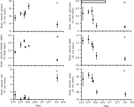
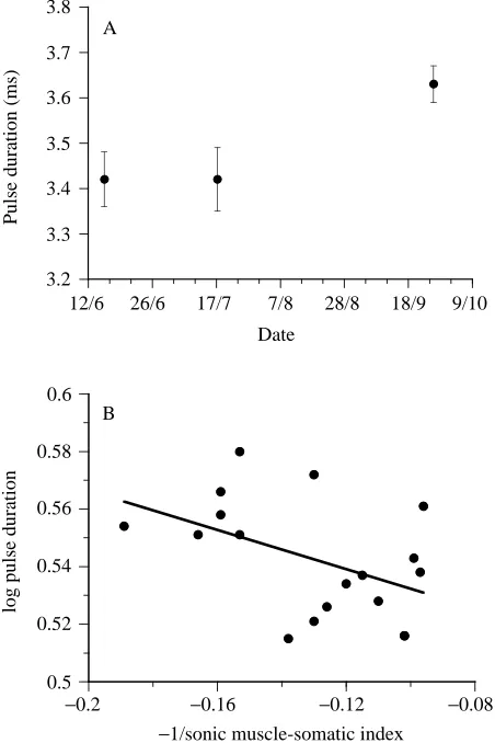
The effects of seasonal hypertrophy and atrophy on fiber morphology, metabolic substrate concentration and sound characteristics of the weakfish sonic muscle
Full text
Figure

Related documents
It was decided that with the presence of such significant red flag signs that she should undergo advanced imaging, in this case an MRI, that revealed an underlying malignancy, which
Twenty-five percent of our respondents listed unilateral hearing loss as an indication for BAHA im- plantation, and only 17% routinely offered this treatment to children with
amarus leaves aqueous extract to castor oil induced diarrhea rats, Charcoal meal test and.. PGE 2 induced enteropooling gastric motility could decrees in activity via
Additionally, our results performed that down- regulation of CXCR3A and up-regulation of CXCR3B brought about the increased expres- sions of PLCβ, MMP-1, and MMP-3 except
Panchakavya + Vermicompost, and sodium azide treated chilli varieties shows a great. influence at all the growth stages of vegetable
The objective is to investigate whether enriched enteral nutrition given shortly before, during and early after colo- rectal surgery, reduces POI and AL via stimulation of the
Note here the problem of the extension to the ultracold atomic collisions in quasi-2D geometry of confining traps [27] needed for resolving the “puzzle” of the 2D CIR [6],
A significant decline in the concentrations of renal reduced glutathione in comparison with the control group (C) were observed in the trained and untrained rats intoxicated with
